
Vitamin A
Vitamin A is not a single molecule but a metabolic hierarchy. Dietary retinol and topical retinoids require irreversible oxidative conversion to all-trans retinoic acid before becoming biologically active, and only ATRA binds the RAR nuclear receptors that drive the downstream gene regulation responsible for every claimed retinoid benefit. The epidermis expresses RARγ/RXRα predominantly in the suprabasal layers, RARα/RXRα in the basal layer, meaning retinoid effects vary with keratinocyte differentiation state. Crucially, ATRA’s effect on epidermal differentiation is not simply “increase cell turnover” – it simultaneously upregulates proliferation and suppresses terminal differentiation markers including filaggrin, which produces the characteristic initial barrier disruption that precedes long-term improvement.
Vitamin A encompasses a family of structurally related fat-soluble compounds – retinol (the dietary storage and transport form), retinaldehyde, all-trans retinoic acid (ATRA), and retinyl esters – collectively termed retinoids. In skin, vitamin A is not biologically active until converted to ATRA through a two-step, largely irreversible oxidative pathway. Retinol is oxidised to retinaldehyde by retinol dehydrogenase (RDH) or alcohol dehydrogenase (ADH); retinaldehyde is then oxidised irreversibly to retinoic acid by retinaldehyde dehydrogenase (RALDH). [2] This irreversibility matters. The conversion commits the molecule to its active form, and the resulting ATRA half-life in tissue is approximately one hour before CYP26 enzymes inactivate it. The skin therefore requires a continuous supply of precursor retinol to maintain active ATRA signalling.
The RAR/RXR Nuclear Receptor System
ATRA exerts the majority of its effects through two families of nuclear receptors: retinoic acid receptors (RARα, RARβ, RARγ) and retinoid X receptors (RXRα, RXRβ, RXRγ). These receptors function as heterodimers – RAR always pairs with RXR before binding to retinoic acid response elements (RAREs) in target gene promoters. In the absence of ATRA, RAR/RXR heterodimers bound to RAREs recruit histone deacetylase (HDAC) complexes that actively repress transcription. ATRA binding displaces the co-repressors and recruits co-activators, decompressing chromatin and driving targeted gene expression. [2]
The receptor subtype distribution in skin is architecturally specific. RARα/RXRα dominates in the basal layer of the epidermis and in dermal fibroblasts – the proliferative cells. RARγ/RXRα dominates in the suprabasal layers – the differentiating cells. [2] This stratified expression means retinoid effects are not uniform across the epidermis: the same ATRA signal produces different transcriptional outputs in basal keratinocytes and differentiating suprabasal cells simultaneously. ATRA regulates over 3,000 genes in keratinocytes – with measurable gene expression changes beginning as early as one hour after exposure – encompassing cell cycle regulation, adhesion, transcription factors, and membrane proteins. [2]
RXRα – the obligate heterodimer partner for RAR – is shared with the vitamin D receptor (VDR/RXRα), the thyroid hormone receptor, and the oestrogen receptor. In keratinocytes where retinoid and vitamin D signalling are simultaneously active, these pathways compete for the available RXRα pool. At clinical concentrations this competitive dynamic is not a documented adverse interaction, but it is a reason to ensure vitamin D adequacy as part of any retinoid protocol: a keratinocyte population with insufficient VDR/RXRα signalling has a compromised differentiation programme regardless of retinoid status. The two pathways are complementary in their outcomes, both driving keratinocyte differentiation and barrier competence, and competitive only in their shared molecular machinery. This interconnection is detailed further in the Vitamin D entity.
RXR also heterodimerises with the vitamin D receptor, the thyroid hormone receptor, and the oestrogen receptor, meaning retinoid signalling intersects directly with multiple other hormone pathways. In post-menopausal skin where oestrogen signalling has declined, the RXR pool available for RAR heterodimerisation changes, and retinoid signalling efficiency may be affected by the same hormonal shift that is driving other structural changes. This interaction is not yet fully characterised but is mechanistically plausible.
Epidermal Effects: Proliferation, Differentiation, and the Filaggrin Paradox
In the epidermis, ATRA drives two simultaneous processes that initially appear contradictory. It increases basal keratinocyte proliferation, thickening the viable epidermis and restoring the rete ridges that flatten with age. [2] It simultaneously suppresses the terminal differentiation programme in suprabasal cells, reducing expression of filaggrin (FLG), loricrin (LOR), transglutaminase-1 (TGM1), and other cornified envelope proteins. [1]
This is not a side effect, it is the mechanism by which retinoids accelerate cell transit time through the epidermis, producing the increased cell turnover that is central to their anti- photoageing effect. The paradox is this: accelerated turnover means earlier filaggrin expression relative to chronological time, but at any given moment, the differentiating cell population has been through its differentiation stages more rapidly and with somewhat suppressed terminal differentiation markers. The net effect observed in clinical studies is a thicker, more actively renewing epidermis with reduced photoageing markers, at the cost of a transiently compromised stratum corneum during adaptation. The “retinoid skin irritation” phase is the clinically visible expression of this suppressed terminal differentiation: a temporarily thinner, less lipid-rich stratum corneum before the increased cell turnover rate establishes a new, faster-cycling epidermal steady state.
Understanding this resolves the common clinical confusion about retinoid-associated barrier disruption. The disruption is not a failure of the treatment but a predictable consequence of the mechanism operating as intended. The adaptation period ends when the new epidermal cycling rate is established.
Dermal Effects: Collagen and MMP Suppression
In the dermis, ATRA’s effects operate through RARα in fibroblasts through two complementary pathways. It upregulates COL1A1 and COL3A1 gene expression, increasing Type I and Type III procollagen synthesis through the TGF-β/CTGF (connective tissue growth factor) pathway. It simultaneously suppresses MMP-1, MMP-3, and MMP-13 through downregulation of c-Jun – the AP-1 transcription factor subunit that drives collagenase gene expression in response to UV and mechanical stress. spandidos-publications.com The RAR specificity of this effect has been confirmed experimentally: RAR agonists reproduce both the procollagen synthesis increase and the MMP suppression; RXR agonists alone do not. spandidos-publications.com
ATRA also upregulates TIMP expression (tissue inhibitors of metalloproteinases) adding a further layer of collagen protection beyond direct AP-1 suppression. [4] The combined effect on the collagen balance – increased synthesis, reduced collagenase expression, increased collagenase inhibition – is why retinoids remain the most comprehensively evidenced topical anti-photoageing intervention after decades of clinical research.
CRABP-II (cellular retinoic acid binding protein II), the intracellular carrier that transports ATRA to the RAR in the nucleus, declines in expression in aged skin. CRABP-II knockout mouse models develop reduced collagen bundle density, reduced epidermal proliferation, and premature severe skin ageing, confirming that the intracellular delivery machinery, not just ATRA availability, is a component of the ageing-related decline in retinoid responsiveness. [2]
The Retinoid Conversion Hierarchy
Not all retinoids are equal, and the conversion hierarchy has practical clinical significance.
Retinoic acid (tretinoin/ATRA) requires no conversion, it binds RAR directly. It is approximately 20 times more potent than retinol at equivalent concentrations but produces proportionally higher irritation, requires prescription in many jurisdictions, and has a more rapid onset of the adaptation phase. [4]
Retinaldehyde requires one oxidation step to become ATRA. It is better tolerated than tretinoin, has antimicrobial properties relevant to acne presentations, and delivers meaningful dermal effects at cosmeceutical concentrations.
Retinol requires two oxidation steps. The majority of applied retinol is esterified and stored rather than converted, only a fraction proceeds to ATRA. At cosmeceutical concentrations (0.0015–0.3%), the effective ATRA dose delivered is substantially lower than equivalent tretinoin concentrations. Retinol is the most widely tolerated form, with the mildest adaptation phase and lowest irritation profile.
Retinyl esters (retinyl palmitate, retinyl acetate) require cleavage before conversion can begin. They are the most stable formulation option but the least potent, with the most attenuated downstream effects. [4]
Dietary Vitamin A and Skin
Dietary vitamin A is obtained as preformed retinol (from animal sources such as liver, eggs, dairy, grass-fed fat) and as provitamin A carotenoids, primarily β-carotene (from plant sources). Preformed retinol has an absorption efficiency of 75–100%; β-carotene conversion to retinol is variable, with a 3.6–28:1 ratio by weight depending on food matrix, dietary fat co-ingestion, and genetic variation in the BCMO1 enzyme responsible for cleavage. [3]
Grass-fed tallow and other traditional animal fats contain retinol and retinyl esters as part of their fat-soluble vitamin profile. Applied topically, retinyl palmitate from these sources is available for esterase-mediated cleavage and subsequent conversion – the same pathway as retinyl ester cosmeceuticals, though at lower, naturally variable concentrations. The skin benefit from traditional fats as a vitamin A vehicle is real but modest relative to formulated retinoid products, reflecting the conversion distance from retinyl ester to ATRA and the concentration differences involved.
Clinical Application
The clinical question Vitamin A creates is not whether to use retinoids – for clients with photoaged, hormonally depleted, or structurally thinning skin, the evidence is clear enough that the question is already answered. The question is which form, at what concentration, introduced at what stage of the client’s current skin state, and how managed through the adaptation phase.
That sequencing question matters because the mechanism that makes retinoids effective – accelerated epidermal turnover with transiently suppressed terminal differentiation – is also the mechanism that produces the adaptation phase. Introducing retinoids into a barrier that is already ceramide-depleted or inflamed adds the retinoid-driven differentiation suppression to an existing barrier deficit. The result is more severe and prolonged irritation, less tolerability, and clients who discontinue before the long-term benefit establishes.
Phase 1: Barrier Before Retinoids
For clients with a reactive, sensitised, or ceramide-depleted barrier – perimenopausal skin, post-treatment recovery, atopic-tendency – retinoids should enter the protocol only after barrier stability has been established. The baseline barrier competence determines how well the skin can tolerate the transient stratum corneum disruption that retinoid adaptation involves.
Niacinamide’s ceramide synthesis upregulation and barrier support is the most directly relevant preparation step. A client using a well-formulated niacinamide product for four to six weeks before introducing retinoids presents with a stronger baseline barrier – the transitional disruption of retinoid adaptation occurs against a more resilient stratum corneum, shortening the adaptation phase and improving tolerability.
For clients with active S. aureus-associated barrier disruption (as established in the Skin Microbiome entity), introducing retinoids while the dysbiosis loop is active compounds the disruption – retinoid-driven differentiation suppression on top of S. aureus-driven FLG suppression is a significant combined barrier challenge. The microbial environment should be addressed first.
Phase 2: Retinoid Introduction and Titration
Start-low-go-slow titration is not conservative caution. It is mechanistically appropriate, because the CYP26 negative feedback loop means that cells exposed to sustained high ATRA concentrations upregulate their own ATRA-inactivating enzymes, reducing effective intracellular ATRA over time. A gradual introduction establishes the epidermal adaptation more sustainably than high-concentration initiation.
For clients new to retinoids, retinol at 0.025–0.1% two to three nights per week, building to nightly over six to eight weeks, allows the epidermal cycling rate to adjust before the full frequency is established. For clients with established retinol tolerance seeking greater efficacy, retinaldehyde provides meaningfully higher ATRA conversion without the prescription requirement of tretinoin, and is worth considering before stepping directly to tretinoin.
Vitamin C in the morning routine pairs directly with the retinoid programme – as established in the Vitamin C entity, vitamin C’s hydroxylation cofactor and MMP-1 suppression mechanisms are complementary to retinoid-driven collagen synthesis. Together they address the synthesis side (retinoid TGF-β/CTGF upregulation), the cofactor side (vitamin C hydroxylation), and the degradation side (both retinoid AP-1/c-Jun suppression and vitamin C ROS scavenging) in a single morning-evening routine.
Retinoids in the Post-Treatment Context
RF microneedling and iPRF both generate a wound-healing collagen synthesis cascade with an associated transient inflammatory response. Introducing retinoids in the days immediately post-treatment adds differentiation suppression to an already inflamed and barrier-disrupted skin state. The appropriate sequencing is: continue retinoids up to three to five days before treatment, pause through the acute recovery phase (typically five to seven days), then resume – allowing retinoids to operate in the recovery and remodelling phase where their MMP suppression and collagen synthesis contributions are most valuable.
The goal retinoids are working toward, understood through their mechanism, is a dermis with genuinely higher collagen density and lower MMP activity, and an epidermis that is thicker, more rapidly renewed, and differentiating through a faster cycle. The adaptation phase is the cost of achieving that steady state. Clients who understand why it happens are more likely to persist through it.
References
Lee DD, Stojadinovic O, Krzyzanowska A, et al. (2009). Retinoid-responsive transcriptional changes in epidermal keratinocytes. J Cell Physiol, 220(2), 427-439 . doi.org/10.1002/jcp.21784
Szymański Ł, Skopek R, Palusińska M, et al. (2020). Retinoic Acid and Its Derivatives in Skin. Cells, 9(12) . doi.org/10.3390/cells9122660
Tang G (2010). Bioconversion of dietary provitamin A carotenoids to vitamin A in humans. Am J Clin Nutr, 91(5), 1468S-1473S . doi.org/10.3945/ajcn.2010.28674g
Zasada M, Budzisz E (2019). Retinoids: active molecules influencing skin structure formation in cosmetic and dermatological treatments. Postepy Dermatol Alergol, 36(4), 392-397 . doi.org/10.5114/ada.2019.87443
Biological Relationships
Influenced By
- this Required by Keratinocyte Evidence: Retinoids (Vitamin A derivatives) regulate keratinocyte differentiation via RAR nuclear receptors; retinoic acid upregulates loricrin and involucrin and modulates keratin expression. Rorke & Eckert Environ Health Perspect 1989 doi:10.1289/EHP.8980109
Learn More
This topic is discussed in 2 articles:
-
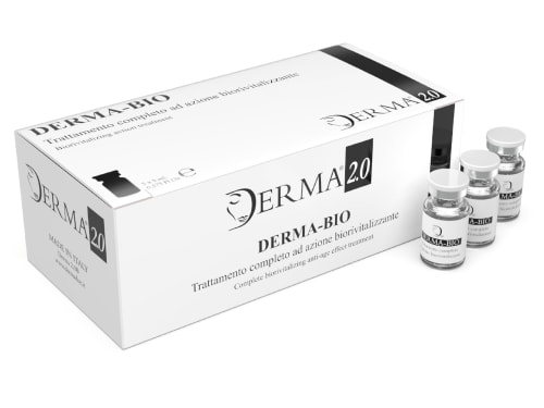
Discover the science behind Derma Bio, a mesotherapy product designed to combat signs of aging and achieve a brighter, smoother, and more youthful appearance.
-

Your 40s bring real hormonal shifts that affect your skin. Discover what’s actually happening and the skincare, nutrition, and treatments that support your skin through perimenopause.