
Epidermis
The epidermis is the skin’s outermost cellular layer, ranging from approximately 0.05mm on the eyelids to 1.5mm on the palms. For clinical and aesthetic purposes, it is best understood not as a static surface but as a continuous production system: a population of actively dividing basal keratinocytes at the base, progressively differentiating upward through distinct layers, eventually completing a terminal transformation into the corneocytes of the stratum corneum. The quality of that differentiation programme – how efficiently it runs, whether it has the lipid precursors and structural proteins it needs, and whether it is suppressed by inflammatory signalling – determines the quality of the barrier at the top.

The Differentiation Journey
Five distinct layers mark the stages of keratinocyte maturation. In the stratum basale, stem cells and basal keratinocytes divide and begin their upward migration. In the stratum spinosum, keratinocytes develop the intercellular bridges (desmosomes) that give skin its mechanical cohesion and begin synthesising structural keratins. In the stratum granulosum, the most metabolically active differentiation events occur: profilaggrin is stored in keratohyalin granules, lamellar bodies are loaded with ceramide precursors and lipid-processing enzymes, and the cell prepares for its final structural transformation. At the stratum granulosum–stratum corneum transition, lamellar bodies are secreted into the extracellular space, profilaggrin is cleaved into filaggrin monomers, and the cell loses its nucleus and organelles. What remains is the corneocyte – a flattened, keratin-filled disc enclosed in a cornified envelope – that forms the structural bricks of the stratum corneum. [6]
Every entity in the ceramide, filaggrin, and barrier lipid cluster of this knowledge base describes events that happen specifically during the granular layer stage and the granulosum-to-corneum transition. Disruption to that transition – whether from inflammatory cytokine activity, UV damage, or nutrient limitation – cascades into barrier deficiency at the surface.
Renewal Rate: Correcting the 28-Day Figure
The widely cited “28-day skin cycle” is an oversimplification that understates how significantly renewal rate declines with age. Keratinocytes complete their journey from basal layer to stratum corneum surface in approximately 40–56 days total. Of this, stratum corneum transit time in young adults is approximately 20 days; in older adults, this transit time lengthens by more than 10 days. Critically, the decline is not gradual or linear throughout adult life – data indicates the renewal rate remains relatively stable through early adulthood and then drops more sharply after the age of approximately 50. For perimenopausal and post-menopausal clients, this represents a biological convergence: the hormonal changes that reduce epidermal ceramide synthesis and filaggrin production arrive at the same time as the structural slowdown in the differentiation cycle itself.
This renewal rate matters clinically for two reasons. Retinoids and AHAs that accelerate the desquamation and renewal cycle are working with a system that already slows with age; their benefit in older skin relates partly to partially compensating for this natural deceleration. Equally, any treatment that disrupts the epidermis should be calibrated against how long recovery will actually take in a given client’s skin – an estimate that should be measured in weeks rather than days for clients over 50. [10]
The Second Barrier: Tight Junctions
A less commonly discussed barrier component exists within the living layers of the epidermis itself. The stratum granulosum contains a belt of tight junctions – protein complexes formed primarily by claudin-1, claudin-4, and occludin – that create a second permeability barrier below the stratum corneum. Claudin-1 null mutation studies confirmed that these junctions prevent the diffusion of molecular tracers toward the skin surface, and that claudin-1 deficiency allows this diffusion to proceed even when the stratum corneum appears structurally intact. Claudin-1 and occludin also play active roles in epidermal wound healing and regeneration, functioning beyond their structural barrier role in the recovery of damaged tissue. [2]
In inflammatory skin conditions including psoriasis and lichen planus, tight junction proteins are mislocalised from the granular to the spinous layer, disrupting their function even when their expression levels are preserved. This means the second barrier can fail in inflammatory skin conditions independently of stratum corneum integrity. Assessing barrier function purely through TEWL or stratum corneum lipid analysis may miss a component of the permeability failure in reactive and inflammatory presentations. [7]
Epidermal Thinning
Epidermal thickness declines measurably with chronological ageing through reduced keratinocyte proliferation, increased keratinocyte apoptosis, and changes in the calcium gradient that regulates proliferation in the basal and spinous layers. The hormonal dimension accelerates this structural thinning: post-menopausal skin shows a measurable 1.13% reduction in skin thickness per year after menopause alongside the lipid synthesis and structural protein changes described in the ceramides and collagen entities. Topical oestrogen has been shown to increase keratinocyte proliferation and epidermal thickness within two weeks of application in oestrogen-deficient skin, confirming that the receptor-mediated proliferation effect is direct and responsive rather than a slow structural adaptation. [8]
Clinically, epidermal thinning affects treatment selection in two directions. Thinned epidermis is more vulnerable to barrier disruption from active ingredients, physical treatments, and environmental stressors, which supports the conservative, layered approach to treatment in perimenopausal clients described elsewhere in this knowledge base. It also means some professional treatments that rely on an intact epidermal layer for the controlled wound healing response (fractional resurfacing in particular) require prior stabilisation of a thinned or fragile epidermis before optimal treatment depth and density can be applied.
Non-Keratinocyte Residents
Two non-keratinocyte cell populations in the epidermis have direct aesthetic and clinical relevance. Melanocytes, found in the basal layer at a ratio of approximately one per ten keratinocytes, produce melanin and transfer it to surrounding keratinocytes through dendritic processes. Their activity is regulated by UV exposure, hormonal signalling, and inflammatory mediators. This makes epidermal melanocyte behaviour central to post-inflammatory hyperpigmentation, melasma, and UV-driven pigmentation concerns. Langerhans cells, the resident antigen-presenting dendritic cells of the epidermis, perform immune surveillance and play a role in initiating and regulating the immune response to both external antigens and treatment-induced signals. Their density and activity are altered by UV exposure and inflammatory conditions, which partly explains why chronically UV-exposed skin has different immune characteristics than sun-protected skin at equivalent chronological age.
Clinical Application
The key distinction for epidermal treatments is between those that act at the epidermis – working on the keratinocyte population, the differentiation cycle, or the surface architecture directly – and those that act through the epidermis to reach their primary target in the dermis. This distinction matters because the epidermis is not a passive conduit; it responds to every treatment signal that passes through it, and understanding those responses determines how treatments are sequenced and combined.
Thulium Fractional Laser: Controlled Epidermal Renewal
Thulium 1927nm fractional laser is the most direct epidermal renewal treatment available. The 1927nm wavelength has a high affinity for water in the epidermis and superficial dermis, creating microscopic thermal zones (MTZs) that cover between 3% and 40% of the treatment area whilst leaving the surrounding tissue intact. This untreated tissue acts as a keratinocyte reservoir: neighbouring epidermal stem cells and basal keratinocytes migrate into the thermally treated zones and begin re-epithelialisation within 24 hours of treatment, completing epidermal recovery significantly faster than ablative resurfacing approaches that remove the entire surface. [5]
The clinical consequence of this mechanism is that thulium laser is simultaneously an epidermal disruption and an epidermal renewal stimulus. The wound-healing signal activates Wnt/β-catenin and TGF-β signalling in migrating keratinocytes, COL17A1 expression is preserved in the surviving follicular reservoir cells, and the differentiation cascade (profilaggrin production, lamellar body loading, lipid secretion) runs at its most active rate in the newly differentiating replacement keratinocytes. For aged, photodamaged, or perimenopausal skin where the renewal cycle has slowed and accumulated solar-damaged keratinocytes are producing poor-quality stratum corneum, this is the treatment most directly targeting the production system itself. It does not just resurface the skin, it resets the quality of the epidermal population generating the barrier. [11]
For pigmentation concerns, thulium’s epidermal precision makes it particularly effective: the MTZs directly target the superficial melanin deposits and accelerate the shedding of hyperpigmented cells, with the characteristic “coffee ground” appearance at 3–7 days reflecting the active clearance of fragmented melanin before it is shed in the renewal process. medicalphysics
LED Photobiomodulation: Driving Renewal from the Dermis Up
LED red light (630–660nm) contributes to epidermal renewal through a mechanism that runs in the opposite direction to most epidermal treatments. Rather than acting on keratinocytes directly, red light penetrates to the dermis and stimulates dermal fibroblasts via cytochrome c oxidase-mediated mitochondrial activation, increasing TGF-β and AKT signalling. The TGF-β stimulates collagen I production in fibroblasts; the accumulated collagen then activates integrin signalling in the overlying basal keratinocytes, which responds with increased proliferation and differentiation. The epidermis renews more actively because the dermal signal it depends on for differentiation cues has been strengthened. [1]
Research confirming that red light specifically accelerates recovery of the epidermal water-impermeable barrier (whilst blue light delays it) is particularly relevant for post-procedure protocols. Following any treatment that disrupts the epidermis, the renewal cycle driving barrier reconstruction is the process that determines recovery timeline and outcome quality. LED red light applied in the post-treatment period is not a passive adjunct; it actively accelerates the mitochondrial energy production and fibroblast-keratinocyte cross-talk that the renewal cycle depends on. [9]
Near-infrared (800–850nm) penetrates more deeply to the dermis and dermal-epidermal junction, while blue light (415nm) acts almost entirely within the epidermis. In the context of epidermal rejuvenation, the most practically relevant LED intervention is red light as a post-procedure recovery accelerant and as a standalone differentiation-normalising treatment for skin with slowed renewal.
Exosomes and Microneedling: Restoring the Epidermal Architecture
One of the structural changes in ageing skin that receives insufficient attention in aesthetic content is the flattening of the rete ridges – the interdigitating projections between the epidermis and dermis that increase dermo-epidermal surface area, facilitate nutrient and signal exchange, and help anchor the epidermis to its dermal substrate. As these flatten with age, the epidermis becomes more fragile, less well-nourished, and increasingly susceptible to the shear forces that cause easier bruising and skin tears.
Microneedling-assisted topical exosome therapy has demonstrated histological restoration of rete ridges alongside enhanced basal keratinocyte proliferation in aged skin biopsies. The mechanism is the combination of microneedling-created epidermal channels, which allow exosome cargo to reach the basal layer and dermo-epidermal junction, and the exosome miRNA and differentiation signals that activate the keratinocyte proliferation and matrix remodelling needed to rebuild the interdigitating architecture. Crucially, topically applied exosomes have been shown to be retained within the epidermis and superficial dermis for several hours post-application, providing a sustained interaction window for the regenerative cargo. [4]
Plant-derived exosome extracts have demonstrated restoration of SPTLC1 expression – the serine palmitoyltransferase subunit that initiates ceramide synthesis – in keratinocytes with inflammation-suppressed lipid metabolism, alongside reduced MMP-1 and increased procollagen expression. This makes exosome treatment, when delivered via microneedling channels, an intervention that simultaneously addresses epidermal architecture, ceramide synthesis capacity, and the collagen-mediated dermal signalling environment. No other single treatment combination addresses all three of these epidermal aging processes simultaneously. [3]
The note from the 2025 histological study warrants transparency: these findings apply specifically to microneedling-assisted topical delivery, not to injectable exosome formulations, which were not evaluated in the same study and whose epidermal effects cannot be inferred from these results. [4]
CAP, Polynucleotides, and Normalising the Epidermal Environment
Cold atmospheric plasma acts directly at the skin surface, with its RONS-mediated cytokine modulation operating primarily in the epidermis and superficial dermis. For the specific epidermal concern of acquired filaggrin suppression, CAP’s reduction of IL-4 and IL-13 at the tissue surface is the most anatomically targeted intervention. It does not stimulate the differentiation cycle directly, but it removes the cytokine signals actively suppressing it.
Polynucleotides, delivered intradermally, influence the epidermis through paracrine signalling: the macrophage reprogramming and fibroblast activation effects described in the collagen entity generate a dermal environment from which the overlying keratinocytes receive stronger differentiation and proliferation cues. Their epidermal benefit is indirect, but the dermal-epidermal cross-talk that red light also exploits means that improving the dermal signalling environment has genuine epidermal renewal consequences.
Treatment Pairings for Epidermal Rejuvenation
Thulium + LED (post-procedure): The most evidenced post-procedure pairing for epidermal renewal. Thulium creates the MTZ injury and activates the keratinocyte reservoir migration; post-procedure red LED accelerates the mitochondrial energy production and fibroblast-keratinocyte TGF-β signalling that completes the renewal cycle. Recovery timeline is shorter and differentiation quality is improved in the replacement keratinocytes compared with thulium alone.
Microneedling + exosomes (topical, same session): Channels created by microneedling allow exosome cargo to reach basal keratinocytes and the dermo-epidermal junction where rete ridge restoration occurs. This pairing addresses architectural renewal (rete ridges, basal proliferation) and biochemical renewal (SPTLC1/ceramide synthesis, MMP-1 reduction) simultaneously. The exosome application should follow immediately after needling whilst channels remain open.
CAP + exosomes: The inflammatory environment that CAP resolves at the tissue surface is the same environment that suppresses the differentiation programme exosomes are designed to restore. Sequencing CAP before exosome application – either in the same session or in a preparatory session – means exosome differentiation signals are delivered into keratinocytes whose FLG and SPTLC1 expression is no longer actively suppressed. For reactive, sensitised, or atopic-tendency skin, this sequencing addresses the epidermal renewal problem from both upstream and downstream simultaneously.
LED as a universal post-procedure adjunct: Across all injury-based epidermal treatments – thulium, microneedling, RF microneedling – post-procedure red LED reduces the inflammatory phase duration and accelerates the transition from inflammation to proliferation in the repair cascade. Given the slowed renewal rate in post-50 skin and the extended recovery timelines this implies, any intervention that accelerates the proliferative phase without compromising repair quality has practical value in this client group.
References
Chang H, Shen Q, Tan Y, et al. (2025). Red light promotes dermis-epidermis remodeling via TGFβ and AKT-mediated collagen dynamics in naturally aging mice. Zool Res, 46(5), 967-982 . doi.org/10.24272/j.issn.2095-8137.2024.405
Furuse M, Hata M, Furuse K, et al. (2002). Claudin-based tight junctions are crucial for the mammalian epidermal barrier: a lesson from claudin-1-deficient mice. J Cell Biol, 156(6), 1099-111 . doi.org/10.1083/jcb.200110122
Kim HR, Lee SH, Bae WB, et al. (2026). Skin Barrier Enhancement and Moisturizing Effects of Exosome Extracts Derived from Pinus densiflora, Zanthoxylum piperitum, and Lagerstroemia indica Plants. Biology (Basel), 15(3) . doi.org/10.3390/biology15030249
Lee YS (2025). Preliminary Histological Evidence of Epidermal and DEJ Remodeling with Microneedling-Assisted Topical Exosome Therapy: A Single-Subject Case Report. Clin Cosmet Investig Dermatol, 18, 2377-2385 . doi.org/10.2147/ccid.s542022
Lu K, Cai S (2022). Efficacy and safety comparison between 1927 nm thulium laser and 2940 nm Er:YAG laser in the treatment of facial atrophic acne scarring: a prospective, simultaneous spilt-face clinical trial. Lasers Med Sci, 37(3), 2025-2031 . doi.org/10.1007/s10103-021-03465-0
Pondeljak N, Lugović-Mihić L, Tomić L, et al. (2023). Key Factors in the Complex and Coordinated Network of Skin Keratinization: Their Significance and Involvement in Common Skin Conditions. Int J Mol Sci, 25(1) . doi.org/10.3390/ijms25010236
Pummi K, Malminen M, Aho H, et al. (2001). Epidermal tight junctions: ZO-1 and occludin are expressed in mature, developing, and affected skin and in vitro differentiating keratinocytes. J Invest Dermatol, 117(5), 1050-8 . doi.org/10.1046/j.0022-202x.2001.01493.x
Thornton MJ (2013). Estrogens and aging skin. Dermatoendocrinol, 5(2), 264-70 . doi.org/10.4161/derm.23872
Umino Y, Denda M (2023). Effect of red light on epidermal proliferation and mitochondrial activity. Skin Res Technol, 29(9), e13447 . doi.org/10.1111/srt.13447
Wang Z, Man MQ, Li T, et al. (2020). Aging-associated alterations in epidermal function and their clinical significance. Aging (Albany NY), 12(6), 5551-5565 . doi.org/10.18632/aging.102946
Wardhani PH, Rahmatika A, Thendria T, et al. (2025). Application of Thulium 1927-nm Laser for Androgenic Alopecia Treatment in Indonesian Patients. J Lasers Med Sci, 16, e42 . doi.org/10.34172/jlms.2025.42
Anatomical Relationships
Structural Connections
- Produces Ceramides Evidence: Entity text describes ceramide synthesis within epidermis via lamellar body secretion. Sakai & Hatano (2025) J Dermatol Sci 118:1 confirm SC ceramides as epidermal barrier biomarkers. doi:10.1016/j.jdermsci.2025.04.001
- Produces Filaggrin Evidence: Entity text: filaggrin suppression disrupts the protein scaffold of the outer epidermis; filaggrin is produced by keratinocytes in stratum granulosum. Epidermis is the producing structure.
- Produces Natural moisturising factor Evidence: Entity text: filaggrin processing produces NMF components; the epidermis is the source organ for NMF. Stratum corneum already produces NMF; epidermis as broader entity is a valid supplementary triple.
- Affects Transepidermal water loss Evidence: TEWL is determined by epidermal barrier integrity; lipid matrix degradation in the epidermis directly increases TEWL. Entity text: preventing desiccation is the primary barrier function.
- Connected to Hair follicle PMID: 29261946 Evidence: ORS is continuous with the interfollicular epidermis at the infundibulum
- Has sub-structure Keratinocyte Evidence: Keratinocytes comprise >90% of epidermal cells and are the structural unit of the epidermal barrier. Entity text: keratinocyte differentiation programme defines epidermal function.
- Has sub-structure Stratum corneum PMID: 29262154
- Has sub-structure Stratum granulosum Evidence: Stratum granulosum is a named sub-layer of the epidermis where profilaggrin-to-filaggrin processing occurs. Roop & Ishitsuka (2022) Antioxidants 11(1):47. PMC8772843
- Has sub-structure Tight Junction Evidence: Tight junctions form at the SG-SC interface and are a structural component of the epidermal barrier. Skin barrier dysfunction associatedAnatomy Tight junction already confirms relevance.
- Part of system Integumentary system Evidence: Epidermis is the outermost layer of skin, the primary organ of the integumentary system. Integumentary system comprisedOf Epidermis already exists; forward direction absent.
- Dermatitis Evidence: Dermatitis manifests primarily as epidermal inflammation and disrupted barrier; entity text discusses allergenic penetration through disrupted epidermis. Dermatitis -> associatedAnatomy -> Epidermis exists.
- Psoriasis Evidence: Psoriasis involves hyperproliferation of epidermal keratinocytes with abnormal differentiation. Zingkou et al. (2021) confirm abnormal epidermal differentiation in psoriasis – Int J Dev Biol 65(10):599. doi:10.1387/ijdb.210161gs
- Skin barrier dysfunction Evidence: Skin barrier dysfunction is intrinsically an epidermal condition; ceramide depletion, filaggrin suppression, and reduced keratinocyte renewal are the stated mechanisms in entity text.
- Cold Atmospheric Plasma therapy Evidence: Entity text: CAP acts at epidermal and papillary dermal level. Dilmaghanian et al. (2021) Sci Rep 11:21792 demonstrate increased epidermal thickness post-CAP. doi:10.1038/s41598-021-01341-z
- LED therapy Evidence: LED photobiomodulation reaches keratinocytes in epidermis; Naharro-Rodriguez et al. (2024) confirm LED-driven cellular effects on epidermal functions – Int J Mol Sci 25(8):4483. doi:10.3390/ijms25084483
- Thulium Fractional Laser Evidence: Entity text explicitly states thulium fractional laser acts at the epidermal and papillary dermal level as a professional treatment for skin quality.
Referenced in Conditions & Treatments
- this Contains Keratinocyte Evidence: Keratinocytes comprise approximately 90% of epidermal cells and are the structural and functional foundation of the skin barrier
- this Affected by Oestrogen decline Evidence: Oestrogen withdrawal reduces keratinocyte proliferation; epidermal thickness declines ~1.13%/year for 19 postmenopausal years; rete ridges flatten. PMC12374573.
- this Affected by Skin ageing Evidence: Skin ageing causes epidermal thinning and reduced keratinocyte turnover – explicitly described as a clinical sign of intrinsic ageing.
- this Connected to Dermis Evidence: The DEJ mediates the bidirectional signalling between dermis and epidermis
- this Connected to Hair follicle PMID: 29261946 Evidence: ORS is continuous with the interfollicular epidermis at the infundibulum
- this Part of Skin Evidence: Skin comprises three layers: epidermis, dermis, subcutaneous tissue.
- this Part of system Integumentary system PMID: 29262154
- this Associated condition Dermatitis Evidence: Atopic dermatitis barrier failure begins in the epidermis (filaggrin loss, tight junction disruption, keratinocyte-driven alarmin release); epidermal pathology is central to all three subtypes (PMC7215310).
- this Associated condition Perimenopausal skin changes Evidence: Epidermal thinning (~1.13%/year), slowed keratinocyte renewal, rete ridge flattening, and impaired wound healing are epidermal consequences of perimenopausal skin change. PMC12374573.
- this Associated condition Psoriasis Evidence: Psoriasis produces keratinocyte hyperproliferation reducing epidermal turnover to 3-4 days; epidermal thickening is the primary structural feature (PMC5796008).
- this Associated condition Skin barrier dysfunction Evidence: Epidermis is the broader anatomical context of barrier dysfunction; all three clinical stages are framed in terms of epidermal keratinocyte events. Entity text explicit.
- this Associated condition Topical steroid withdrawal Evidence: TSW mechanism centres on suppression of keratinocyte (epidermal) local cortisol synthesis via 11beta-HSD1 downregulation; the epidermis is the primary site of mechanistic dysfunction (PMC8481181).
Learn More
This topic is discussed in 7 articles:
-

The outer layer of skin containing the stratum corneum barrier
-
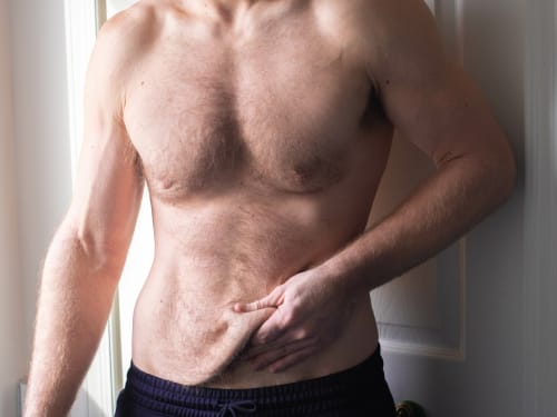
Don’t let loose skin overshadow your weight loss success. Learn why skin sags after significant weight loss and explore our advanced skin tightening and volume restoration treatments.
-

The skin’s outer cellular layer, containing both the stratum corneum barrier and the keratinocyte differentiation programme that builds it.
-

Getting adequate rest is the best investment you can make into your beauty. We take a look at some of your body’s natural skin rejuvenation processes that occur during sleep.
-
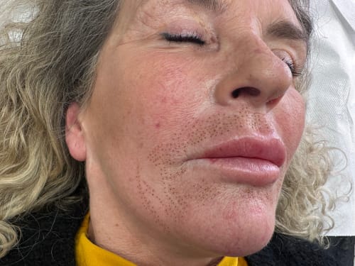
Find out how Plasma Fibroblast Therapy can rejuvenate your skin by stimulating collagen and elastin. Learn what areas can be treated, how the procedure works, and what to expect after treatment.
-

-
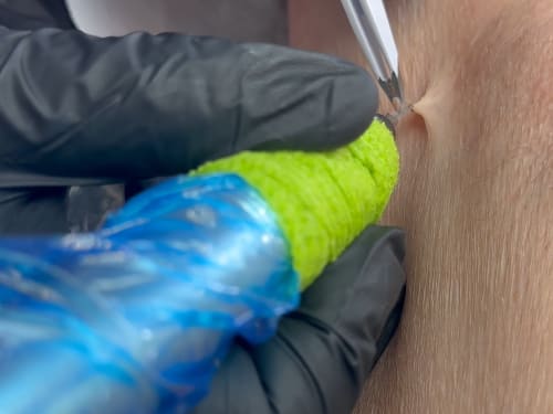
Not liking those pesky skin tags? Learn about causes, blemish removal options at Creative Touch, and prevention tips for smoother, bump-free skin.