
Collagen
Collagen provides the skin’s tensile scaffolding, but its clinical value lies in its structural organisation rather than simple quantity. Our focus is the balance between synthesis and MMP-driven degradation, specifically managing the Type I:III ratio which shifts during perimenopause. To achieve durable results, we must resolve the “maturation bottleneck”, ensuring that procollagen is successfully hydroxylated and cross-linked into functional fibrils rather than being degraded intracellularly.
Collagen is the most abundant protein in the human body and the primary structural component of the dermis, forming the fibrillar scaffold that gives skin its tensile strength, firmness, and capacity for resilience. In skin, the total collagen content decreases by approximately 1% per year from early adulthood, a decline that accelerates significantly with UV exposure, hormonal transition, and chronic stress. What makes this decline clinically interesting is that it does not operate through a single mechanism: reduced synthesis, increased enzymatic degradation, and impaired procollagen maturation are three independently occurring processes that can compound independently of each other. Understanding which is dominant in a given client determines which approach to collagen support is most relevant.
The Collagen Types in Skin
Human skin contains numerous collagen types, but two dominate the dermis. Collagen Type I comprises approximately 70% of the skin’s dry weight and provides the primary tensile strength. Collagen Type III makes up 8–11%, contributing elasticity and the capacity for tissue responsiveness. The adult skin maintains a Type I:III ratio of approximately 4:1, compared to the 1:1 ratio found in foetal and actively healing skin. This ratio is not simply a compositional detail: research on hybrid I/III collagen fibrils has shown that fibrils containing both types are significantly more effective at activating fibroblasts, stimulating cell polarisation, and driving new collagen synthesis than fibrils composed of either type alone. The ratio between these two collagens therefore feeds back into the capacity for collagen production – a lower Type III proportion progressively reduces the signalling that drives fibroblast activity. [2]
Collagen Type VII forms the anchoring fibrils that connect the basement membrane to the underlying dermis, maintaining the structural integrity of the dermo-epidermal junction. Its loss is particularly relevant in fragile, thinning, or photo-damaged skin where the junction’s mechanical stability is compromised. These less-discussed collagen types are where the conversation between barrier repair, skin thinning, and professional treatment effects becomes structurally precise. [5]
How Collagen Is Made
Collagen synthesis is a multi-step intracellular and extracellular process, each stage of which can independently become a limiting factor.
Fibroblasts transcribe procollagen mRNA, with TGF-β1 being the primary growth factor signal that drives procollagen gene expression. TGF-β1 signals through SMAD2 and SMAD3 transcription factors, which translocate to the nucleus and directly activate the COL1A1 and COL1A2 promoters driving Type I procollagen chain transcription – the same SMAD pathway that polynucleotides activate through the macrophage-mediated A2AR route, reaching fibroblast procollagen transcription through a paracrine mechanism when direct TGF-β responsiveness is reduced by senescence. Procollagen chains are then hydroxylated intracellularly: prolyl 4-hydroxylase and lysyl hydroxylase modify specific proline and lysine residues, a process that requires vitamin C as an obligate cofactor for enzyme activity. Without adequate hydroxylation, procollagen chains fail to form the stable triple helix structure and are instead degraded intracellularly before secretion. Vitamin C depletion does not simply reduce collagen synthesis speed; it produces structurally defective collagen that is degraded before it contributes to the extracellular matrix at all. Beyond its cofactor role, ascorbic acid also directly stimulates procollagen mRNA expression in fibroblasts, making it active at two independent points in the synthesis pathway. [1]
Once secreted, procollagen is cleaved to tropocollagen by specific extracellular proteinases – ADAMTS-2 and ADAMTS-3 remove the N-terminal propeptide; BMP-1 and tolloid-family enzymes cleave the C-terminal propeptide – yielding mature tropocollagen that self-assembles into fibrils. The cleaved propeptides do not simply disperse: the C-propeptide circulates systemically and acts as an autoregulatory feedback inhibitor of further procollagen synthesis in fibroblasts, providing a self-limiting mechanism that prevents runaway collagen overproduction under sustained TGF-β drive. Clinically, circulating procollagen C-propeptide (PICP) is a usable biomarker of fibroblast synthetic activity – and the feedback mechanism is a useful counterpoint to the assumption that maximal TGF-β stimulation produces maximal collagen output.
Cross-linking is catalysed by lysyl oxidase (LOX), a copper-dependent amine oxidase secreted by fibroblasts into the extracellular space, where it drives the oxidative deamination of lysine and hydroxylysine residues on adjacent tropocollagen molecules – generating the covalent aldehyde crosslinks that lock mature fibrils into mechanically stable structures. LOX activity is absolutely dependent on dietary copper, and deficiency produces connective tissue fragility equivalent to impaired cross-linking by any other mechanism. LOX is additionally suppressed by elevated homocysteine – the amino acid that accumulates with insufficient B12, B6, or folate – providing a specific nutritional pathway to impaired collagen maturation entirely independent of the synthesis and hydroxylation steps. This extracellular maturation step is the one most often overlooked in conversations about collagen support – a treatment that increases procollagen synthesis but does not support the maturation and cross-linking steps produces less functional extracellular collagen than the gene expression data alone would suggest.
Why Collagen Declines
Chronological Ageing
Fibroblasts produce less TGF-β1 over time, progressively reducing the signal for procollagen transcription. Alongside this, MMP activity increases with age independently of UV exposure, meaning basal collagen turnover gradually shifts toward net loss. The Type I:III ratio typically shifts toward higher Type I dominance as Type III production is disproportionately reduced, further impairing the fibroblast activation feedback loop described above. [1]
UV Radiation
UV exposure operates through two parallel mechanisms. A single minimal erythema dose (2 MED) produces near-complete suppression of procollagen synthesis that persists for 24 hours, mediated by TGF-β pathway impairment. Simultaneously, UV exposure activates AP-1 transcription through reactive oxygen species, which drives MMP-1, MMP-3, and MMP-9 upregulation. MMP-1 cleaves fibrillar collagen Types I and III at a single site within the triple helix; MMP-3 and MMP-9 then degrade the cleaved fragments. Chronic UV exposure therefore creates compounding fragmented collagen accumulation alongside progressive synthesis suppression. Which is why photoaged skin feels and behaves structurally differently from intrinsically aged skin of equivalent chronological age. [9]
Hormonal Transition
The perimenopause mechanism is more nuanced than the commonly cited statistic of “30% collagen loss in five years” suggests. Primary fibroblast research using in vitro menopausal hormone conditions produced a counterintuitive finding: procollagen gene expression actually increased under menopausal conditions in the short term, representing a compensatory fibroblast response. However, this increased synthesis was insufficient to prevent net collagen loss because of simultaneous upregulation of MMP-1 and MMP-3 activity, and because of impaired procollagen protein maturation downstream of the transcription stage. The problem in menopausal skin is not primarily that fibroblasts stop trying to make collagen; it is that the degradation and maturation environment changes such that what they make is increasingly lost before contributing to the structural matrix. [10]
This distinction is clinically important. Treatments that increase procollagen mRNA expression alone are working on a step that is already being compensated for. Approaches that simultaneously reduce MMP activity, support the maturation pathway, and restore the Type III/Type I balance are more comprehensively addressing what is actually failing.
For clients where fibroblast senescence is the prominent limiting factor – where the fibroblast population has genuinely reduced capacity rather than simply reduced stimulation – PLLA addresses this at a more fundamental level than treatments that provide a synthesis signal alone. Its direct PI3K/AKT activation suppresses the p53/p21 senescence axis in fibroblasts, partially restoring their synthetic capacity rather than asking cells with diminished function to perform more. [7] For the post-menopausal presentation where previous stimulation treatments have underdelivered, this mechanism distinction is worth considering in treatment planning.
Chronic Stress
Cortisol reduces fibroblast activity and impairs TGF-β1 signalling directly, suppressing both procollagen transcription and the growth factor environment that sustains fibroblast viability. The same HPA axis elevation that suppresses cholesterol and ceramide synthesis in the stratum corneum is simultaneously reducing the dermal collagen production rate. Stress-related skin changes operate at both the epidermal barrier and the dermal structural layers concurrently.
Vitamin C: Two Mechanisms, One Priority
Given collagen synthesis’s absolute dependency on vitamin C at the hydroxylation step, and vitamin C’s direct stimulation of procollagen mRNA independently of that cofactor role, it occupies a unique position among topical actives. Topical vitamin C at stable, bioavailable concentrations reaches the dermis sufficiently to support both functions. It also reduces the ROS-driven AP-1 signalling that upregulates MMPs, giving it a simultaneous synthesis-supporting and degradation-reducing action. For clients whose collagen support strategy includes a single topical active alongside professional treatments, vitamin C addresses the mechanism more completely than any other cosmeceutical. [8]
Retinoids and Collagen Remodelling
Retinoids support collagen production through a complementary but distinct mechanism: they upregulate TGF-β1 and reduce MMP expression, working on the growth factor signalling environment rather than the synthesis machinery directly. Long-term retinoid use consistently increases measurable dermal collagen density and improves the Type I:III ratio by supporting the fibroblast activation environment. The combination of retinoids and vitamin C addresses the TGF-β/MMP regulatory level and the procollagen hydroxylation/maturation level simultaneously, which is why this pairing is so consistently effective in collagen-focused homecare protocols.
Clinical Application
The framework that makes most sense for collagen treatments is not the synthesis-versus-inflammatory-removal distinction used for barrier lipids. For collagen, the clinically meaningful separation is between treatments that address synthesis only, treatments that address both synthesis and MMP degradation simultaneously, and treatments that work through the structural mechanics of the dermal environment rather than the cellular signalling pathways directly.
This distinction matters particularly for the perimenopausal and photo-damaged presentations where – as the entity description establishes – the primary failure is not that fibroblasts have stopped trying to make collagen, but that increased MMP activity is degrading it faster than the maturation pathway can complete. A treatment that increases procollagen mRNA without reducing MMP activity is accelerating synthesis into a still-unfavourable degradation environment.
iPRF: The Most Complete Collagen Signal
Injectable platelet-rich fibrin (iPRF) is the treatment whose mechanism most directly matches what the collagen entity describes as the core perimenopause and photo-ageing problem. In UV-irradiated human dermal fibroblast research, iPRF demonstrated stronger activation of the TGF-β/Smad signalling pathway than PRP – driving procollagen mRNA upregulation – whilst simultaneously producing greater suppression of MMP-1 expression. The same paper found iPRF reduced ROS generation more effectively, addressing one of the primary upstream drivers of AP-1-mediated MMP transcription. This dual action on synthesis and degradation is clinically distinct from treatments that operate on only one side of the equation. [6]
The fibroblast response to iPRF also demonstrates quantitative advantages over PRP for collagen synthesis specifically. Dermal fibroblast research comparing fluid PRF and PRP directly found that TGF-β, collagen I, and fibronectin mRNA levels were all significantly highest in the iPRF group, with fibroblast migration over 350% greater than control and 200% greater than PRP, and measurably superior collagen matrix synthesis across the study period. For clients where MMP-driven degradation is the dominant mechanism, iPRF’s ability to address both ends of the collagen balance makes it the most mechanistically targeted treatment in our portfolio. [12]
Polynucleotides: Collagen Synthesis Through the Macrophage Axis
Polynucleotides influence collagen production through a pathway that is distinct from every other treatment here and particularly relevant for older or post-menopausal skin. Their mechanism operates through macrophage reprogramming rather than direct fibroblast stimulation: polynucleotide treatment activates the A2AR/AC/PKA/CREB/PCK1 cascade in macrophages, increasing M2 macrophage polarisation and subsequent secretion of IL-10 and TGF-β. These cytokines then activate SMAD2 and STAT3 in adjacent fibroblasts, simultaneously increasing collagen I and III synthesis and inhibiting NF-κB-mediated collagen degradation. [3]
In senescent fibroblasts specifically – cells that have reduced responsiveness to direct TGF-β stimulation – this indirect pathway through macrophage reprogramming restores collagen synthesis that direct approaches may struggle to replicate. For the perimenopausal and post-menopausal presentation, where cellular senescence in the dermis is part of the mechanism of decline, polynucleotides are not simply an anti-inflammatory treatment but an active collagen restoration strategy working through a route that ages less poorly than direct fibroblast stimulation alone. The simultaneous NF-κB suppression also addresses MMP-driven degradation, giving polynucleotides the same dual synthesis-and-preservation advantage as iPRF, through a completely different molecular pathway. [3]
Microneedling and RF Microneedling: Controlled Injury, Two Collagen Signals
Standard microneedling creates a wound-healing cascade whose primary collagen signal is TGF-β1 released from platelets and activated fibroblasts at the injury site, driving procollagen I and III synthesis over the following weeks. RF microneedling adds a second, distinct signal: the controlled thermal injury from radiofrequency energy at the needle tips creates localised collagen denaturation in the mid-to-deep dermis, which triggers a heat-shock protein response and a separate wave of fibroblast activation and collagen remodelling that operates on a longer timeline than the needling-driven cascade alone. [4]
The practical consequence is that RF microneedling produces two temporally separated collagen responses: the needling-driven synthesis peak at approximately 2–4 weeks, and the RF-thermal remodelling peak extending to 3–6 months. For clients where dermal thinning and loss of structural depth are primary concerns alongside surface texture, the deeper thermal remodelling that RF energy generates reaches tissue layers that needling-only treatments do not address as effectively.
Neither standard nor RF microneedling has the MMP-suppressive dual action of iPRF or polynucleotides. Their mechanism is synthesis-primary. For perimenopausal clients in whom MMP-driven degradation is significant, combining microneedling with iPRF or polynucleotides in the same treatment session addresses both sides of the collagen equation more completely than either modality alone.
Hyaluronic Acid Skin Boosters: Structural TGF-β Activation
HA skin boosters influence collagen through a mechanism that is structurally mechanical rather than growth-factor or wound-healing driven. Injected cross-linked hyaluronic acid creates a physical volume and structural support in the dermis that changes the mechanical environment experienced by surrounding fibroblasts. Within one week of CL-HA injection, fibroblasts stretch and elongate in response to this structural support, with the mechanical force activating TGF-β signalling through mechanoreception rather than direct growth factor delivery. The result is new Type I procollagen synthesis that is converted to intact collagen bundles, detectable at four weeks and still measurable at twelve months in biopsied tissue. [11]
This is a clinically distinct signal from any other treatment in this group. The fibroblast activation is maintained for six to nine months after injection, driven not by the presence of growth factors but by the ongoing structural support the HA provides whilst it remains in the tissue. It also makes HA boosters particularly appropriate for photoaged skin, where TGF-β signalling is specifically reduced – the mechanical activation pathway bypasses the impaired signalling environment that limits the response to direct TGF-β stimulation.
Matching Treatment to Mechanism
| Treatment | Collagen synthesis signal | MMP suppression | Relevant presentation |
|---|---|---|---|
| iPRF | TGF-β/Smad, PDGF direct | Yes – MMP-1 suppression | Photo-ageing, perimenopause, UV-damaged skin |
| Polynucleotides | Indirect via macrophage TGF-β/IL-10 | Yes – NF-κB inhibition | Senescent/perimenopausal fibroblast environment |
| RF microneedling | TGF-β (needling) + thermal remodelling | Partial, via NF-κB | Dermal thinning, structural depth loss |
| Microneedling | TGF-β (needling cascade) | Minimal | Surface texture, early decline |
| HA skin boosters | Mechanical TGF-β activation | Minimal direct | Photoaged skin, structural support deficit |
| PLLA | Indirect: M2 macrophage → TGF-β/SMAD2/3 + direct PI3K/AKT fibroblast activation | Yes – TIMP-1/2 upregulation reducing MMP activity | Significant structural decline; post-menopausal; senescent fibroblast environment; long-horizon presentations |
For most collagen-focused presentations at Creative Touch, the highest-yield approach combines a dual-action treatment (iPRF or polynucleotides) with a synthesis-stimulating modality (microneedling or RF microneedling) in a protocol designed around the client’s specific pattern of decline – which is determined as much by the hormonal and inflammatory context as by the visible surface presentation.
References
Boo YC (2022). Ascorbic Acid (Vitamin C) as a Cosmeceutical to Increase Dermal Collagen for Skin Antiaging Purposes: Emerging Combination Therapies. Antioxidants (Basel), 11(9) . doi.org/10.3390/antiox11091663
Boraldi F, Lofaro FD, Bonacorsi S, et al. (2024). The Role of Fibroblasts in Skin Homeostasis and Repair. Biomedicines, 12(7) . doi.org/10.3390/biomedicines12071586
Byun KA, Park HJ, Oh S, et al. (2025). Polynucleotides Enhance Collagen Synthesis via Modulating Phosphoenolpyruvate Carboxykinase 1 in Senescent Macrophages: Experimental Evidence. Int J Mol Sci, 26(17) . doi.org/10.3390/ijms26178720
Chandra S, Mysore V, Shah S, et al. (2024). Physics of fractional microneedle radiofrequency – A review. J Cutan Aesthet Surg, 17(3), 177-183 . doi.org/10.25259/jcas_98_23
Conradt G, Hausser I, Nyström A (2024). Epidermal or Dermal Collagen VII Is Sufficient for Skin Integrity: Insights to Anchoring Fibril Homeostasis. J Invest Dermatol, 144(6), 1301-1310.e7 . doi.org/10.1016/j.jid.2023.11.003
Li Y, Song P, He J, et al. (2022). Comparison Between Injectable Platelet-rich Fibrin and Platelet-rich Plasma in Ameliorating UVA-induced Photoaging in Human Dermal Fibroblasts via the Activation of TGF-β/Smad Signaling Pathway. Photochem Photobiol, 98(6), 1395-1401 . doi.org/10.1111/php.13628
Oh S, Lee JH, Kim HM, et al. (2023). Poly-L-Lactic Acid Fillers Improved Dermal Collagen Synthesis by Modulating M2 Macrophage Polarization in Aged Animal Skin. Cells, 12(9) . doi.org/10.3390/cells12091320
Pullar JM, Carr AC, Vissers MCM (2017). The Roles of Vitamin C in Skin Health. Nutrients, 9(8) . doi.org/10.3390/nu9080866
Quan T, Qin Z, Xia W, et al. (2009). Matrix-degrading metalloproteinases in photoaging. J Investig Dermatol Symp Proc, 14(1), 20-4 . doi.org/10.1038/jidsymp.2009.8
Remoué N, Molinari J, Andres E, et al. (2013). Development of an in vitro model of menopause using primary human dermal fibroblasts. Int J Cosmet Sci, 35(6), 546-54 . doi.org/10.1111/ics.12075
Wang F, Do TT, Smith N, et al. (2024). Implications for cumulative and prolonged clinical improvement induced by cross-linked hyaluronic acid: An in vivo biochemical/microscopic study in humans. Exp Dermatol, 33(1), e14998 . doi.org/10.1111/exd.14998
Wang X, Yang Y, Zhang Y, et al. (2019). Fluid platelet-rich fibrin stimulates greater dermal skin fibroblast cell migration, proliferation, and collagen synthesis when compared to platelet-rich plasma. J Cosmet Dermatol, 18(6), 2004-2010 . doi.org/10.1111/jocd.12955
Biological Relationships
Biological Interactions
- Located in Dermis Evidence: Collagen is the primary structural component of the dermis; ~70% of skin dry weight. Type I and III fibres form the fibrillar scaffold that gives skin tensile strength and resilience.
- Affects Fibroblast Evidence: Hybrid I/III collagen fibrils activate fibroblasts via integrin-mediated mechanosensing; Type I:III ratio feeds back into fibroblast synthetic activity (PMC5842211; entity full_description).
- Affects Glycation-related skin changes Evidence: Dermal collagen is the primary substrate for AGE formation in skin; AGE-modified collagen directly drives the structural and cellular changes defining glycation-related skin ageing (PMC9655929).
- Affects Perimenopausal skin changes Evidence: Perimenopause shifts MMP/TIMP balance and impairs procollagen maturation; net collagen loss despite compensatory synthesis increase is the primary menopausal skin structural change (Wiley doi:10.1111/ics.12075).
- Affects Skin ageing Evidence: Collagen content declines ~1%/year from early adulthood; fragmentation by MMP-1 is a key driver of age-related skin functional decline. All three failure modes (synthesis, degradation, maturation) compound independently.
- Requires Vitamin C Evidence: Vitamin C is obligate cofactor for prolyl 4-hydroxylase and lysyl hydroxylase; depletion produces structurally defective procollagen degraded intracellularly before secretion (PMC9495646).
Influenced By
- this Stimulated by Cold Atmospheric Plasma therapy
- this Stimulated by Injectable Platelet-Rich Fibrin Evidence: iPRF activates TGF-beta/Smad pathway more strongly than PRP, drives procollagen mRNA upregulation, suppresses MMP-1, and produces fibroblast migration >350% greater than control; dual synthesis+degradation action (Wiley doi:10.1111/php.13628).
- this Stimulated by Interleukin-13 Evidence: IL-13 induces type I collagen transcription in human dermal fibroblasts via STAT6 dose-dependently. Ihn et al. 2004 JBC doi:10.1074/JBC.M406951200
- this Stimulated by Interleukin-4 Evidence: IL-4 promotes profibrotic collagen synthesis in fibroblasts via STAT6/Smad pathways in dermal fibrosis contexts. Mamalis et al. 2019 Arch Dermatol Res doi:10.1007/s00403-019-01972-3
- this Stimulated by Microneedling Evidence: Microneedling wound cascade releases platelet/fibroblast TGF-beta1, driving procollagen I and III synthesis over subsequent weeks; collagen response documented in biopsy studies (PMC11497551; Collagen entity).
- this Stimulated by Oestrogen
- this Stimulated by Peptides
- this Stimulated by Platelet-derived growth factor Evidence: PDGF+IGF-1 synergistically stimulate connective tissue/collagen deposition via TGF-beta1 cascade (PDGF brings workforce; TGF-beta activates collagen synthesis). PMC1886289 Pierce 1991; PMC2115493 Pierce 1989.
- this Stimulated by Poly-L-Lactic Acid Evidence: PLLA microspheres activate PI3K/AKT to suppress p53/p21 senescence axis in fibroblasts, restoring synthetic capacity; M2 macrophage-mediated TGF-beta/SMAD2/3 pathway provides indirect collagen synthesis signal (PMC10177436).
- this Stimulated by Radiofrequency Microneedling Evidence: RF thermal injury creates controlled collagen denaturation triggering heat-shock protein response and a second fibroblast collagen remodelling wave extending to 3-6 months post-treatment (PMC11497551; Collagen entity).
- this Stimulated by Retinoid Evidence: Retinoids upregulate TGF-beta1 and reduce MMP expression; long-term use increases measurable dermal collagen density and improves Type I:III ratio via fibroblast activation environment restoration (Collagen entity clinical_context_summary).
- this Stimulated by Transforming growth factor beta Evidence: TGF-beta1 drives procollagen transcription via Smad2/Smad3 activation of COL1A1 and COL1A2 promoters; the primary upstream signal for fibroblast collagen synthesis (PMC9495646; entity full_description).
- this Stimulated by Vitamin C Evidence: Ascorbic acid directly stimulates procollagen mRNA expression in fibroblasts AND serves as obligate cofactor for hydroxylation; acts at two independent synthesis pathway points (PMC5579659; entity full_description).
- this Inhibited by Cortisol
- this Inhibited by Interleukin-6 Evidence: Chronic IL-6 trans-signalling via SASP drives MMP upregulation and impairs fibroblast collagen synthesis in aged skin. Yu et al. 2023 Aging Cell doi:10.1111/acel.14054
- this Inhibited by Matrix metalloproteinase
- this Inhibited by Oestrogen decline Evidence: Post-menopausal skin loses up to 30% collagen in first 5 years; Type I and III collagen both decline with Type I:III ratio shift accelerating fibroblast senescence. PMC12374573; PMC10991793.
- this Inhibited by Tumour necrosis factor Evidence: TNF-α drives type I collagen degradation via MMP-1/MMP-3 upregulation; net collagen turnover weighted toward degradation. PMID:25457675
- this Produced by Dermis Evidence: Fibroblast, responsible for synthesising… collagen Types I, III, and V
- this Produced by Fibroblast PMID: 34297930 Evidence: Procollagen I and III are transcribed in response to TGF-β1 signalling and secreted into the extracellular space for maturation and cross-linking
- this Affected by Glycation Evidence: AGE accumulation on dermal collagen cross-links alter fibril mechanical properties, cause stiffness and roughness, shorten fibrils, and impair MMP accessibility and fibroblast signalling (PMC10707495; PMC9655929).
- this Affected by Hallmarks of ageing Evidence: Multiple hallmarks – mitochondrial dysfunction, cellular senescence, genomic instability, deregulated nutrient sensing – converge to impair fibroblast collagen synthesis; collagen declines ~1%/year via hallmarks mechanisms (PMC10676801).
- this Affected by Inflammageing Evidence: Inflammageing-derived TNF-alpha and IL-6 activate NF-kappaB, upregulating MMP-1, MMP-3, and MMP-9, which degrade dermal collagen; SASP-driven chronic MMP activation causes progressive collagen fragmentation (PMC10178737).
- this Required by Tissue regeneration Evidence: Collagen synthesis by fibroblasts is the primary structural output of tissue regeneration; Type I:III ratio determines regenerative vs fibrotic outcome; inadequate collagen produces non-functional granulation tissue (PMC3663196; PMC5831781).
Learn More
This topic is discussed in 22 articles:
-

Discover the detailed science behind PRP and iPRF therapies. Learn how growth factors, platelets, and cellular mechanisms drive hair restoration and skin rejuvenation at a molecular level.
-

Don’t let loose skin overshadow your weight loss success. Learn why skin sags after significant weight loss and explore our advanced skin tightening and volume restoration treatments.
-

Primary structural protein in dermis. Women experience approximately 30% loss in first five years after menopause. Collagen supplements have mixed evidence with high-quality studies showing no significant effect.
-
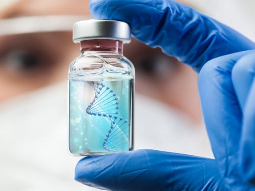
The science behind polynucleotides – a revolutionary treatment that offers a comprehensive approach to skin rejuvenation. How does it compare?
-

Structural protein providing skin firmness and elasticity, production directly regulated by oestrogen levels
-
-

Getting adequate rest is the best investment you can make into your beauty. We take a look at some of your body’s natural skin rejuvenation processes that occur during sleep.
-
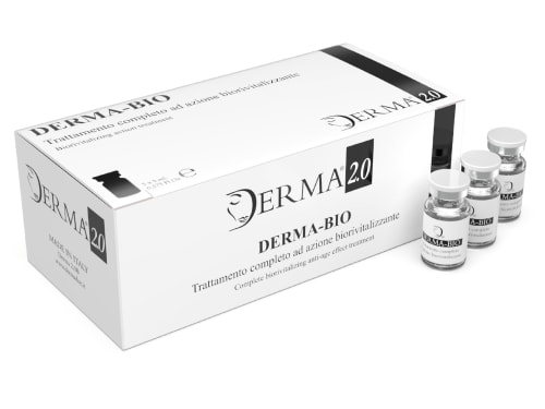
Discover the science behind Derma Bio, a mesotherapy product designed to combat signs of aging and achieve a brighter, smoother, and more youthful appearance.
-

Elevate your autumn skincare routine with our expert advice and innovative treatments: achieve a radiant complexion as the seasons change and the leaves fall.
-
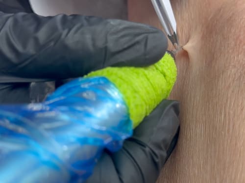
Not liking those pesky skin tags? Learn about causes, blemish removal options at Creative Touch, and prevention tips for smoother, bump-free skin.