
Oestrogen
Oestrogen is a systemic steroid hormone and a primary regulator of skin homeostasis across all compartments. Operating through nuclear receptors (ERα and ERβ) in both fibroblasts and keratinocytes, it sustains collagen synthesis, inhibits Matrix Metalloproteinase (MMP) degradation, and maintains the hyaluronic acid ground substance. Critically, oestrogen withdrawal at menopause triggers a multi-system failure: a 30% loss of dermal collagen in five years, epidermal thinning, and a specific impairment of ceramide hydrolysis. In the clinic, post-menopausal skin is defined by a “maturation deficit” where the primary issue is not necessarily a lack of production signals, but a failure in protein maturation and lipid processing that requires direct substrate supply and enzymatic support.
Oestrogen is not a skin hormone in the narrow sense, it is a systemic steroid hormone whose skin effects are extensive and well-characterised because skin is a highly oestrogen-sensitive organ. Dermal fibroblasts express both oestrogen receptor alpha (ERα) and oestrogen receptor beta (ERβ), with ERβ expressed at higher levels in female skin fibroblasts. [2] Epidermal keratinocytes also express oestrogen receptors, giving oestrogen direct regulatory access to both the dermal structural compartment and the epidermal barrier production chain simultaneously. The skin effects of oestrogen are not mediated through a single downstream pathway; they operate through several independent mechanisms in parallel, which is why the decline at menopause produces changes across multiple aspects of skin biology at once rather than through one primary route.
The Oestrogen Receptor (ER) Map
| Cell Target | Primary Receptor | Biological Effect | Result of Withdrawal |
|---|---|---|---|
| Fibroblast | ERβ (Dominant) | Stimulates Collagen I & III; suppresses MMP-1. | Net collagen loss; fragmented dermal matrix. |
| Keratinocyte | ERα / ERβ | Drives proliferation and EGF signaling. | Epidermal thinning; slower transit time. |
| Corneocyte | Metabolic Action | Regulates ceramide hydrolysis enzymes. | Brittle barrier; elevated precursor lipids. |
| Macrophage | ERα | Suppresses MIF (Pro-inflammatory cytokine). | Prolonged healing; chronic low-grade inflammation. |
Oestrogen and Collagen
The collagen effect is the most consistently cited and quantitatively best-characterised oestrogen–skin relationship. Type I and III dermal collagen decreases by up to 30% in the first five years after menopause, with an ongoing loss of approximately 2% per year thereafter; skin thickness decreases by approximately 1.1% per year. [6] The mechanism operates at the level of fibroblast ERβ – oestradiol upregulates collagen synthesis in fibroblasts through ERβ-mediated pathways, and ERβ-deficient fibroblasts lose responsiveness to oestradiol’s stimulatory collagen effects despite higher baseline collagen production. [3] Oestrogen also suppresses MMP-1 expression in fibroblasts, reducing collagen degradation alongside its synthesis-stimulating role – meaning that oestrogen withdrawal affects both sides of the collagen balance simultaneously. [5]
The clinical precision that matters here – and that the Collagen entity establishes in detail – is that menopausal fibroblasts increase procollagen gene transcription as a compensatory response, yet net collagen still declines because of simultaneous MMP upregulation and impaired procollagen maturation. Oestrogen withdrawal does not simply turn off collagen production. It shifts the balance of an already-complex multi-step process in ways that require treatment strategies addressing multiple points in the pathway rather than transcription alone.
Oestrogen and Ceramide Production
Research published in 2022 established a specific and previously underappreciated oestrogen–ceramide relationship at the stratum corneum level. Analysis of stratum corneum lipids from pre-menopausal, post-menopausal, and HRT-treated women found significantly reduced total ceramide levels post-menopause, with HRT normalising ceramide profiles toward pre-menopausal values. Critically, post-menopausal women showed elevated sphingomyelin levels – a ceramide precursor – suggesting the deficit sits at the hydrolysis step rather than upstream synthesis. [10] Oestradiol treatment of primary human keratinocytes directly increased production of CER[NS] and CER[NDS] ceramide subtypes, confirming that oestrogen exerts a direct effect on keratinocyte ceramide metabolism rather than an indirect one mediated through other hormonal pathways.
This is a distinct mechanism from the inflammatory suppression of ceramide synthesis described in the Ceramides entity. Where IL-4 and IL-13 suppress the synthesis enzymes (SPT, ELOVL elongases), oestrogen withdrawal appears to impair the hydrolysis of ceramide precursors – a different point of failure in the same overall production chain. The clinical implication is that post-menopausal ceramide deficiency may not respond to the same interventions as inflammation-driven ceramide deficiency, and that restoring the inflammatory environment alone is insufficient when the hormonal substrate for ceramide hydrolysis has also been lost.
The Post-Menopausal “Maturation Gap”
| Biological Step | Youthful (Oestrogen+) | Post-Menopausal (Oestrogen-) | Clinical Requirement |
|---|---|---|---|
| Collagen | Efficient hydroxylation & cross-linking. | High transcription; failed maturation. | Vitamin C (cofactor) + MMP inhibitors. |
| Ceramides | Rapid hydrolysis of precursors to active lipids. | Precursor accumulation (Sphingomyelin). | Topical Ceramide Triad (Direct substrate). |
| Hyaluronic Acid | HAS3-driven ground substance volume. | Collapse of HA reservoir; low tension. | HA Skin Boosters (Physical tension). |
Oestrogen and Hyaluronic Acid
Oestrogen stimulates hyaluronic acid synthesis in skin through a pathway that involves EGF signalling: oestrogen induces EGF expression in keratinocytes, which then stimulates hyaluronic acid synthase 3 (HAS3) and versican expression in dermal fibroblasts. [7] HAS3 knockdown in this model inhibited fibroblast proliferation, indicating that the HA matrix component is not merely a hydration reservoir but part of the proliferative signalling environment of the dermis – consistent with the mechanosensitivity framework established in the Fibroblast entity. Earlier animal research confirmed that topical oestradiol increases hyaluronic acid synthase activity in skin in a receptor-mediated, anti-oestrogen-blockable manner. [9] The post-menopausal decline in dermal HA therefore reflects not simply reduced production but the loss of the hormonal signal that was sustaining HAS3 expression and the ground substance environment that fibroblasts depend on for mechanosensory input.
Oestrogen and Wound Healing
Oestrogen is a significant regulator of cutaneous wound repair. Oestrogen-deficient skin shows excessive early-phase leukocyte recruitment, reduced matrix deposition at wound sites, and impaired re-epithelialisation. [1] The mechanism involves oestrogen’s suppression of macrophage migration inhibitory factor (MIF), a proinflammatory cytokine whose elevated activity in oestrogen-deficient wounds prolongs the inflammatory phase and impairs the transition to repair. ERα and ERβ mediate distinct aspects of the healing response – ERα agonism increases fibroblast migration and keratinocyte proliferation through TGF-β1-dependent pathways, whilst ERβ agonism accelerates re-epithelialisation through a TGF-β1-independent route. [4]
The clinical relevance extends beyond injury response to treatment recovery. Professional treatments at Creative Touch – microneedling, RF microneedling, thulium laser, iPRF – all initiate controlled wound-healing cascades. In post-menopausal clients, the same oestrogen-deficiency mechanisms that impair natural wound healing apply to treatment-induced healing responses, potentially extending recovery timelines and attenuating the fibroblast activation that these treatments depend on for their collagen synthesis outcomes.
Oestrogen and Epidermal Thickness
Oestrogen directly influences keratinocyte proliferation through epidermal oestrogen receptors. In oestrogen-deficient women, skin thickness reduces by approximately 1.13% per year; topical oestrogen administration restores keratinocyte proliferation and increases epidermal thickness within two weeks – a direct receptor-mediated effect. [8] This epidermal thinning is independent of and additive to the dermal collagen loss. It is a separate oestrogen-withdrawal effect operating in the epidermal compartment whilst collagen loss operates in the dermal compartment. The convergence of thinner epidermis, reduced ceramides, depleted dermal collagen, reduced HA, and impaired wound healing in post-menopausal skin reflects the simultaneous withdrawal of oestrogen’s regulatory effects across multiple skin compartments, not a single cascade from one primary change.
Clinical Application
Oestrogen’s clinical context is not about oestrogen as a treatment – Creative Touch does not prescribe HRT. It is about understanding oestrogen deficiency as the context in which perimenopausal and post-menopausal clients are presenting, so that treatment selection addresses the specific mechanisms that oestrogen withdrawal has disrupted rather than applying generic anti-ageing protocols.
The central clinical insight is that post-menopausal skin faces multiple concurrent independent deficits – collagen synthesis–degradation imbalance, ceramide hydrolysis impairment, HA depletion, wound healing attenuation, and epidermal thinning – that do not share a single therapeutic solution. A treatment protocol that addresses one or two of these whilst ignoring the others will produce partial and less durable outcomes than one designed around the full picture.
Addressing the Collagen Deficit
As the Collagen page establishes, the collagen problem in post-menopausal skin is not primarily a transcription failure – fibroblasts are already compensating at the transcription level. The treatment priority is MMP suppression alongside synthesis stimulation, and support for the procollagen hydroxylation step that oestrogen withdrawal impairs.
Polynucleotides address the MMP suppression and macrophage-mediated TGF-β restoration route that bypasses the reduced direct TGF-β receptor responsiveness of senescent post-menopausal fibroblasts. iPRF delivers the growth factor payload – TGF-β, PDGF – alongside MMP-1 suppression. RF microneedling reaches the reticular dermis where the structural collagen deficit is greatest. Vitamin C is non-negotiable as a cofactor for the hydroxylation step that post-menopausal fibroblasts are failing at downstream of transcription. [5] Together, these address the collagen balance from synthesis stimulation, MMP suppression, and maturation support simultaneously.
Addressing the Ceramide and Barrier Deficit
The post-menopausal ceramide deficit – with elevated sphingomyelin suggesting a hydrolysis-step failure – means that treatments targeting ceramide synthesis upstream may not fully correct the deficit when the hydrolysis step converting precursors to active ceramides is also impaired. [10] This makes direct substrate supply via topical ceramide- cholesterol-FFA formulations more important in this group than in younger clients with inflammation-driven barrier dysfunction – supplementing the end product rather than only stimulating a pathway whose later steps are compromised. Phytoceramides orally provide additional ceramide via the salvage pathway, bypassing the compromised de novo and hydrolysis routes. CAP and polynucleotides addressing the inflammatory environment remain relevant, but they are not sufficient alone when the hormonal substrate for ceramide hydrolysis has been withdrawn.
Addressing the HA and Ground Substance Deficit
HA skin boosters are the most direct treatment response to the post-menopausal HA depletion and the fibroblast mechanosensitivity collapse it drives. They restore the physical tension environment that sustains fibroblast synthetic activity independently of the HAS3 signalling that oestrogen was providing – and the biopsy-confirmed 6–9 month fibroblast activation period provides a sustained dermal environment improvement that supplements what the depleted hormonal signal can no longer maintain.
The Wound Healing Dimension for Treatment Recovery
For post-menopausal clients undergoing professional treatments that rely on controlled wound-healing cascades, the oestrogen-deficiency impairment of healing response is a practical protocol consideration. Extended recovery timelines should be anticipated and communicated. iPRF’s direct growth factor delivery – including EGF, which supports the EGFR–COL17A1 axis for keratinocyte stem cell motility – partially compensates for the reduced intrinsic healing environment. Timing treatment series with appropriate recovery intervals rather than pushing frequency is more productive than compressing protocols in a population whose healing capacity has been genuinely reduced.
Homecare for the Post-Menopausal Client
The homecare priorities follow directly from the mechanism: vitamin C (procollagen hydroxylation, MMP suppression), retinoids (TGF-β upregulation, MMP reduction, differentiation support), niacinamide (ceramide synthesis, barrier lipid support), and a full-triad topical ceramide formulation (direct substrate supply for the compromised lipid matrix). This is not a generic anti-ageing stack – each active addresses a specific oestrogen-withdrawal mechanism. For clients where oral supplementation is part of the programme, phytoceramides complement topical barrier support through the salvage pathway.
Treatment Context for the Mature Client
| Challenge | Mechanism | CT Treatment Strategy |
|---|---|---|
| Dermal Fragility | 1.1% thickness loss per year. | RF Microneedling (Deep remodeling). |
| Barrier Reactivity | Ceramide hydrolysis failure. | Full Triad Topicals + Phytoceramides. |
| Dullness/Texture | Slower epidermal turnover (30+ days). | Retinoids + Lactic Acid (Mild desquamation). |
| Slow Recovery | Impaired re-epithelialisation. | Extended gaps between treatments + iPRF. |
References
Ashcroft GS, Mills SJ, Lei K, et al. (2003). Estrogen modulates cutaneous wound healing by downregulating macrophage migration inhibitory factor. J Clin Invest, 111(9), 1309-18 . doi.org/10.1172/jci16288
Haczynski J, Tarkowski R, Jarzabek K, et al. (2002). Human cultured skin fibroblasts express estrogen receptor alpha and beta. Int J Mol Med, 10(2), 149-53 . doi.org/10.3892/ijmm.10.2.149
Markiewicz M, Znoyko S, Stawski L, et al. (2013). A role for estrogen receptor-α and estrogen receptor-β in collagen biosynthesis in mouse skin. J Invest Dermatol, 133(1), 120-7 . doi.org/10.1038/jid.2012.264
Peržeľová V, Sabol F, Vasilenko T, et al. (2016). Pharmacological activation of estrogen receptors-α and -β differentially modulates keratinocyte differentiation with functional impact on wound healing. Int J Mol Med, 37(1), 21-8 . doi.org/10.3892/ijmm.2015.2351
Philips N, Devaney J (2003). Beneficial regulation of type I collagen and matrixmetalloproteinase-1 expression by estrogen, progesterone, and its combination in skin fibroblasts. J Am Aging Assoc, 26(3-4), 59-62 . doi.org/10.1007/s11357-003-0006-7
Rzepecki AK, Murase JE, Juran R, et al. (2019). Estrogen-deficient skin: The role of topical therapy. Int J Womens Dermatol, 5(2), 85-90 . doi.org/10.1016/j.ijwd.2019.01.001
Röck K, Meusch M, Fuchs N, et al. (2012). Estradiol protects dermal hyaluronan/versican matrix during photoaging by release of epidermal growth factor from keratinocytes. J Biol Chem, 287(24), 20056-69 . doi.org/10.1074/jbc.m112.353151
Thornton MJ (2013). Estrogens and aging skin. Dermatoendocrinol, 5(2), 264-70 . doi.org/10.4161/derm.23872
Uzuka M, Nakajima K, Ohta S, et al. (1981). Induction of hyaluronic acid synthetase by estrogen in the mouse skin. Biochim Biophys Acta, 673(4), 387-93 . doi.org/10.1016/0304-4165(81)90470-0
Unknown Author. PMC: PMC9755298. PMC9755298
Also Known As
- Estrogen
Biological Relationships
Biological Interactions
- Stimulates Collagen
- Stimulates Elastin Evidence: Academic: 17β-estradiol stimulates tropoelastin and elastic fibre proteins in fibroblasts; biorxiv.org/lookup/doi/10.1101/728865
- Stimulates Fibroblast Evidence: Text: Oestrogen stimulates fibroblast migration and proliferation via ERα/ERβ; pmc.ncbi.nlm.nih.gov/articles/PMC4687436/
- Stimulates Filaggrin Evidence: Academic: Oestrogen supports filaggrin expression; oestrogen deficiency reduces filaggrin; pmc.ncbi.nlm.nih.gov/articles/PMC3772914/
- Stimulates Hyaluronic acid
- Stimulates Keratinocyte Evidence: Oestrogen has direct effects on keratinocyte proliferation through epidermal oestrogen receptors
- Stimulates Sebaceous gland
- Stimulates Tissue regeneration Evidence: Text: Oestrogen regulates cutaneous wound repair and re-epithelialisation; pmc.ncbi.nlm.nih.gov/articles/PMC154440/
- Inhibits Matrix metalloproteinase
- Inhibits Skin ageing Evidence: Text: Oestrogen inhibits skin ageing; withdrawal causes 30% collagen loss in 5 years; pmc.ncbi.nlm.nih.gov/articles/PMC6451761/
- Affects Ceramides
- Affects Hair follicle Evidence: Academic: Oestrogen modulates hair follicle cycling; pmc.ncbi.nlm.nih.gov/articles/PMC2685269/
- Affects Melanocyte Evidence: Academic: Oestrogen modulates melanocyte activity via ER expression; pmc.ncbi.nlm.nih.gov/articles/PMC3772914/
- Affects Perimenopausal skin changes Evidence: Text: Oestrogen withdrawal triggers perimenopausal skin changes across all compartments
- Affects Sebaceous gland Evidence: Academic: Oestrogen modulates sebaceous gland activity; pmc.ncbi.nlm.nih.gov/articles/PMC2685269/
Learn More
This topic is discussed in 9 articles:
-

A steroid sex hormone acting directly on dermal fibroblasts and epidermal keratinocytes to regulate collagen synthesis, ceramide production, hyaluronic acid content, and barrier integrity.
-
A steroid sex hormone acting directly on dermal fibroblasts and epidermal keratinocytes to regulate collagen synthesis, ceramide production, hyaluronic acid content, and barrier integrity.
-

-

A steroid sex hormone acting directly on dermal fibroblasts and epidermal keratinocytes to regulate collagen synthesis, ceramide production, hyaluronic acid content, and barrier integrity.
-

Hormone that influences lipid synthesis, natural oil production, and skin hydration. Declining oestrogen during perimenopause directly affects ceramide production.
-
Primary female sex hormone that regulates collagen production, sebum secretion, and skin moisture retention
-
Not liking those pesky skin tags? Learn about causes, blemish removal options at Creative Touch, and prevention tips for smoother, bump-free skin.
-
That sudden 4pm carb craving after a proper lunch isn’t a lack of willpower. It’s three biological systems colliding at exactly the wrong moment. Discover the metabolic science behind it and what actually helps.
-

Getting adequate rest is the best investment you can make into your beauty. We take a look at some of your body’s natural skin rejuvenation processes that occur during sleep.
-
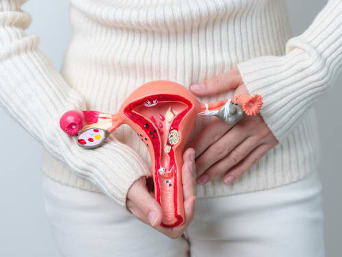
As we age, our reproductive health undergoes significant changes. Find out how our EMS Therapy can help slow ovarian ageing.