
Radiofrequency Microneedling
RF microneedling (MNRF) combines the wound healing cascade of standard microneedling with a second, independent mechanism: controlled delivery of bipolar radiofrequency energy at insulated needle tips. The thermal component generates radiofrequency thermal zones (RFTZs) in the dermis, triggering HSP47-mediated collagen synthesis and HSP70-mediated NF-κB suppression – a pathway that simultaneously drives collagen production and reduces MMP-driven degradation. A 2025 Seoul National University split-face RCT demonstrated a mechanism that distinguishes MNRF from all other collagen stimulation treatments in the portfolio: RF thermal energy induces apoptosis of senescent fibroblasts and their replacement by freshly proliferating non-senescent fibroblasts, with the degree of senescent fibroblast clearance correlating directly with improvements in collagen density. The treatment also produces measurable increases in tropoelastin and fibrillin – the elastic fibre scaffolding components – making it the primary tool for skin laxity and elastic fibre fragmentation as well as structural collagen restoration. MNRF is the most structurally complete treatment in the portfolio for clients where ageing-environment remodelling, rather than environment preparation alone, is the clinical priority.
RF microneedling (MNRF) is a minimally invasive procedure that combines fine-needle percutaneous micro-injury with the controlled delivery of bipolar radiofrequency energy. It is architecturally and mechanistically distinct from standard microneedling: where standard microneedling initiates healing through physical tissue disruption alone, MNRF adds a thermal energy component that generates independent biological effects in the dermis – effects that include senescent fibroblast clearance, heat shock protein-mediated collagen chaperone activity, and elastic fibre precursor upregulation – none of which standard needling produces to a clinically significant degree. The insulated needle design is central to how the treatment delivers these effects: the shaft is electrically non-conducting, so RF energy is released only at the exposed needle tips in the dermis, preserving the overlying epidermis from thermal spread. This architecture is what allows precise reticular dermal targeting whilst maintaining a surface recovery profile significantly shorter than ablative laser alternatives. [1]
The Dual Mechanism
Component 1 – Wound healing cascade: Needle penetration creates controlled micro-injuries that initiate the same three-phase wound healing response as standard microneedling – inflammation, proliferation, and remodelling – releasing TGF-β, PDGF, and VEGF, recruiting fibroblasts, stimulating Type I and III collagen synthesis, and driving fibronectin matrix formation. This component of MNRF is mechanistically identical to standard microneedling and produces the same basket-weave collagen architecture through TGF-β3 isoform predominance. [4]
Component 2 – RF thermal mechanism: Bipolar RF energy delivered at needle tips generates resistive heating in the surrounding dermal tissue, creating radiofrequency thermal zones (RFTZs) of precisely calibrated thermal injury. This thermal component produces a cascade that is entirely independent of the wound healing route: immediate collagen denaturation and contraction; HSP47 and HSP70 upregulation; tropoelastin and fibrillin induction; and, as 2025 evidence now confirms, apoptosis of senescent dermal fibroblasts followed by their replacement by newly proliferating non-senescent fibroblasts. [1] [2] These two components operate simultaneously, but they activate different cell populations, different molecular pathways, and produce different structural outcomes. That is why MNRF is not simply an enhanced version of microneedling – it is a treatment that contains microneedling as one of its two mechanisms.
The Senescent Fibroblast Mechanism
The most clinically significant mechanistic distinction MNRF holds over every other treatment in the portfolio is its selective effect on fibroblast senescence – a finding confirmed in a 2025 Seoul National University split-face RCT that, for the first time, directly compared MNRF to standard microneedling on the same patients with biopsy analysis. [2]
Senescent fibroblasts – cells that have exited the cell cycle due to cumulative UV damage, oxidative stress, or telomere shortening – do not produce collagen. They secrete instead a toxic inflammatory mix collectively termed the senescence-associated secretory phenotype (SASP): pro-inflammatory cytokines, MMPs, and other factors that actively degrade the ECM whilst suppressing collagen synthesis in neighbouring cells. The accumulation of senescent fibroblasts in aged and photodamaged skin is one of the central reasons why other collagen-stimulating treatments – including standard microneedling – produce less pronounced results in older or significantly photodamaged clients than the mechanism alone would predict. You are applying the stimulus to a fibroblast population that is, in part, constitutively non-functional and actively hostile to collagen preservation.
RF thermal energy addresses this through a mechanism that no topical, injectable, or standard needling treatment can replicate: cleaved caspase-3 and TUNEL evidence confirms that thermal energy at needle tips induces apoptosis of dermal fibroblasts non-specifically – affecting both senescent and non-senescent cells in the RFTZs. The body then repopulates these zones through proliferation. Crucially, the cells that proliferate to replace the apoptosis-cleared population are predominantly non-senescent: p16^INK4A^-negative, HSP47-positive, synthetically active fibroblasts. [2] The 2025 RCT confirmed that the proportion of senescent fibroblasts decreased significantly on the MNRF-treated side, the absolute count of non-senescent, collagen-producing fibroblasts increased simultaneously, and – critically – the degree of this senescent fibroblast clearance correlated directly with improvements in collagen density and hydration. This is not a peripheral observation; it establishes the clearance of senescent fibroblasts as an active mechanism of outcome, not merely a bystander effect.
Senescent Fibroblast Clearance: Why It Matters Clinically
| Fibroblast State | Behaviour | Treatment Response |
|---|---|---|
| Active (non-senescent) | Synthesises collagen I/III; responds to TGF-β | Responds well to all stimulatory treatments |
| Senescent (p16^INK4A^+) | Secretes SASP (MMPs, IL-6, TNF-α); does not synthesise collagen | Unresponsive to growth factors, PN, or needling alone |
| Post-MNRF (repopulated) | Cleared by thermal apoptosis; replaced by non-senescent proliferators | RF creates a new collagen-competent fibroblast population |
HSP47 and HSP70: The Two Heat Shock Pathways
The thermal component of MNRF activates two heat shock protein pathways with distinct downstream effects.
HSP47 – the collagen chaperone: HSP72 (the acute stress response protein) peaks rapidly after RF treatment and then diminishes. HSP47 – a collagen-specific chaperone protein that stabilises procollagen triple helices in the endoplasmic reticulum – increases progressively through ten weeks. [1] This extended HSP47 expression is responsible for the sustained collagen synthesis that follows MNRF well beyond the initial wound healing window, and the 2009 foundational bipolar RF histology study confirmed complete replacement of all RFTZs with new collagen by ten weeks post-treatment – driven by this HSP47 chaperone activity. HSP47 expression tripling with higher RF energy settings confirms that energy calibration directly scales the collagen synthesis response.
HSP70 – the NF-κB suppressor: A 2025 study examining RF irradiation in senescent facial ligaments established a distinct and clinically important second pathway. RF enhances the interaction between HSP70 and IKKγ (a component of the IκB kinase complex), reducing IκBα phosphorylation and thereby decreasing NF-κB activation in senescent fibroblasts. [3] NF-κB suppression via HSP70 simultaneously downregulates MMP expression and SMAD7 (an inhibitory SMAD that suppresses TGF-β signalling) in the treated tissue – creating a dermal environment that is simultaneously more synthetically active and less degradative. The clinical significance: MNRF does not simply stimulate collagen production whilst leaving the MMP-driven degradation environment intact. The HSP70-NF-κB pathway directly reduces the enzymatic degradation that would otherwise erode whatever new collagen is being produced.
Tropoelastin, Fibrillin, and the Elastic Fibre Dimension
RF microneedling is the most effective treatment in the portfolio for elastic fibre restoration – a dimension of dermal ageing that standard microneedling, polynucleotides, and skin boosters address partially or indirectly, but that MNRF addresses through a direct molecular mechanism. The foundational bipolar RF histology study confirmed marked induction of both tropoelastin and fibrillin – the two principal structural proteins of mature elastic fibres – at 28 days post-treatment, alongside procollagens I and III. [1] The 2025 split-face RCT confirmed a significant increase in elastin fluorescence intensity in the treated dermis at three and five months. [2] This is clinically relevant because skin laxity – the loss of the skin’s ability to recoil against gravity – is primarily an elastic fibre fragmentation problem rather than a pure collagen deficit. Restoring collagen density in skin with severely fragmented elastic fibres improves texture and quality but does not restore recoil. Tropoelastin and fibrillin upregulation through the thermal HSP mechanism is the route to genuine laxity correction, and it is specific to energy-based treatments.
Depth, Tissue Zone Targeting, and an Important Nuance
MNRF protocols use insulated needles at adjustable depths (typically 1.5–4mm for facial applications; up to 7mm for body) with the RF energy deposited at the tip. Standard protocols for skin quality and laxity target the mid-to-deep reticular dermis – the tissue zone where structural collagen architecture, fibrillin scaffolding, and elastic fibre networks are located. At depths of 3.5–4mm and beyond, RF energy reaches the superficial fat compartment, inducing adipocyte remodelling and contributing to the contouring and submental fat reduction effects that fall outside standard microneedling’s capability.
One nuance from the 2025 split-face RCT deserves honest acknowledgement: the study’s quantitative histological analysis found that the significant increases in collagen deposition and elastin fluorescence were measured in the papillary dermis rather than the reticular zone. [2] This does not contradict the reticular dermis targeting rationale – the study’s settings and the particular biopsy analysis zones used may not have captured the full reticular depth effect – but it does suggest that a binary papillary/reticular framework may be a simplification of where the measurable cellular changes actually occur. The tissue zone targeting model is a useful clinical organising principle, but it should be held as a framework rather than a rigid boundary.
Evidence Base
The histological evidence base for RF microneedling is established and meaningful. Biopsy studies across multiple controlled investigations show: collagen increase of up to 25% at three months; elastin increase of 33.3% with improved fibre organisation; dermal thickness increase of 15–20% at three months; reduced inflammatory infiltration and reduced elastin fragmentation. The 247-patient retrospective study reported a 1.4-point improvement in lower face and neck laxity on the Baker Face/Neck Classification, with 93% patient satisfaction. A 2024 Morpheus8 histological study of patients aged 40+ confirmed particularly pronounced elastogenesis stimulation in older patients – an age-dependent benefit that aligns directly with the senescent fibroblast clearance mechanism: the clients with the most accumulated senescent burden potentially benefit most from the thermal replacement pathway.
Clinical Application
RF microneedling is the most structurally complete treatment in the Creative Touch portfolio for clients where the clinical goal is genuine structural remodelling rather than environment preparation, substrate provision, or skin quality maintenance. That distinction shapes everything about when to use it, what to precede it with, and what to tell clients to expect.
The three-mechanism ageing model from maps MNRF clearly: it is the primary tool for net collagen loss (through the wound healing cascade and HSP47 synthesis pathway) and elastic fibre fragmentation (through tropoelastin and fibrillin upregulation), and it contributes to the ground substance dimension through endogenous HA stimulation within the RFTZs. It does not replace the environment-preparation role that polynucleotides fulfil – and using MNRF as the opening intervention in a chronically inflamed, MMP-elevated, or significantly senescent tissue environment is asking the treatment to work against the grain of the biology. The sequencing logic matters.
The senescent fibroblast mechanism is also the reason MNRF produces results in older and post-menopausal clients that other collagen-stimulating treatments plateau on. Growth factor delivery (iPRF), direct synthesis stimulation (skin boosters via mechanoreception), and substrate provision ( mesotherapy) all require a fibroblast population that is capable of responding. MNRF creates that population by clearing the senescent fraction and repopulating the dermis with functionally active cells – which is why it is often most clinically productive as a treatment in the 50s and 60s rather than something to defer until later.
Who benefits most
Clients with visible skin laxity – loss of jawline definition, jowl formation, neck laxity, crepiness under the eyes or on the décolletage – where elastic fibre fragmentation is a primary component. Of the treatments in our portfolio with confirmed biopsy evidence, MNRF provides the most direct and quantified upregulation of tropoelastin and fibrillin specifically – the molecular scaffolding that gives skin its recoil. PE Face’s synchronized RF component also produces thermal elastin synthesis, though via surface delivery at lower thermal intensity; for clients where structural laxity is the primary concern, MNRF’s deeper thermal profile and confirmed elastic fibre precursor induction make it the more targeted choice.”
Post-menopausal and significantly photodamaged clients with plateauing results from other treatments. The senescent fibroblast burden in this population is the most common explanation for a growth factor or biostimulatory treatment that produces initial improvement but then stalls – those treatments are stimulating cells that are no longer fully capable of responding. MNRF’s thermal fibroblast clearance and repopulation pathway addresses this at the source, creating a biologically younger fibroblast environment that then responds more fully to the treatments they are being combined with.
Clients in their 40s–50s with early-to-moderate structural decline. Earlier intervention intercepts the progressive accumulation of senescent cells and elastic fibre fragmentation before it becomes the dominant clinical picture. A structured MNRF protocol at this stage preserves the collagen and elastic architecture that later decline would otherwise erode.
Acne scarring, particularly atrophic and rolling subtypes, where the combination of mechanical collagen induction from needling and the RF thermal remodelling produces more consistent scar depth reduction than standard microneedling alone. The 2025 split-face RCT confirmed statistically superior improvements in surface roughness on the MNRF side vs. standard MN at both three and five months. [2]
Setting expectations honestly
RF microneedling produces visible improvement on a timeline that requires managing: immediate mild tightening from thermal collagen contraction, progressive quality improvement from four to six weeks as the wound healing cascade completes, and the most significant structural changes – including the benefits of senescent fibroblast repopulation – developing from three to six months. Peak improvement from a three-session course is typically measurable at six months and is sustained well beyond that as the new collagen and elastic fibre networks integrate.
Downtime is real but modest: redness and mild swelling for 24–72 hours, small punctate crusting at needle sites that resolves within three to five days. This is not an ablative recovery, but clients should be advised against intensive exercise, heat exposure, and active skincare ingredients in the first 48–72 hours. The treatment is not appropriate for FST V–VI without careful assessment and conservative settings – the insulated needle design minimises epidermal RF spread, but the mechanical injury component still carries PIH risk in darker skin types, and Fitzpatrick assessment should inform depth, spacing, and energy calibration.
Where the combinations are most productive
MNRF + polynucleotides is the most complete protocol for post-menopausal clients or those with significant photodamage and senescent burden. Polynucleotides establish the MMP-suppressive, pro-regenerative environment through the macrophage-fibroblast axis; MNRF then delivers the thermal remodelling and senescent fibroblast clearance into a tissue that is no longer actively degrading whatever is produced. Sequencing: polynucleotides two to four weeks prior, or as a concurrent course.
MNRF + skin boosters addresses structural remodelling (MNRF) alongside ground substance restoration and mechanical TGF-β activation (skin boosters). The two mechanisms are complementary at the level of the ECM – MNRF drives collagen and elastin structural synthesis; skin boosters restore the HA-rich ground substance environment that maintains dermal tension and supports fibroblast mechanoreception. Sequencing: skin boosters two to four weeks before MNRF (to restore the ground substance environment first) or two to four weeks after (as recovery-phase structural support).
MNRF + iPRF – the combination pairing where both are applied in the same session or in rapid sequence. iPRF’s growth factor delivery is amplified in the MNRF environment because the inflammatory and repair signalling cascade is already active; PDGF, TGF-β, and IGF-1 from iPRF enter a tissue in the midst of the wound healing phase where fibroblast responsiveness to these signals is at its peak. The timing window matters: iPRF applied at the end of the MNRF session or in the 24-hour post-treatment window when microchannels remain partially open.
MNRF + microneedling (as preparation or maintenance). A course of standard microneedling before introducing MNRF – improving skin quality, reducing surface-level photodamage, and priming the dermal environment – produces a more responsive dermal starting point for the more intensive RF remodelling. In maintenance protocols, standard microneedling sessions between MNRF courses sustain skin quality and extend the interval before the next full MNRF course is needed.
The Protocol Reference
| Treatment Goal | Recommended Approach | Combination Logic |
|---|---|---|
| Structural laxity (face/neck) | 3 sessions, 4–6 week intervals | + PN pre-course; + skin boosters concurrent or post |
| Post-menopausal remodelling | 3 sessions + maintenance every 9–12 months | + PN (environment prep); + iPRF same session |
| Acne scarring (atrophic/rolling) | 4–6 sessions, 4–6 week intervals | + subcision for rolling scars; + PN for inflamed skin |
| Body (laxity, cellulite, submental) | 3 sessions (deep settings); device-dependent | + PLLA for complementary fat/volume layer |
| Maintenance (post-course) | Single session every 9–12 months | + standard microneedling between courses |
References
Hantash BM, Ubeid AA, Chang H, et al. (2009). Bipolar fractional radiofrequency treatment induces neoelastogenesis and neocollagenesis. Lasers Surg Med, 41(1), 1-9 . doi.org/10.1002/lsm.20731
Hwang JM, Lee SH, Baek EJ, et al. (2025). Comparison of the effects of fractional microneedle radiofrequency and microneedling on modulating the senescent fibroblast milieu in aged skin. Sci Rep, 15(1), 18296 . doi.org/10.1038/s41598-025-02545-3
Oh S, Kim HM, Cheon GW, et al. (2025). Radiofrequency-Induced Thermal Modulation Reduces Senescence-Induced Collagen Fiber Degradation in Facial Ligaments of Animal Models. Cells, 14(22) . doi.org/10.3390/cells14221757
Tehrani L, Tashjian M, Mayrovitz HN (2025). Physiological Mechanisms and Therapeutic Applications of Microneedling: A Narrative Review. Cureus, 17(3), e80510 . doi.org/10.7759/cureus.80510
Also Known As
- FMRF
- fractional microneedle radiofrequency
- fractional radiofrequency microneedling
- FRM
- microneedling radiofrequency
- MNRF
- RF microneedling
Therapeutic Relationships
Therapeutic Context
- Stimulates Collagen Evidence: RF thermal injury creates controlled collagen denaturation triggering heat-shock protein response and a second fibroblast collagen remodelling wave extending to 3-6 months post-treatment (PMC11497551; Collagen entity).
- Stimulates Elastin Evidence: RF microneedling drives elastic fibre restoration: biopsy shows 75.9% elastin increase at 1 month, 110.8% at 2 months. PMC10833192; PMC11965193.
Indications & References
- this Adipocyte Evidence: RF targets adipocyte thermal selectivity (lower thermal conductivity of lipid-filled cells); concurrent skin tightening via collagen. Entity text.
- this Dermis Evidence: RF microneedling… target this zone specifically, delivering thermal signals where the structural remodelling opportunity is greatest
- this Fibroblast Evidence: RF Microneedling activates fibroblasts through the controlled injury cascade
- this Skin Evidence: RF Microneedling targets reticular dermis for deep collagen stimulation.
- this May treat Perimenopausal skin changes Evidence: RF microneedling addresses structural laxity, reduces p16INK4A senescent fibroblasts, restores IGF-1 mRNA; clinically recommended for post-menopausal structural decline. Entity text; PMC12106790.
Learn More
This topic is discussed in 3 articles:
-
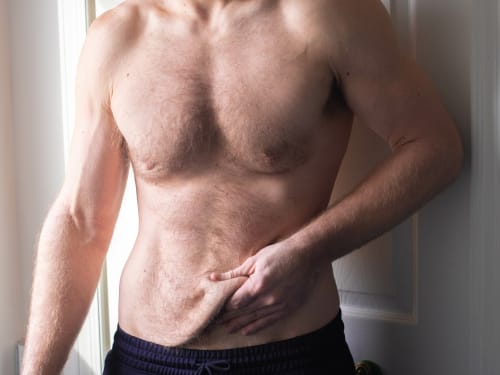
Don’t let loose skin overshadow your weight loss success. Learn why skin sags after significant weight loss and explore our advanced skin tightening and volume restoration treatments.
-

Fine lines and wrinkles? Say hello to your skin’s new BFFs: microneedling and RF microneedling! Discover the difference and find your perfect treatment at Creative Touch.
-

Enhanced microneedling combining radiofrequency energy for collagen stimulation. For use after barrier stabilisation.