
Fibroblast
Fibroblasts are the primary “engineers” of the extracellular matrix, but their productivity is dictated by their environment. Clinical success depends on addressing three biological hurdles: Signal Reception ( TGF-β responsiveness), Cellular Senescence (clearing the “toxic” SASP environment), and Mechanotransduction. A fibroblast cannot build a structural scaffold if it is floating in a “slack” environment; we must restore physical matrix tension and quench inflammatory noise to transition these cells from a destructive, senescent state back into a productive, synthetic one.
Fibroblasts are the primary synthetic cells of the dermis, responsible for producing and maintaining the extracellular matrix (ECM) – the structural framework of collagen fibres, elastic fibres, proteoglycans, and ground substance in which they are embedded. They are not passive factories waiting for instructions. Fibroblasts continuously sense their mechanical and biochemical environment through surface receptors, adjusting their synthetic output in response to physical tension, growth factor signals, inflammatory cytokines, and the structural state of the matrix around them. That responsiveness is central to understanding both how fibroblasts decline with age and how treatments restore their activity.
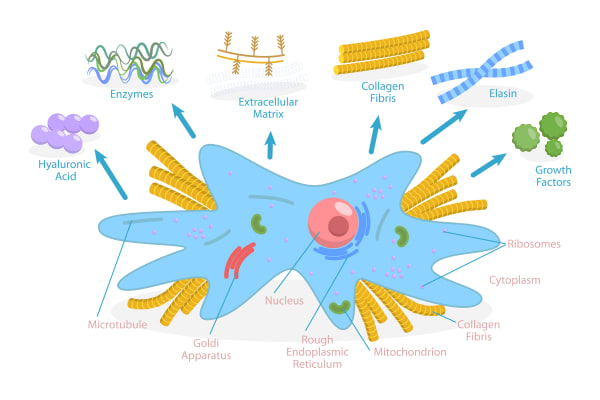
What Fibroblasts Produce
The collagen and elastin entities describe the synthesis of these proteins in detail, fibroblasts are the cells performing those processes. Procollagen I and III are transcribed in response to TGF-β1 signalling and secreted into the extracellular space for maturation and cross-linking. Tropoelastin is assembled onto a fibrillin microfibril scaffold to form functional elastic fibres. Alongside these structural proteins, fibroblasts produce hyaluronic acid and other proteoglycans, including decorin, which regulates the lateral spacing of collagen fibrils. These form the ground substance filling the spaces between fibres.
The proteoglycan output is less often discussed than collagen, but it is not peripheral. Decorin does not simply occupy space, it governs how collagen fibrils organise laterally, determining whether the resulting matrix is fine and even or coarse and disordered. UV exposure degrades decorin via MMPs, which is why photoaged skin displays abnormal collagen architecture even before quantitative collagen loss is measurable. Ground substance production also directly governs fibroblast mechanosensitivity: as hyaluronic acid in the dermis declines, fibroblasts lose the mechanical tension they require to maintain normal synthesis rates. They reduce output even when TGF-β signalling is otherwise intact.
How Fibroblasts Receive and Transmit Signals
TGF-β1 is the primary growth factor signal driving fibroblast procollagen transcription; but “primary” should not be read as “only.” Fibroblasts integrate signals from multiple sources simultaneously: growth factors delivered through the ECM or by platelets during wound healing (EGF, PDGF, FGF), inflammatory cytokines that can either stimulate or suppress their activity depending on type and concentration, and mechanical forces transmitted through integrin receptors embedded in the cell membrane.
The mechanoreception pathway is particularly clinically relevant and frequently underestimated. When a fibroblast is embedded in a structurally supported matrix, with adequate collagen, HA, and ground substance providing physical tension, integrin-mediated mechanoreception actively drives TGF-β signalling and synthetic output. When the matrix loses volume and structural integrity, fibroblasts experience reduced mechanical tension and reduce their production accordingly, even in the absence of any direct growth factor deficit. This is not a failure of the cells. It is an appropriate mechanosensory response to an environment signalling that less matrix maintenance is needed. The clinical implication is that restoring the physical environment around fibroblasts – the ground substance, the structural tension, the ECM architecture – can restore synthesis capacity without requiring direct growth factor delivery.
Fibroblast Decline: Three Converging Problems
The commonly cited picture of fibroblast decline – fewer cells producing less collagen – understates what actually happens, and that understatement has clinical consequences.
The first problem is reduced TGF-β1 production with age, which progressively reduces the primary signal for procollagen transcription. This is the straightforward synthesis decline most anti-ageing discussions focus on. [7]
The second is cellular senescence. Fibroblasts do not simply become less active with age; a proportion enter a senescent state in which they stop dividing, reduce their synthetic output, and begin secreting a pro-inflammatory mix of cytokines, proteases, and growth factors collectively known as the senescence-associated secretory phenotype (SASP). The structural consequence is quantifiable: senescence-associated changes produce a 68% reduction in Type I procollagen content and a 30% decrease in overall collagen-synthetic capacity, independent of chronological collagen loss measured through other mechanisms. [9] Senescent fibroblasts are not neutral bystanders, they actively upregulate MMP-1, MMP-3, and MMP-9, contributing to collagen and elastin degradation whilst simultaneously reducing synthesis. The accumulation of senescent fibroblasts in ageing dermis therefore creates a local environment that is simultaneously less productive and more destructive.
A compounding factor that is rarely discussed is the macrophage environment shift that accompanies dermal ageing. The ratio of pro-inflammatory M1 macrophages to repair-oriented M2 macrophages increases with age, and conditioned media from M1 macrophages measurably increases the number of SA-β-gal-positive senescent fibroblasts. Conditioned media from M2 macrophages does the opposite: it actively inhibits fibroblast senescence. [10] In aged skin, the tissue is not only producing fewer repair signals; the macrophage environment itself is actively maintaining and propagating the fibroblast senescence it has created. This is a self-reinforcing cycle, and it is why treatments that shift the macrophage balance – polynucleotides and PLLA – have clinical relevance that extends beyond their direct fibroblast effects.
The third is the mechanosensitivity collapse described above. As ground substance depletes and ECM architecture deteriorates, the structural signals that sustain normal fibroblast activity progressively weaken. By the time visible laxity and thinning are evident, the fibroblast population is likely responding to a biomechanical environment that is actively suppressing synthesis through multiple routes simultaneously.
These three mechanisms are not sequential, they compound. A fibroblast population that is simultaneously losing TGF-β1 drive, accumulating senescent cells with pro-inflammatory secretory profiles, and experiencing reduced mechanosensory input is in a fundamentally different state from one that is simply “slowing down.” Treatments that address only one of these three components produce more limited outcomes than protocols designed around all three.
The Perimenopause Dimension
The research from primary fibroblast studies is counterintuitive enough to be worth stating clearly here: post-menopausal fibroblasts actually increase procollagen gene expression in vitro, representing a compensatory response to oestrogen withdrawal. This is not a sign that menopausal fibroblasts are functioning normally. The increased transcription is insufficient to prevent net collagen loss because of simultaneous MMP-1 and MMP-3 upregulation and impaired procollagen maturation downstream of the transcription stage – of which the vitamin C-dependent hydroxylation step is a key component.
The clinical precision this demands is important. In perimenopausal and post-menopausal skin, the fibroblast problem is not that the cells have stopped trying to produce collagen. It is that the maturation and degradation environment has shifted such that what they produce is increasingly lost before contributing to the structural matrix. A treatment strategy that increases procollagen mRNA without addressing MMP activity or supporting the hydroxylation pathway is, in effect, accelerating synthesis into an unfavourable environment. It is working on a step the fibroblasts are already compensating for.
The Fibroblast Intervention Matrix
| Decline Mechanism | Clinical Presentation | Fibroblast Status | Strategic Priority | Recommended Modalities | Homecare Support |
|---|---|---|---|---|---|
| Reduced Signalling (TGF-β Drive) | Early fine lines, slight loss of glow, 30s to mid-40s | Responsive but under-stimulated; receptors intact | Direct stimulation: deliver growth factor signals to trigger synthesis | iPRF, Microneedling, Chemical Peels | Retinoids (signalling), Vitamin C (cofactor) |
| Cellular Senescence (SASP) | Thinning skin, chronic redness, inflammageing, post-menopause | Non-productive; secreting destructive MMPs and cytokines; p16/p21 axis active | Environmental reset and senescence suppression: quieten SASP, restore M2 macrophage environment, suppress p53/p21 axis | Polynucleotides (macrophage M2 route, SMAD2/STAT3); PLLA (direct PI3K/AKT senescence suppression) | Niacinamide (anti-inflammatory), high-potency Vitamin C |
| Mechanosensitivity Collapse | Sagging, deep folds, crepey texture, volume loss | Shrunken and stellate; unable to sense structural tension via integrins | Structural tension restoration: rebuild the physical environment to reactivate mechanoreceptors | HA Skin Boosters (Profhilo, Seventy Hyal) | Topical HA (surface hydration support), Ceramides |
| Maturation Bottleneck | Slow treatment recovery, skin that won’t respond | Transcribing procollagen but unable to hydroxylate or cross-link it | Metabolic support: ensure biochemical tools for collagen assembly are present | iPRF (fibrin scaffold), gentle heat-based therapies | Mandatory Vitamin C, Copper Peptides, Amino Acids |
Note on PLLA row placement: PLLA is sometimes grouped with volume-based treatments for sagging or crepey presentations. Its primary mechanism is senescence suppression via PI3K/AKT and M2 macrophage polarisation – placing it in the Cellular Senescence row reflects where it does its most meaningful work. The gradual structural improvement it produces is a consequence of restored fibroblast capacity, not a direct mechanical signal to existing cells.
UV Irradiation and Extrinsic Fibroblast Damage
Whilst intrinsic ageing steadily erodes fibroblast function through the three mechanisms outlined above, ultraviolet irradiation imposes a qualitatively distinct and more aggressive threat. The UV-driven pathway simultaneously increases collagen degradation and decreases collagen synthesis – a dual insult that cannot be replicated by chronological ageing alone and that fundamentally explains why photoaged skin displays more dramatic structural collapse per decade than chronologically aged, UV-protected skin.
The AP-1/MMP Cascade: UV as a Collagenase Switch
Within minutes of UV irradiation, keratinocytes and dermal fibroblasts respond to reactive oxygen species (ROS) by activating mitogen-activated protein (MAP) kinase signalling cascades – specifically ERK, JNK, and p38 pathways. These kinases converge in the nucleus to phosphorylate c-Jun, which heterodimerises with constitutively expressed c- Fos to form the transcription factor activator protein-1 (AP-1). AP-1 then drives transcription of matrix metalloproteinases, principally MMP-1 (interstitial collagenase), MMP-3 (stromelysin), and MMP-9 (92-kDa gelatinase), all of which degrade the fibrillar type I and type III collagen that constitutes the dermis.
This mechanism was characterised in human skin in vivo by [4], who demonstrated that MMP mRNAs, proteins, and enzymatic activities were induced within hours of a single suberythemal UVB exposure – a dose well below the threshold for visible skin reddening. A subsequent landmark study in the New England Journal of Medicine by [6] quantified the functional consequence: a single UV exposure increased endogenous type I collagen fibril degradation by 58%, whilst four exposures delivered over eight days maintained collagenase activity at 4.4 times baseline for the entire interval. The upstream MAP kinase → c-Jun → AP-1 → MMP pathway was mapped in detail by [5], confirming that the dermal fibroblast, alongside the keratinocyte, is an active participant in UV-induced collagen degradation – not merely a passive bystander.
Simultaneous Suppression of Procollagen Synthesis
UV irradiation does not only stimulate collagen degradation – it independently and simultaneously suppresses new collagen synthesis through a separate molecular pathway. The TGF-β/Smad signalling axis is the primary driver of procollagen gene transcription in fibroblasts; UV irradiation impairs this axis by transcriptionally down-regulating the TGF-β type II receptor (TβRII) in human skin fibroblasts. Loss of TβRII prevents TGF-β from binding to cell-surface receptors, which in turn blocks downstream phosphorylation of Smad2 and Smad3, reducing type I and type III procollagen gene expression. This is distinct from the age-related decline in TGF-β1 ligand production outlined above; in photoaged skin, both ligand availability and receptor expression are compromised, compounding the signalling deficit. [11] demonstrated that TβRII down-regulation occurs within 8 hours of UV irradiation in human skin in vivo, preceding measurable procollagen loss, and that overexpression of TβRII is sufficient to protect fibroblasts against UV-induced procollagen suppression. A complementary mechanism was identified by [12]: UV irradiation reduces expression of connective tissue growth factor (CTGF), a downstream mediator of TGF-β action in fibroblasts, and disrupts TGF-β/Smad-responsive elements in the CTGF gene promoter – providing a second route by which UV uncouples TGF-β signalling from procollagen output.
The Double Hit: Why Photoageing is Mechanistically Distinct
[3], publishing in the Journal of Clinical Investigation, formally characterised the dual assault: “UV irradiation, therefore, damages human skin connective tissue by simultaneously inhibiting procollagen synthesis and stimulating collagen breakdown.” This double hit – degradation accelerated whilst synthesis is suppressed – is not reproduced by intrinsic ageing mechanisms alone. Cellular senescence and reduced TGF-β responsiveness associated with chronological ageing shift the balance away from synthesis, but they do not simultaneously activate high-level MMP transcription via AP-1 in the way UV irradiation does. The net consequence is that sun-exposed skin accumulates what the literature describes as ‘solar scars’ with each UV exposure: episodes of imperfect collagen repair that, as Fisher et al. 2000 characterised, compound over a lifetime to produce the pronounced fibril fragmentation, volume loss, and textural irregularity characteristic of photoageing.
The Photoageing Fibroblast Phenotype
Dermal fibroblasts in UV-damaged skin do not simply produce less collagen – they adopt a functionally distinct morphology. In normal dermis, fibroblasts attach to intact collagen fibrils and achieve a stretched, mechanically engaged state that maintains synthetic output. In photoaged dermis, cumulative MMP-mediated collagen fibril fragmentation undermines this attachment, leaving fibroblasts in a collapsed, mechanically slack conformation with reduced cytoplasmic area, disassembled actin cytoskeleton, and impaired TGF-β pathway responsiveness. [14] reproduced this phenotype experimentally by expressing constitutively active MMP-1 in young fibroblast cultures: MMP-mediated collagen fragmentation alone was sufficient to recapitulate the morphological and functional features of aged/photoaged fibroblasts, including reduced collagen production. [2], from the same Michigan group, demonstrated that restoring mechanical tension to photoaged dermis via cross-linked hyaluronic acid injection was sufficient to re-stretch fibroblasts, restore collagen gene expression, and deposit structurally mature collagen bundles resembling those of young skin – confirming that the “collapsed phenotype” in photoaged fibroblasts is an adaptive, environmentally mediated state rather than irreversible cellular damage.
This distinction has important implications for treatment sequencing: the photoaged fibroblast phenotype arises principally from the extracellular environment (fragmented matrix, impaired TGF-β signalling, ongoing UV-driven AP-1 activation) rather than from intrinsic cellular senescence. Addressing the environmental inputs – quenching ongoing MMP activity, restoring matrix tension, protecting against further UV insult – can partially reverse fibroblast functional collapse even when the cells themselves retain proliferative capacity.
Photoprotection as Fibroblast Protection
UV irradiation is not merely a surface-level epidermal concern. Each unprotected UV exposure activates AP-1 in dermal fibroblasts, induces MMP transcription, degrades fibrillar collagen, and simultaneously suppresses TGF-β-driven procollagen synthesis. The cumulative consequence is progressive loss of the structural matrix upon which fibroblast function depends. Broad-spectrum photoprotection – by attenuating UV-induced MAP kinase activation before it reaches the dermis – is mechanistically a fibroblast-preservation strategy, upstream of any collagen-stimulating treatment.
Clinical Application
The fibroblast occupies a specific position in our knowledgebase that needs to be handled deliberately. Collagen, elastin, and the ground substance framework (established in the dermis page) each describe what fibroblasts produce and how those products decline. The clinical context here should not repeat that treatment logic. What the fibroblast uniquely contributes is the cellular-level framing of why treatments work on fibroblasts differently, depending on which of the three decline mechanisms is dominant: reduced TGF-β drive, senescent fibroblast accumulation, or mechanosensitivity collapse. That distinction is what this clinical context is built around.
Treatments That Work With Remaining Fibroblast Responsiveness
For fibroblasts that retain TGF-β sensitivity – which is most fibroblasts in clients who are not significantly post-menopausal and whose skin has not been heavily photodamaged – the treatment question is straightforward: what delivers the right signal most effectively?
iPRF is the most complete answer to that question from the growth factor delivery category. It is not simply that iPRF contains TGF-β and PDGF; it is that the fibroblast migration response to iPRF is measurably superior to PRP at 350% greater than control and 200% greater than PRP in direct comparison research, with TGF-β, collagen I, and fibronectin mRNA all highest in the iPRF group. For clients whose fibroblasts are responsive but under-stimulated – the early decline presentation in their 30s and 40s, or post-inflammatory skin that has calmed – iPRF delivers a growth factor environment that the fibroblasts can respond to with measurable productive output.
Microneedling activates fibroblasts through the controlled injury cascade: platelet degranulation at the needle sites releases TGF-β1, PDGF, and EGF, creating a localised fibroblast activation event whose collagen synthesis peak occurs at approximately 4–6 weeks post-treatment. This is a reliable, well-characterised signal. What microneedling cannot do is bypass the reduced responsiveness of senescent fibroblasts. It works well when the fibroblast population is intact, but in skin where senescent cells have accumulated significantly, the wound-healing signal reaches a population that is less able to respond to it.
Treatments That Work Around, and Directly On, Senescent Fibroblasts
This is where polynucleotides and PLLA each occupy a clinically distinct position, working on fibroblast senescence through different routes that are complementary rather than interchangeable.
Polynucleotides reach senescent fibroblasts through an indirect pathway that bypasses the direct TGF-β receptor responsiveness those cells have lost. Their mechanism – activating the A2AR→ cAMP→PKA→CREB→PCK1 cascade in macrophages, increasing M2 polarisation, and driving IL-10 and TGF-β secretion that then activates SMAD2 and STAT3 in adjacent fibroblasts – has been specifically confirmed in senescent fibroblast populations. [1] This is not simply that polynucleotides are anti-inflammatory. They restore collagen synthesis through a macrophage-mediated route that ages less poorly than direct fibroblast stimulation, and their M2 polarisation effect directly counteracts the pro-senescent macrophage environment that aged tissue sustains. [8] For post-menopausal clients in particular, polynucleotides are the upstream environment treatment, quietening the SASP-driven inflammatory context and restoring a macrophage balance in which direct stimulation then produces a better response.
PLLA addresses fibroblast senescence more directly. Where polynucleotides work through the macrophage axis to reach fibroblasts indirectly, PLLA’s direct activation of the fibroblast PI3K/AKT pathway suppresses the p53/p21 senescence axis at the cellular level, measurably reducing SA-β-gal, p16, and p21 senescence markers in aged fibroblasts. [10] This is mechanistically significant: PLLA is not simply providing a synthesis signal and asking a senescent cell to respond. It is partially restoring that cell’s capacity to function by suppressing the pathways maintaining its senescent state. PLLA simultaneously initiates its own M2 macrophage polarisation via the foreign body response, reinforcing the anti-senescent macrophage environment that polynucleotides also create, through a completely different route. [15]
The practical distinction: polynucleotides are the appropriate first step and ongoing maintenance treatment for the senescent fibroblast environment – resolving the inflammatory and SASP-driven suppression promptly, with a manageable treatment schedule. PLLA is the longer-horizon structural investment for clients where fibroblast senescence is a significant contributor and who are willing to commit to the three-session course and gradual development timeline it requires. For clients appropriate for both, they are sequenced rather than competing: polynucleotides prepare and maintain the tissue environment; PLLA rebuilds fibroblast capacity within it.
Clinical Pearl For clients where previous collagen-stimulating treatments have underperformed expectations, fibroblast senescence is worth considering as the limiting factor. When direct stimulation is working into a population with reduced responsiveness, results plateau quickly. Addressing the senescence environment first – with polynucleotides, or with PLLA’s PI3K/AKT route – changes what the stimulation step can achieve.
Restoring Mechanosensitivity: The Often-Missed Route
If fibroblast mechanosensitivity collapse is a genuine contributor to output decline – and the evidence that HA loss creates mechanical slack that reduces fibroblast synthesis even with intact TGF-β signalling is well-characterised – then HA skin boosters are not simply a hydration treatment. They are a fibroblast mechanosensory restoration treatment.
The biopsy-confirmed mechanism is worth restating in this context: within one week of cross-linked HA injection into the mid-dermis, fibroblasts stretch and elongate in response to the restored physical tension, with integrin-mediated TGF-β activation driving new Type I procollagen synthesis measurable at four weeks and still present at twelve months. This is a fibroblast activation signal that does not rely on growth factor delivery, does not require an intact wound-healing cascade, and does not ask anything of the direct TGF-β receptor pathway that senescent fibroblasts struggle with. It works through the structural environment rather than the cellular signalling pathways directly. That makes it particularly relevant for older skin where multiple signalling routes have been compromised.
The six-to-nine month duration of effect, whilst the HA remains structurally integrated, is also clinically meaningful: it is a sustained fibroblast activation rather than a pulse. Most professional treatments produce a synthesis peak and then return to baseline. HA skin boosters create a period of elevated fibroblast activity that extends well beyond what any growth factor stimulus maintains independently.
The Homecare Layer: Vitamin C and Retinoids
These two actives address the fibroblast environment from different angles that are worth being precise about here. Vitamin C is not primarily a fibroblast stimulator. It is the cofactor without which hydroxylation of procollagen chains cannot complete, meaning fibroblasts that are synthesising procollagen in a vitamin C-depleted environment produce structurally defective collagen that is degraded intracellularly before secretion. In post-menopausal skin, where the procollagen hydroxylation step is specifically compromised, topical vitamin C at bioavailable concentrations is addressing the maturation bottleneck that is causing net collagen loss despite adequate transcription. It also reduces the ROS-driven AP-1 signalling that upregulates MMP-1, giving it simultaneous synthesis-support and degradation-reduction activity. [13]
Retinoids work on the signalling environment: upregulating TGF-β1 expression and reducing MMP activity, which addresses the reduced growth factor drive and the elevated degradation simultaneously. Long-term retinoid use increases measurable dermal collagen density and improves the Type I:III ratio by maintaining the fibroblast activation environment rather than delivering a single stimulus. The pairing of retinoids and vitamin C addresses TGF-β signalling, MMP degradation, and the hydroxylation maturation step concurrently. This is precisely why it remains the most consistently effective collagen-focused homecare protocol despite decades of newer alternatives.
Treatment Sequencing for the Fibroblast
For younger clients or early decline (30s–mid 40s, no significant photodamage): fibroblasts are responsive and direct stimulation is appropriate from the outset. iPRF or microneedling delivers a clear growth factor signal to a population that can act on it. Vitamin C and retinoids in homecare maintain the environment between professional treatments.
For perimenopausal and post-menopausal clients: the senescent fibroblast accumulation, mechanosensitivity collapse, and pro-senescent macrophage environment warrant a more layered approach. Polynucleotides first to reduce the SASP-driven inflammatory environment, restore M2 macrophage balance, and re-establish the macrophage-mediated signalling route to fibroblasts. HA skin boosters to restore mechanosensory input to the fibroblast population and activate the structural TGF-β route. Then iPRF or RF microneedling to deliver direct synthesis stimulation to a population now operating in a recovered environment. For clients where senescence is the prominent driver – where structural decline is significant, the presentation suggests long-standing fibroblast underperformance, or where previous stimulation treatments have underdelivered – PLLA’s direct PI3K/AKT senescence suppression is the appropriate longer-horizon addition to the protocol. Vitamin C is non-negotiable in this group: it addresses the hydroxylation bottleneck that is causing net collagen loss despite apparently adequate transcription.
For heavily photodamaged skin regardless of age: the MMP-driven collagen and decorin degradation that has disorganised the ECM needs to be addressed alongside fibroblast stimulation. Polynucleotides’ NF-κB suppression protecting decorin and elastin, combined with the TGF-β and mechanosensory signals from iPRF and HA boosters, addresses both the structural environment and the fibroblast population simultaneously. Stimulating fibroblasts in a still-disorganised matrix produces new collagen that is deposited into a compromised architectural environment – the new synthesis contributes less than it would in a matrix where decorin has been protected and ground substance partially restored.
The unifying principle across all presentations is that fibroblasts respond to their environment as much as to direct signalling. Restoring the physical tension, the cytokine balance, the matrix architecture, is not preparatory work before the “real” treatment. It is part of the treatment.
An additional and qualitatively distinct pathway of fibroblast decline – operating through cumulative UV irradiation rather than intrinsic cellular ageing – compounds the three mechanisms outlined above. Whereas intrinsic mechanisms erode fibroblast productivity gradually and relatively symmetrically across sun-protected and sun-exposed sites, extrinsic UV damage creates a qualitatively different biochemical environment: sustained AP-1 activation drives ongoing MMP transcription whilst simultaneously impairing TGF-β type II receptor expression to suppress procollagen synthesis. The result is a more aggressive and compound collagen deficit that accumulates with each unprotected UV exposure across decades.
For the practitioner, this pathway demands a focused patient history. Cumulative UV exposure – including occupational history, geographical latitude, recreational habits (gardening, sport, outdoor work), and decades of intermittent sun exposure before SPF awareness became routine – is a material determinant of which fibroblast decline pathway predominates. A patient with a primarily photoaged presentation and a patient of the same chronological age with largely UV-protected skin may have equivalent biological age at the cellular level yet present with dramatically different dermal architecture. Identifying the predominant pathway informs treatment prioritisation: for predominantly photoaged presentations, ongoing AP-1-driven MMP activity creates a persistent degradative environment in which collagen biostimulation is working against an active headwind. Pre-treatment or co-treatment with a topical retinoid is mechanistically rational here, since retinoids (all-trans retinoic acid) inhibit UV induction of c-Jun protein and consequently reduce AP-1-mediated MMP transcription, as demonstrated by [6] – inhibition of approximately 70–80% for MMP-1 and total collagenase activity – with the upstream MAP kinase mechanism confirmed by [5]. Until ongoing collagen degradation is attenuated, neosynthesis stimulated by microneedling, RF microneedling, or PLLA will be partially offset by concurrent matrix breakdown, reducing net treatment yield.
References
Byun KA, Park HJ, Oh S, et al. (2025). Polynucleotides Enhance Collagen Synthesis via Modulating Phosphoenolpyruvate Carboxykinase 1 in Senescent Macrophages: Experimental Evidence. Int J Mol Sci, 26(17) . doi.org/10.3390/ijms26178720
Cui Y, Wang F, Voorhees JJ, et al. (2021). Rejuvenation of Aged Human Skin by Injection of Cross-linked Hyaluronic Acid. Plast Reconstr Surg, 147(1S-2), 43S-49S . doi.org/10.1097/prs.0000000000007620
Fisher GJ, Datta S, Wang Z, et al. (2000). c-Jun-dependent inhibition of cutaneous procollagen transcription following ultraviolet irradiation is reversed by all-trans retinoic acid. J Clin Invest, 106(5), 663-70 . doi.org/10.1172/jci9362
Fisher GJ, Datta SC, Talwar HS, et al. (1996). Molecular basis of sun-induced premature skin ageing and retinoid antagonism. Nature, 379(6563), 335-9 . doi.org/10.1038/379335a0
Fisher GJ, Talwar HS, Lin J, et al. (1998). Retinoic acid inhibits induction of c-Jun protein by ultraviolet radiation that occurs subsequent to activation of mitogen-activated protein kinase pathways in human skin in vivo. J Clin Invest, 101(6), 1432-40 . doi.org/10.1172/jci2153
Fisher GJ, Wang ZQ, Datta SC, et al. (1997). Pathophysiology of premature skin aging induced by ultraviolet light. N Engl J Med, 337(20), 1419-28 . doi.org/10.1056/nejm199711133372003
Ganceviciene R, Liakou AI, Theodoridis A, et al. (2012). Skin anti-aging strategies. Dermatoendocrinol, 4(3), 308-19 . doi.org/10.4161/derm.22804
Lee KWA, Chan KWL, Lee A, et al. (2024). Polynucleotides in Aesthetic Medicine: A Review of Current Practices and Perceived Effectiveness. Int J Mol Sci, 25(15) . doi.org/10.3390/ijms25158224
Nan L, Guo P, Hui W, et al. (2025). Recent advances in dermal fibroblast senescence and skin aging: unraveling mechanisms and pioneering therapeutic strategies. Front Pharmacol, 16, 1592596 . doi.org/10.3389/fphar.2025.1592596
Oh S, Lee JH, Kim HM, et al. (2023). Poly-L-Lactic Acid Fillers Improved Dermal Collagen Synthesis by Modulating M2 Macrophage Polarization in Aged Animal Skin. Cells, 12(9) . doi.org/10.3390/cells12091320
Quan T, He T, Kang S, et al. (2004). Solar ultraviolet irradiation reduces collagen in photoaged human skin by blocking transforming growth factor-beta type II receptor/Smad signaling. Am J Pathol, 165(3), 741-51 . doi.org/10.1016/s0002-9440(10)63337-8
Quan T, He T, Kang S, et al. (2002). Connective tissue growth factor: expression in human skin in vivo and inhibition by ultraviolet irradiation. J Invest Dermatol, 118(3), 402-8 . doi.org/10.1046/j.0022-202x.2001.01678.x
Telang PS (2013). Vitamin C in dermatology. Indian Dermatol Online J, 4(2), 143-6 . doi.org/10.4103/2229-5178.110593
Xia W, Hammerberg C, Li Y, et al. (2013). Expression of catalytically active matrix metalloproteinase-1 in dermal fibroblasts induces collagen fragmentation and functional alterations that resemble aged human skin. Aging Cell, 12(4), 661-71 . doi.org/10.1111/acel.12089
Zhu W, Dong C (2023). Poly-L-Lactic acid increases collagen gene expression and synthesis in cultured dermal fibroblast (Hs68) through the TGF-β/Smad pathway. J Cosmet Dermatol, 22(4), 1213-1219 . doi.org/10.1111/jocd.15571
Also Known As
- fibroblasts
Anatomical Relationships
Structural Connections
- Produces Collagen PMID: 34297930 Evidence: Procollagen I and III are transcribed in response to TGF-β1 signalling and secreted into the extracellular space for maturation and cross-linking
- Produces Decorin Evidence: Fibroblasts are the predominant producers of dermal decorin; decorin mRNA expressed by papillary and reticular fibroblasts. Li et al. Sci Rep 2013 doi:10.1038/srep02422
- Produces Elastin PMID: 34297930 Evidence: Tropoelastin is assembled onto a fibrillin microfibril scaffold to form functional elastic fibres
- Produces Hyaluronic acid PMID: 34297930 Evidence: Fibroblasts produce hyaluronic acid and other proteoglycans
- Produces Matrix metalloproteinase Evidence: Senescent fibroblasts… actively upregulate MMP-1, MMP-3, and MMP-9, contributing to collagen and elastin degradation
- Located in Dermis Evidence: Fibroblasts are the primary resident synthetic cells of the dermis embedded within ECM of papillary and reticular zones. Yu et al. Aging Cell 2023 doi:10.1111/acel.14054
- Located in Skin PMID: 34297930 Evidence: Fibroblasts are the primary synthetic cells of the dermis
- Affects Keratinocyte Evidence: Papillary fibroblasts signal to keratinocytes via KGF/FGF7 TGF-β; fibroblast senescence impairs epidermal differentiation through breakdown of DEJ crosstalk. Costello et al. J Cell Physiol 2024 doi:10.1002/jcp.31463
- Requires Glycine Evidence: Glycine is most abundant amino acid in collagen (every third Gly-X-Y residue); fibroblast collagen synthesis is directly limited by glycine availability. Standard collagen biochemistry.
- Requires Proline Evidence: Proline is obligate substrate for collagen synthesis; fibroblasts hydroxylate proline to hydroxyproline via prolyl hydroxylase in procollagen maturation. Standard collagen biochemistry.
- Requires Vitamin C Evidence: Fibroblasts that are synthesising procollagen in a vitamin C-depleted environment produce structurally defective collagen that is degraded intracellularly before secretion
- Has sub-structure Mitochondria Evidence: Fibroblasts are particularly mitochondria-rich; hundreds to thousands of mitochondria per cell described as integral to fibroblast energy production and synthetic function (PMC10693346).
- Dermatitis Evidence: Fibroblast IL-6 and SASP factors contribute to dermatitis; fibroblast-keratinocyte crosstalk drives both barrier restoration and inflammatory amplification. Stevenson et al. Biomedicines 2020 doi:10.3390/biomedicines8050101
- Skin ageing Evidence: Fibroblast decline – fewer cells producing less collagen – leads to skin ageing
- Injectable Platelet-Rich Fibrin Evidence: iPRF produces 350% greater fibroblast migration vs control and 200% vs PRP; TGF-β collagen I and fibronectin mRNA highest in iPRF group. doi:10.1111/php.13628
- Microneedling Evidence: Microneedling activates fibroblasts through the controlled injury cascade
- Poly-L-Lactic Acid Evidence: PLLA activates fibroblast PI3K/AKT suppressing p53/p21 senescence markers and restoring synthetic capacity in aged fibroblasts. PMC10177436
- Polynucleotides Evidence: Polynucleotides activate macrophage M2 polarisation; IL-10/TGF-β activates SMAD2/STAT3 in senescent fibroblasts bypassing impaired direct TGF-β responsiveness. Pubmed 40943641; PMC11311621
- Radiofrequency Microneedling Evidence: RF Microneedling activates fibroblasts through the controlled injury cascade
- Skin Boosters Evidence: HA skin boosters are a fibroblast mechanosensory restoration treatment
Referenced in Conditions & Treatments
- this Stimulated by Dermis Evidence: Fibroblasts are mechanosensitive cells that respond to physical tension in the surrounding matrix
- this Stimulated by Epidermal growth factor Evidence: Platelet degranulation at microneedling sites releases TGF-β1, PDGF, and EGF, creating a localised fibroblast activation event
- this Stimulated by Fibroblast growth factor
- this Stimulated by Insulin-like growth factor 1 Evidence: IGF-1 stimulates dermal fibroblast proliferation and collagen synthesis; delivered via iPRF activates fibroblasts across both dermis zones. Yu et al. Aging Cell 2023
- this Stimulated by Interleukin-6 Evidence: IL-6 activates fibroblast proliferation and collagen production in the reparative phase of cutaneous wound healing. Stevenson et al. Biomedicines 2020 doi:10.3390/biomedicines8050101
- this Stimulated by Oestrogen Evidence: Text: Oestrogen stimulates fibroblast migration and proliferation via ERα/ERβ; pmc.ncbi.nlm.nih.gov/articles/PMC4687436/
- this Stimulated by Platelet-derived growth factor Evidence: Platelet-Derived Growth Factor (PDGF) acts directly on fibroblasts, stimulating them to proliferate and produce the essential structural components needed for tissue repair and rejuvenation
- this Stimulated by Polynucleotides Evidence: PN stimulates fibroblast activity via M2 macrophage-derived IL-10 and TGF-β; pSMAD2/3 and pSTAT3 increase in fibroblasts, driving collagen I and III synthesis. PMC12429772.
- this Stimulated by Transforming growth factor beta Evidence: TGF-β1 is the primary growth factor signal driving fibroblast procollagen transcription
- this Inhibited by Cortisol Evidence: Cortisol suppresses fibroblast TGF-β responsiveness and collagen synthesis; chronic elevation drives net collagen loss. Entity text PMC12374573
- this Inhibited by Tumour necrosis factor Evidence: TNF-α suppresses collagen synthesis in dermal fibroblasts and increases MMP-1/collagen I ratio. Huuskonen et al. Microorganisms 2023 doi:10.3390/microorganisms11061465
- this Affected by Adipocyte Evidence: dWAT fibroblast adipogenic shift reduces matrix-producing fibroblasts; lipid infiltration disrupts collagen. FASEB doi:10.1096/fj.202400653R.
- this Affected by Cellular senescence Evidence: High proportion of senescent fibroblasts raises SASP-driven MMP burden and suppresses adjacent stem cell activation, impairing tissue environment.
- this Affected by Collagen Evidence: Hybrid I/III collagen fibrils activate fibroblasts via integrin-mediated mechanosensing; Type I:III ratio feeds back into fibroblast synthetic activity (PMC5842211; entity full_description).
- this Affected by Deoxycholic acid Evidence: Post-adipocytolysis fibroblast recruitment and collagen deposition remodel cleared space. Entity text histological timeline.
- this Affected by Inflammageing Evidence: Inflammageing from SASP cytokines IL-6 IL-8 TNF-α creates self-reinforcing cycle impairing non-senescent fibroblast function. Front Cell Dev Biol 2022 doi:10.3389/fcell.2022.835675
- this Affected by Keratinocyte Evidence: Keratinocytes signal to fibroblasts via integrin-mediated and paracrine routes regulating fibroblast collagen and proteoglycan synthesis through bidirectional DEJ crosstalk. Costello et al. J Cell Physiol 2024 doi:10.1002/jcp.31463
- this Affected by Oestrogen decline Evidence: Post-menopausal fibroblasts increase procollagen gene expression in vitro, representing a compensatory response to oestrogen withdrawal
- this Affected by Psychological stress Evidence: Psychological stress elevates cortisol and catecholamines which suppress fibroblast synthetic activity and increase MMP activity impairing dermal ECM. Entity text PMC12374573
- this Affected by Senescence-associated secretory phenotype Evidence: SASP propagates senescence to neighbouring fibroblasts via paracrine signalling; M1 macrophage conditioned media increases SA-β-gal-positive senescent fibroblasts. PMC10177436; Front Pharmacol 2025 doi:10.3389/fphar.2025.1592596
- this Required by Tissue regeneration Evidence: Fibroblasts are the principal ECM-producing cells in dermal tissue regeneration; fibroblast proliferation, migration, and collagen/fibronectin synthesis are obligate steps in the proliferative healing phase (PMC3663196).
- this Connected to Skin Evidence: Fibroblasts are the primary structural cell population of the skin dermis; entity text describes fibroblast senescence as central to skin ageing. Fibroblast locatedIn Skin already exists.
Learn More
This topic is discussed in 10 articles:
-

Primary collagen-producing cells in dermis responsible for synthesizing extracellular matrix components including collagen, elastin, and hyaluronic acid
-
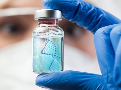
The science behind polynucleotides – a revolutionary treatment that offers a comprehensive approach to skin rejuvenation. How does it compare?
-

Getting adequate rest is the best investment you can make into your beauty. We take a look at some of your body’s natural skin rejuvenation processes that occur during sleep.
-
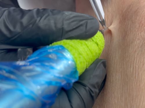
Not liking those pesky skin tags? Learn about causes, blemish removal options at Creative Touch, and prevention tips for smoother, bump-free skin.
-

Searching for the best lip balm for dry lips? Discover which ingredients have real evidence behind them, why your lips get dry, and how to choose products that actually work.
-

Don’t let loose skin overshadow your weight loss success. Learn why skin sags after significant weight loss and explore our advanced skin tightening and volume restoration treatments.
-

Dermal connective tissue cells that synthesise collagen, elastin, hyaluronic acid, and the broader extracellular matrix.
-

Elevate your autumn skincare routine with our expert advice and innovative treatments: achieve a radiant complexion as the seasons change and the leaves fall.
-
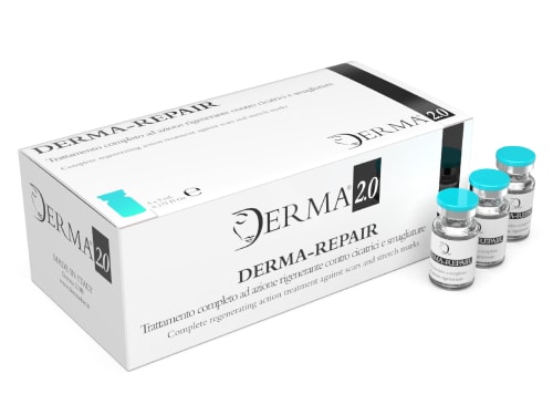
Scars and stretch marks can be a source of self-consciousness. Derma-Repair, a medical-grade mesotherapy solution, offers a powerful approach to address these concerns.
-

If you can’t stop thinking about food, it might not be addiction. Discover why chronic dieting and restriction create a biological backlash, and how to fix it.