
Dexpanthenol
Panthenol’s three mechanisms in skin are genuinely distinct rather than variations on a single theme. As a coenzyme A (CoA) precursor, it supports the metabolic pathways that drive both keratinocyte and fibroblast function – pantothenic acid deficiency measurably suppresses keratinocyte proliferation and skews differentiation, and calcium pantothenate modulates fibroblast gene expression in a direction consistent with active repair rather than maintenance. As a humectant, it reduces TEWL at concentrations as low as 1% with effects measurable at 30 weeks of consistent use. As an anti-inflammatory, dexpanthenol has a clinical evidence base in atopic dermatitis sufficient to earn a consensus recommendation as both a flare-reduction and topical corticosteroid-sparing agent; a clinical status that positions it above most cosmeceutical “soothing” ingredients.
Panthenol is the provitamin, alcohol form of pantothenic acid (vitamin B5), used in topical formulations because it is more stable and better absorbed through the stratum corneum than pantothenic acid itself. Once inside keratinocytes and fibroblasts, panthenol is oxidised to pantothenic acid, which is then incorporated into coenzyme A (CoA) – the central metabolic cofactor involved in fatty acid synthesis, the citric acid cycle, and the acetylation reactions that regulate gene expression. [3] This intracellular conversion is the foundation of all of panthenol’s downstream skin effects: it is not acting topically on the stratum corneum surface but metabolically within the cells that build and maintain it.
The alias situation is worth clarifying directly, as it causes consistent confusion in formulation literature. Panthenol and dexpanthenol refer to the same molecule (D-panthenol, the biologically active enantiomer) used interchangeably in skincare and pharmaceutical formulations. DL-panthenol is the racemic mixture. Pantothenic acid is the vitamin form after oxidation. Calcium pantothenate is the dietary supplement salt form. All convert to pantothenic acid in tissue; all are biologically equivalent at the point of CoA synthesis. The distinction matters for reading research literature accurately – clinical studies use these names inconsistently – but not for formulation or clinical outcome purposes.
Keratinocyte Proliferation and the Differentiation Balance
Pantothenic acid is not optional for normal epidermal function. A 2011 study in the Journal of Pharmacological Sciences (Kobayashi et al.) demonstrated that depleting pantothenic acid from keratinocyte culture medium suppressed proliferation, promoted premature differentiation, and increased apoptosis – effects reversed by pantothenic acid reintroduction. [4] The mechanism operates through CoA ; without adequate CoA, keratinocytes cannot sustain the energy metabolism and acetylation reactions that drive active cell division, and the balance tips from proliferation toward differentiation.
This is a clinically meaningful finding. In barrier-compromised skin – post-procedure, atopic-tendency, perimenopausal – the epidermal renewal rate is already reduced, and the differentiation programme is often dysregulated in directions that produce a thinner, less regularly structured stratum corneum. Panthenol supports the proliferative capacity of the basal keratinocyte population that is responsible for producing the replacement cells the barrier needs. It is not accelerating turnover artificially; it is maintaining the metabolic conditions under which normal turnover can proceed.
Fibroblast Activation and Wound Healing
In the dermis, calcium pantothenate modulates fibroblast gene expression in a specific direction. A 2009 gene expression study (Biro et al.) established that calcium pantothenate (20 µg/mL) in proliferating human dermal fibroblasts downregulated fibronectin, transferrin receptor, and several MHC Class I-associated genes, whilst upregulating metabolic and synthetic pathways – a pattern consistent with active repair-phase fibroblast behaviour rather than quiescent maintenance. [6] Pantothenate also measurably increased fibroblast proliferation and Ki67 expression (the cell cycle marker), confirming genuine mitogenic activity rather than simply altered gene expression without functional consequence. [3]
The wound-healing parallel in the current content is therefore mechanistically accurate, not merely a useful metaphor. Barrier damage produces the same cellular repair demands as a superficial wound: keratinocyte re-epithelialisation, fibroblast migration into the repair zone, ECM deposition. Panthenol supports both cell types through the CoA pathway at exactly the stages where these processes are most metabolically demanding.
A 2023 study in Advanced Healthcare Materials (Panthenol Citrate biomaterial formulation) confirmed that panthenol derivatives promote keratinocyte and dermal fibroblast migration and proliferation through integrin α3 upregulation – the cell-surface receptor that guides keratinocyte movement during re-epithelialisation – producing improved re-epithelialisation and granulation tissue formation in wound models. [5] The integrin α3 mechanism specifically – regulating epidermal growth, differentiation, and fibroblast migration – gives panthenol’s wound-healing activity a more precise cellular address than “supports healing.”
Humectant Function and TEWL Reduction
Topically applied panthenol is a humectant: it attracts and retains water within the stratum corneum, reducing transepidermal water loss through a film-forming mechanism at the skin surface. A concentration-dependent clinical study measuring panthenol across 0.5%, 1.0%, and 5.0% formulations found that significant TEWL reduction required at least 1.0% concentration, with effects measurable at 30 weeks of daily use and evident within two hours of a single application in barrier-disrupted skin (post-SLES wash test). library.scconline.org The 0.5% concentration produced no statistically significant TEWL reduction.
This concentration threshold matters for reading product labels. Panthenol appears in many formulations at concentrations below 1%, often as a label-presence ingredient rather than a functionally effective one. Formulations where barrier repair is the stated goal should contain panthenol at 1% or above to deliver the documented TEWL-reduction benefit.
The humectant mechanism is distinct from the cellular CoA mechanism: one operates at the stratum corneum surface, the other within viable keratinocytes. Both contribute to barrier function through different routes, which is why panthenol’s overall barrier benefit is greater than either mechanism alone would suggest.
Dexpanthenol in Atopic Dermatitis: A Clinical Evidence Standard
The most robust clinical evidence base for panthenol is in atopic dermatitis. A 2022 consensus review (Use of Dexpanthenol for Atopic Dermatitis, Seoul National University) established that dexpanthenol improves skin barrier function, reduces acute flare frequency, and has a significant topical corticosteroid (TCS)-sparing effect in mild to moderate AD. [1]
The TCS-sparing finding is clinically significant. Topical corticosteroids are effective for acute AD flares but carry risks of skin atrophy, barrier thinning, and rebound flare with prolonged use. An ingredient that reduces the frequency with which corticosteroids are needed – by maintaining barrier function and reducing flare frequency between acute episodes – has a clinical utility well beyond the cosmeceutical “soothing” category. The consensus review positions dexpanthenol as an appropriate maintenance treatment for AD, not merely a supportive adjunct.
This evidence level – a consensus recommendation with TCS-sparing data – makes dexpanthenol one of the few non-prescription topical ingredients with a defined clinical role in a diagnosed inflammatory skin condition rather than a general cosmeceutical claim.
Clinical Application
The clinical question panthenol creates is a positioning question: where in a barrier repair protocol does it fit, and what is it doing that other barrier ingredients are not? The answer depends on which of its three mechanisms is most relevant to the client’s current skin state.
For a client in acute barrier disruption – post-procedure, post-inflammatory flare, or in an atopic tendency presentation – the wound-healing mechanism is primary: supporting keratinocyte proliferation and fibroblast migration at a point where the skin is actively trying to re-epithelialise. For a client managing chronic barrier insufficiency – perimenopausal ceramide decline, sensitised skin maintaining daily exposure to desiccating environments – the TEWL-reduction humectant mechanism is primary. For a client with inflammatory tendency or AD-pattern reactivity, the anti-inflammatory and TCS-sparing mechanism is primary.
In practice, all three apply simultaneously. But the sequencing emphasis changes.
Phase 1: Acute Recovery – Post-Procedure and Disrupted Barrier
In the immediate post-procedure window – after microneedling, laser, chemical peel, or any treatment involving barrier disruption – the skin is performing the same cellular tasks as wound healing: keratinocyte proliferation, fibroblast migration into the repair zone, and re-epithelialisation. Panthenol in the post-procedure homecare formulation supports all three through the CoA and integrin α3 mechanisms directly relevant to this phase.
Cold atmospheric plasma (CAP) treatments produce controlled RONS-mediated cell stress and immune activation. Post-CAP, the keratinocyte and fibroblast populations are in an activated state that the CoA-supported proliferation and migration pathways panthenol supports are exactly positioned for. Including panthenol in the post-CAP recovery formulation is not precautionary soothing, it is providing metabolic support to the cell types that are doing the repair work the treatment has initiated.
A 2025 RCT of a panthenol-enriched mask applied after non-ablative laser treatment confirmed significant improvement in post-procedure skin hydration, barrier function, erythema reduction, and melanin index at days 3, 7, and 14 compared to controls – with the melanin index reduction particularly relevant for FST III–VI clients where post-procedure dyspigmentation is the primary recovery concern. [2]
The practical formulation consideration for post-procedure use is concentration: at least 1% for TEWL reduction, ideally within a formulation that also provides ceramides and FFAs for structural barrier repair. Panthenol is genuinely compatible with ceramide- cholesterol-FFA barrier repair formulations; the mechanisms are complementary without interaction.
Phase 2: Maintenance – Managing Chronic Barrier Insufficiency
For clients managing ongoing barrier fragility – the perimenopausal skin state with reduced ceramide synthesis, the sensitised skin that never fully settles, the AD-tendency client managing between flares – panthenol at 1%+ in a daily moisturiser or targeted serum provides sustained TEWL reduction that complements but does not replace the structural ceramide support.
The niacinamide + panthenol pairing is particularly well-matched for this client group. Niacinamide addresses the ceramide synthesis rate through SPT upregulation, restoring structural barrier lipid production. Panthenol reduces TEWL at the surface and supports the keratinocyte proliferation that is building new stratum corneum from below. The two mechanisms are operating at different points in the same barrier maintenance cycle (synthesis and surface retention) without overlap.
For clients with AD-tendency presentations where the ceramide suppression pathway ( IL-4/ IL-13 driven FLG and SPT suppression established in the Filaggrin and Ceramides entities) is active, panthenol’s anti-inflammatory contribution through the dexpanthenol AD mechanism is a relevant addition. It is not replacing CAP or polynucleotides as professional cytokine-burden reduction tools, it is a daily maintenance anti-inflammatory contribution that sustains the improvements the professional treatments have created.
Concentration and Formulation Guidance
The 1% minimum concentration for TEWL reduction is the practical clinical threshold for recommending panthenol-containing products. Below 1%, panthenol contributes to the formulation’s overall skin feel and humectant blend but does not deliver the documented barrier function benefit independently. This is worth reviewing when recommending specific products. Many widely-used barrier repair formulations contain panthenol at well below 1%, relying on the ceramide and NMF components for functional barrier repair rather than panthenol’s independent contribution.
Panthenol is stable, odourless, non-sensitising, and compatible with essentially all other actives in barrier formulations. It has no known significant interactions with retinoids, vitamin C, niacinamide, or ceramide preparations, and its safety in pregnancy and breastfeeding is well-established; relevant given the frequency with which pregnancy prompts an interest in clean, evidence-supported skincare.
References
Cho YS, Kim HO, Woo SM, et al. (2022). Use of Dexpanthenol for Atopic Dermatitis-Benefits and Recommendations Based on Current Evidence. J Clin Med, 11(14) . doi.org/10.3390/jcm11143943
Gao M, Gao N, Wang L, et al. (2025). Evaluation of the Efficacy and Safety of a Panthenol-Enriched Mask for Skin Barrier Recovery After Facial Laser Treatment: Results of a Double-Blind Randomized Controlled Study. J Cosmet Dermatol, 24(7), e70223 . doi.org/10.1111/jocd.70223
Gorski J, Proksch E, Baron JM, et al. (2020). Dexpanthenol in Wound Healing after Medical and Cosmetic Interventions (Postprocedure Wound Healing). Pharmaceuticals (Basel), 13(7) . doi.org/10.3390/ph13070138
Kobayashi D, Kusama M, Onda M, et al. (2011). The effect of pantothenic acid deficiency on keratinocyte proliferation and the synthesis of keratinocyte growth factor and collagen in fibroblasts. J Pharmacol Sci, 115(2), 230-4 . doi.org/10.1254/jphs.10224sc
Wang H, Duan C, Keate RL, et al. (2023). Panthenol Citrate Biomaterials Accelerate Wound Healing and Restore Tissue Integrity. Adv Healthc Mater, 12(31), e2301683 . doi.org/10.1002/adhm.202301683
Wiederholt T, Heise R, Skazik C, et al. (2009). Calcium pantothenate modulates gene expression in proliferating human dermal fibroblasts. Exp Dermatol, 18(11), 969-78 . doi.org/10.1111/j.1600-0625.2009.00884.x
Molecular Structure
- Formula
- C₉H₁₉NO₄
- Weight
- 205.25 g/mol
- IUPAC
- (2R)-2,4-dihydroxy-N-(3-hydroxypropyl)-3,3-dimethylbutanamide
Computational Identifiers
| InChI | InChI=1S/C9H19NO4/c1-9(2,6-12)7(13)8(14)10-4-3-5-11/h7,11-13H,3-6H2,1-2H3,(H,10,14)/t7-/m0/s1 | |
|---|---|---|
| InChIKey | SNPLKNRPJHDVJA-ZETCQYMHSA-N | |
| Canonical SMILES | CC(C)(CO)C(C(=O)NCCCO)O | |
| Isomeric SMILES | CC(C)(CO)[C@H](C(=O)NCCCO)O | |
Data sourced from: PubChem (NCBI) ↗ | ||
Also Known As
- D-Panthenol
- Panthenol
- Pro-vitamin B5
- Provitamin B5
Biological Relationships
Biological Interactions
- Inhibits Transepidermal water loss Evidence: Dexpanthenol reduces TEWL – explicitly listed in entity executive summary as a treatment. Clinical evidence in atopic dermatitis. DOI:10.1111/jocd.16171
Influenced By
- this May treat Dermatitis Evidence: 2022 consensus review (Seoul National Univ.) established dexpanthenol improves barrier function, reduces AD flare frequency, and has TCS-sparing effect in mild-to-moderate atopic dermatitis (PMID 35887707).
Learn More
This topic is discussed in 3 articles:
-

Has anti-inflammatory properties and supports wound healing, relevant because barrier damage is essentially a form of microscopic wounding
-
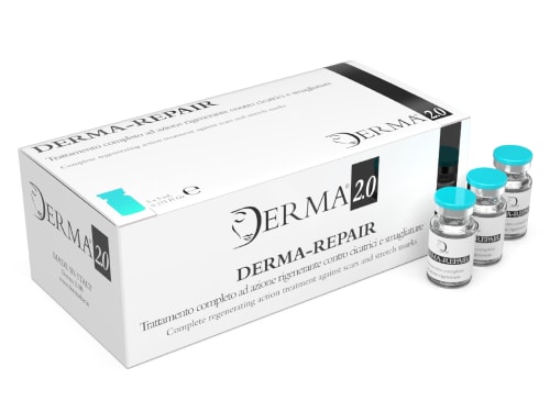
Scars and stretch marks can be a source of self-consciousness. Derma-Repair, a medical-grade mesotherapy solution, offers a powerful approach to address these concerns.
-

Provitamin form of pantothenic acid (B5) that converts to pantothenic acid within skin cells, supporting keratinocyte proliferation, dermal fibroblast migration, and barrier function.