
Polynucleotides
Polynucleotides (PN) are highly purified, DNA-derived fragments that act as regenerative “environment shifters.” Unlike traditional biostimulators that demand a response from increasingly unresponsive senescent fibroblasts, PN operates primarily through Adenosine A2A Receptor (A2AR) activation on macrophages. This triggers a phenotypic shift from pro-inflammatory (M1) to reparative (M2) states, indirectly restoring the fibroblast environment. Clinically, this translates to a simultaneous “Tap and Plug” effect: upregulating collagen synthesis via SMAD2/3 while suppressing the destructive NF-κB/MMP pathway. This makes PN the non-negotiable “priming” step for post-menopausal skin and chronically inflamed barriers.
Polynucleotides (PN) and their closely related subclass polydeoxyribonucleotides (PDRN) are injectable preparations of highly purified, fragmented DNA derived from salmon or trout sperm cells. [6] The source material is chosen for its high degree of structural similarity to human DNA. The base pair composition and backbone chemistry of salmon DNA is sufficiently close to human DNA that the preparations are well-tolerated without triggering inflammatory or immunogenic responses when purified to medical-grade standard. The key distinction in terminology: PDRN refers specifically to preparations in the 50–2000 base pair molecular weight range; PN is used more broadly and encompasses larger fragment preparations. Both act through the same primary receptor pathway. [4]
The Primary Mechanism: A2AR Activation and the Macrophage-Fibroblast Axis
The central mechanism of polynucleotide action in skin is not direct fibroblast stimulation but macrophage reprogramming that then indirectly drives fibroblast activity. [1] This distinction is clinically important and is what makes polynucleotides particularly relevant for aged and post-menopausal skin where direct TGF-β responsiveness in fibroblasts has declined.
Polynucleotides, degraded by endogenous nucleases to nucleotide fragments, bind to the adenosine A2A receptor (A2AR) on macrophages. A2AR activation increases adenylate cyclase (AC) activity, elevating intracellular cAMP levels, which activates protein kinase A (PKA). PKA phosphorylates the transcription factor CREB (cAMP response element-binding protein), which upregulates PCK1 (phosphoenolpyruvate carboxykinase 1). [1] A 2025 study provided the first direct experimental evidence that PCK1 acts as the downstream mediator of adenosine receptor activation in this pathway – a mechanistic detail previously inferred but not directly demonstrated. PCK1 upregulation reduces ROS production in macrophages, suppressing M1 polarisation and promoting M2 polarisation. M2-polarised macrophages then secrete IL-10 and TGF-β into the surrounding tissue.
These M2-derived cytokines act on adjacent dermal fibroblasts through SMAD2/3 and STAT3 signalling: pSMAD2/3 increases procollagen I and III synthesis; pSTAT3 reduces NF-κB activity in fibroblasts, simultaneously suppressing MMP-1, MMP-3, and MMP-9 expression. [1] The net result is a tissue environment that is simultaneously more synthetically active and less degradative – not through a single on/off signal but through a cascading reprogramming of the macrophage population that then reshapes the fibroblast environment.
The A2AR Signaling Cascade
| Stage | Action | Downstream Effect |
|---|---|---|
| Binding | PN Fragments → A2A Receptor | Increases Adenylate Cyclase (AC) activity. |
| Signaling | ↑ cAMP → PKA Phosphorylation | Activates CREB and PCK1 expression. |
| Polarization | ROS Reduction | Shifts Macrophage from M1 (Destructive) to M2 (Reparative). |
| Secretions | Release of IL-10 & TGF-β | Triggers Fibroblast SMAD2/3 signaling. |
| Net Result | NF-κB Suppression | Inhibits MMPs 1, 3, 9, and 12. |
Why This Pathway Matters for Aged Skin
The macrophage-mediated route is not simply an alternative to direct TGF-β delivery – it is a route that ages less poorly than direct fibroblast stimulation. In aged and post-menopausal skin, dermal fibroblasts accumulate senescence, reducing their direct TGF-β receptor responsiveness. Direct growth factor delivery (through iPRF, wound-healing cascades, or exogenous TGF-β) reaches a fibroblast population whose capacity to respond has been diminished. The macrophage-fibroblast axis that polynucleotides activate involves a different cell type as the primary responder, and macrophage A2AR responsiveness declines less with age than fibroblast TGF-β sensitivity. [1] This is why polynucleotides are not simply a “gentler” version of growth factor treatments. They access a collagen synthesis restoration route that is specifically better preserved in the tissue environment where other routes have been compromised.
Clinical Logic: PN vs. Traditional Stimulators
| Feature | Traditional Stimulators (RFM/iPRF) | Polynucleotides (PN) |
|---|---|---|
| Primary Responder | Fibroblasts (Direct) | Macrophages (Indirect relay) |
| Aged Skin Barrier | Fibroblast senescence reduces response. | A2AR pathway is better preserved in age. |
| Tissue Impact | Adds stimulus to current environment. | Changes the environment itself. |
| Degradation | Variable impact on MMPs. | Directly silences NF-κB/MMP cascade. |
MMP Suppression: The Degradation Side of the Balance
Beyond synthesis stimulation, the NF-κB suppression in fibroblasts driven by M2 macrophage-derived signals reduces the three primary collagenolytic and stromelytic MMPs (MMP-1, MMP-3, and MMP-9) simultaneously with collagen synthesis activation. [4] This dual action on the synthesis–degradation balance is what distinguishes polynucleotides from treatments that stimulate only one side of that equation. MMP-12 (macrophage metalloelastase) suppression through the same NF-κB pathway also protects elastin fibres and the fibrillin-1 scaffold they assemble on, making polynucleotides relevant to elastin preservation alongside collagen restoration.
The proteoglycan decorin – which governs collagen fibril organisation and is degraded by MMPs in photoaged skin – is also protected by this NF-κB/MMP suppression, preserving the architectural order of the collagen matrix in addition to its quantity.
VEGF-Mediated Angiogenesis
A mechanistic dimension that is distinct from the collagen synthesis and MMP suppression pathways is polynucleotides’ stimulation of VEGF ( vascular endothelial growth factor) expression through A2AR activation. PDRN treatment in wound models increases VEGF mRNA and protein content, stimulates angiogenesis, and increases CD31 immunostaining (a marker of new capillary formation) alongside improved tissue remodelling. [2] In the context of skin rejuvenation, VEGF-driven capillary neovascularisation in treated dermis improves vascular supply to fibroblasts and keratinocytes, supporting the nutrient and oxygen delivery that sustains the tissue environment improvements the treatment produces. This makes polynucleotides a simultaneously regenerative and vascular treatment – a combination that most other biostimulators do not replicate through a single mechanism.
Barrier Function Effects
A 2025 study investigating polynucleotides in an atopic dermatitis model found that PN treatment significantly reduced TEWL, decreased mast cell and eosinophil infiltration in the dermis, and reduced abnormal collagen deposition in inflamed tissue – whilst separately, the anti-inflammatory pathway reduced the epidermal and dermal thickening characteristic of atopic inflammation. [3] This is not a direct ceramide synthesis effect but a downstream consequence of the NF-κB and M2-driven inflammatory resolution that removes the cytokine signals suppressing barrier function. For clients with reactive or atopic-tendency skin, this means polynucleotides address barrier dysfunction from the inflammatory environment side rather than the structural lipid side; complementary to topical barrier support rather than an alternative to it.
Source, Preparation, and Biocompatibility
The salmon or trout DNA source material is processed through rigorous purification (removal of proteins, lipids, and cellular debris) to produce a preparation that contains only the DNA fragments and carrier solution. [6] The biocompatibility arises from the structural similarity of fish and human DNA at the base pair level, not from any special property of salmon DNA itself. The purification removes everything that would distinguish the source species. Allergic reactions are rare but not impossible; documented contraindications include known fish allergy and autoimmune conditions.
Clinical Application
Polynucleotides sit in an unusual position in the Creative Touch treatment portfolio, not because they’re the most dramatic treatment, but because they’re the most environmentally complete one. Every other treatment we offer that stimulates collagen synthesis is entering the tissue and asking fibroblasts to produce more. Polynucleotides are doing something different first: they’re changing what kind of tissue environment those fibroblasts are living in. That distinction shapes everything about when to use them, what to pair them with, and what to tell clients to expect.
The clearest way to understand the clinical positioning is through a simple framework: treatments that restore a favourable tissue environment need to precede or accompany treatments that deliver direct stimulation, precisely because direct stimulation into an unfavourable environment produces attenuated results. Polynucleotides are the most complete environment-restoring treatment in the portfolio. They suppress the MMP activity that’s actively degrading whatever new collagen is being produced, they reprogramme the macrophage population from inflammatory to reparative, and they do this through a pathway (the A2AR–macrophage axis) that is specifically better preserved in aged tissue than the direct TGF-β responsiveness those same fibroblasts have lost. [1] Using them after RF microneedling has already been applied to a compromised environment is working backwards.
Who benefits most
Post-menopausal clients – for whom polynucleotides aren’t simply a good choice among several equivalent options, but the most mechanistically appropriate first intervention. Oestrogen withdrawal has simultaneously increased MMP activity, reduced TIMP counter-regulation, and diminished direct TGF-β responsiveness in fibroblasts that are accumulating senescence alongside hormonal change. Polynucleotides reach fibroblasts through macrophages as the primary responder, and macrophage A2AR responsiveness declines less with age than fibroblast sensitivity to direct growth factor signals. [1] This is the practical reason why post-menopausal clients who go straight to RF microneedling or iPRF without polynucleotides often see results that plateau earlier and fade faster than the treatment mechanism alone would predict.
Significantly photodamaged skin – where MMP activity is the dominant concern. A single UV exposure induces MMP-1 and MMP-3 mRNA at several-thousand-fold above baseline at 24 hours. [5] Decades of cumulative exposure means the NF-κB and AP-1 pathways driving that response have been chronically activated, and the TIMP counter-system progressively eroded. Stimulating collagen synthesis in that environment without simultaneously suppressing MMP activity is like filling a bath with the plug out. Polynucleotides’ NF-κB suppression (reaching MMP-1, MMP-3, MMP-9, and MMP-12 simultaneously) addresses the plug rather than just the tap.
Reactive or chronically inflamed skin – where persistent NF-κB activation and type 2 cytokines are suppressing ceramide synthesis, elevating MMPs, and keeping fibroblasts in a low-output state. Polynucleotides quiet the source rather than working around it. The anti-inflammatory mechanism produces real downstream barrier effects, confirmed in a 2025 study showing measurable TEWL reduction and reduced mast cell infiltration [3] This makes them complementary to topical barrier repair rather than a replacement for it.
Pre-treatment environment preparation – for any client where the dermal environment is unfavourable for direct stimulation: high MMP activity, significant senescent cell burden, or post-inflammatory suppression. A course of polynucleotides before introducing iPRF, RF microneedling, or microneedling produces better outcomes than leading with the stimulating modality into unprepared tissue.
Setting expectations honestly
The timeline is the thing that most requires careful communication, because it runs counter to what clients have been conditioned to expect. Polynucleotides produce no immediate visual change; no volume, no visible lifting, nothing to see in the mirror that afternoon. Improvement develops from around three to four weeks as collagen density increases and the inflammatory environment resolves, with progressive change through eight to twelve weeks.
That timeline isn’t a limitation but rather it’s the biology of macrophage polarisation and fibroblast collagen synthesis, which operates over weeks rather than hours. The honest framing in consultation is: this treatment is changing the environment your skin cells work in. That takes time to show. By your eight-week review, the changes in skin quality, texture, and resilience will be visible and measurable. It’s a different kind of result to anything that fills or lifts immediately, but one that’s actually restoring something rather than masking it.
Where the combinations are most productive
Polynucleotides + iPRF is the most complementary pairing in the portfolio because the two mechanisms are genuinely independent. iPRF delivers growth factors directly to fibroblasts; polynucleotides create the sustained anti-inflammatory environment that improves how effectively those signals land. For post-menopausal clients, the combination reaches fibroblasts through two routes where neither alone is fully sufficient. Sequencing matters: polynucleotides first or concurrent, not after iPRF has already been applied into an unprepared environment.
Polynucleotides + HA skin boosters addresses complementary deficits that happen to coexist in the same tissue. Skin boosters restore the physical ground substance volume and mechanical tension that drives fibroblast mechanoreception; polynucleotides restore the signalling environment and suppress degradation. There’s no mechanistic conflict, and the combination should be the default protocol for post-menopausal clients presenting with both structural volume loss and skin quality concerns – which is most of them.
Polynucleotides + RF microneedling – the most complete protocol for active structural collagen loss. RF microneedling provides thermal remodelling and direct TGF-β stimulus in the reticular dermis; polynucleotides provide the MMP-suppressive, pro-regenerative environment in which that stimulus produces lasting structural change rather than transient improvement. Polynucleotides first to establish the environment, then RF microneedling to deliver active remodelling into it.
The “Priming” Protocol
| Companion Treatment | The Synergistic Logic | Recommended Sequencing |
|---|---|---|
| RF Microneedling | PN silences MMPs so RFM-induced collagen survives. | PN 2–4 weeks prior to RFM. |
| iPRF | PN creates the “soil”; iPRF provides the “seeds” (GFs). | Concurrent or PN first. |
| HA Skin Boosters | PN fixes the signal; HA fixes the physical tension. | Concurrent. |
| Topical Vitamin C | PN suppresses MMP transcription; Vit C matures the protein. | Daily homecare during PN course. |
References
Byun KA, Park HJ, Oh S, et al. (2025). Polynucleotides Enhance Collagen Synthesis via Modulating Phosphoenolpyruvate Carboxykinase 1 in Senescent Macrophages: Experimental Evidence. Int J Mol Sci, 26(17) . doi.org/10.3390/ijms26178720
Galeano M, Bitto A, Altavilla D, et al. (2008). Polydeoxyribonucleotide stimulates angiogenesis and wound healing in the genetically diabetic mouse. Wound Repair Regen, 16(2), 208-17 . doi.org/10.1111/j.1524-475x.2008.00361.x
Ha YJ, Tak KH, Lee JL, et al. (2025). Polynucleotides Enhance Skin Barrier Function and Reduce Inflammation in a 2,4-Dinitrochlorobenzene-Induced Mouse Model of Atopic Dermatitis. Skin Res Technol, 31(6), e70189 . doi.org/10.1111/srt.70189
Lee KWA, Chan KWL, Lee A, et al. (2024). Polynucleotides in Aesthetic Medicine: A Review of Current Practices and Perceived Effectiveness. Int J Mol Sci, 25(15) . doi.org/10.3390/ijms25158224
Quan T, Qin Z, Xia W, et al. (2009). Matrix-degrading metalloproteinases in photoaging. J Investig Dermatol Symp Proc, 14(1), 20-4 . doi.org/10.1038/jidsymp.2009.8
Squadrito F, Bitto A, Irrera N, et al. (2017). Pharmacological Activity and Clinical Use of PDRN. Front Pharmacol, 8, 224 . doi.org/10.3389/fphar.2017.00224
Also Known As
- PN
- polynucleotide
Therapeutic Relationships
Therapeutic Context
- Stimulates Elastin Evidence: Polynucleotides drive M2 macrophage-fibroblast axis activation supporting elastin synthesis; in vivo study showed significant skin elasticity improvement. PMC12429772.
- Stimulates Fibroblast Evidence: PN stimulates fibroblast activity via M2 macrophage-derived IL-10 and TGF-β; pSMAD2/3 and pSTAT3 increase in fibroblasts, driving collagen I and III synthesis. PMC12429772.
- Stimulates Hair follicle Evidence: PN stimulates dermal papilla fibroblast paracrine signals for follicular regeneration, extends anagen via VEGF vascularisation, reduces follicular inflammatory burden. PMID 39951159.
- Stimulates Heparan sulfate proteoglycans
- Stimulates Tissue regeneration Evidence: PN promotes cellular proliferation, migration and differentiation across skin cell types; review of 35 studies confirmed tissue regeneration as primary effect. PMC11311621; PMC6299200.
- Stimulates Transforming growth factor beta Evidence: Polynucleotides activate A2AR/AC/PKA/CREB/PCK1 in macrophages, increasing M2 polarisation and subsequent TGF-beta secretion; this paracrine TGF-beta then activates SMAD2/STAT3 in fibroblasts (PMC12429772; Collagen entity).
- Stimulates Vascular endothelial growth factor Evidence: PDRN increases VEGF mRNA and protein, stimulates angiogenesis, increases CD31 immunostaining (new capillary formation) via A2AR activation. PMC6299200, Cell Transplantation 2018.
- Inhibits Cellular senescence Evidence: PN modulates macrophage-fibroblast interactions via CREB/PCK1 axis in senescent cell models, counteracting age-related ECM changes and restoring collagen synthesis capacity. PMC12429772.
- Inhibits Inflammageing Evidence: PN activates A2AR→PKA/CREB→PCK1, reducing oxidative stress and shifting macrophages M1→M2, directly suppressing the chronic low-grade inflammation defining inflammageing. PMC12429772.
- Inhibits Matrix metalloproteinase
- Treats Androgenetic alopecia Evidence: Prospective study n=28: PN injections at 4-week intervals produced significant improvement in hair diameter and density; 82.1% patient satisfaction. PMID 39951159, Arch Dermatol Res 2025.
- Treats Hyperpigmentation Evidence: PDRN demonstrates anti-melanogenesis activity; PN HPT improved pigmentation category significantly over 6-month follow-up in Asian cohort n=30. PMC10874187; PMC11311621.
- Treats Rosacea Evidence: PN NF-κB suppression and M2 macrophage polarisation directly address the neurogenic/vascular inflammatory environment of rosacea. Entity text; PMC12145983.
- Treats Skin ageing Evidence: Multiple RCTs and cohort studies confirm PN/PDRN improves skin texture, reduces wrinkle depth, enhances elasticity; A2AR-macrophage-fibroblast axis restores collagen synthesis. PMC11311621.
- Treats Telogen effluvium Evidence: PN anti-inflammatory and vascular effects address chronic inflammatory scalp environment contributing to prolonged telogen effluvium; documented in scalp treatment protocols. PMID 39951159.
- Affects Decorin Evidence: PN NF-κB suppression reduces MMP-mediated degradation of decorin (governs collagen fibril organisation), preserving collagen matrix architecture in aged/photoaged skin. Entity text.
- Affects Dermal Papilla Evidence: PN stimulates dermal papilla fibroblast activity; paracrine signalling from PN-activated fibroblasts initiates and sustains follicular regeneration and hair follicle neogenesis. PMID 39951159.
- Affects Keratinocyte Evidence: PN- HA combination enhanced HaCaT keratinocyte migration, proliferation, and VEGF/MMP gene expression in vitro; PN affects keratinocyte function. PMID 40009152, 2025.
Indications & References
- this Dermal Papilla Evidence: We view the DP as the primary target for iPRF and Polynucleotides: if we can restore the DP signalling output, we can override the suppressive veto of DHT and Cortisol.
- this Dermis Evidence: Polynucleotides specifically valuable for older or significantly photo-aged dermis where the proportion of responsive fibroblasts has fallen
- this Fibroblast Evidence: Polynucleotides activate macrophage M2 polarisation; IL-10/TGF-β activates SMAD2/STAT3 in senescent fibroblasts bypassing impaired direct TGF-β responsiveness. Pubmed 40943641; PMC11311621
- this Hair follicle Evidence: Polynucleotides stimulate FGF and tissue regeneration pathways in follicular environment. Gessese et al. (2024) Scientific World J 2024:5259055 confirm FGF as essential for follicular morphogenesis. doi:10.1155/tswj/5259055
- this Keratinocyte Evidence: Polynucleotides complement this through their A2AR-mediated anti-inflammatory pathway, calming the tissue environment in which keratinocyte differentiation is occurring
- this Skin Evidence: Polynucleotides deliver signals across papillary and reticular dermis; calm inflammation.
- this May treat Perimenopausal skin changes Evidence: Polynucleotides: A2AR-macrophage pathway circumvents senescent fibroblast TGF-β resistance; primary environment-preparation treatment for oestrogen-depleted skin. Entity text; PMC12429772.
- this May treat Skin barrier dysfunction Evidence: Polynucleotides suppress MMP activity, reprogramme macrophages, reduce cytokine burden at dermal level – explicitly cited as treatment for chronic barrier dysfunction. PMC12429772
Learn More
This topic is discussed in 5 articles:
-
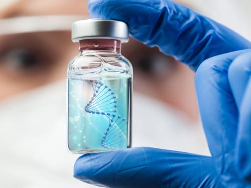
The science behind polynucleotides – a revolutionary treatment that offers a comprehensive approach to skin rejuvenation. How does it compare?
-

DNA-derived injectable treatment that stimulates tissue regeneration whilst calming inflammation, particularly beneficial for reactive or sensitised skin.
-

DNA-derived molecules that stimulate regeneration with notable anti-inflammatory properties. Research shows polynucleotides can improve barrier function even in conditions like atopic dermatitis.
-

Getting adequate rest is the best investment you can make into your beauty. We take a look at some of your body’s natural skin rejuvenation processes that occur during sleep.
-
