
Tissue regeneration
Tissue regeneration describes the biological process by which damaged or depleted tissue is restored through coordinated cellular activity – but regeneration is not a single outcome. It exists on a continuum from true like-for-like tissue replacement at one end, through functional-but-imperfect repair, to fibrotic scar replacement at the other. Which outcome a given healing event produces is determined by three principal variables: the balance of TGFβ isoforms in the repair environment, the transition timing of macrophages from pro-inflammatory to pro-regenerative phenotype, and the degree to which inflammageing has impaired the tissue’s stem and progenitor cell capacity. Regenerative aesthetics treatments – iPRF, polynucleotides, growth factor therapies – intervene in the proliferation and remodelling phases of tissue repair with the aim of shifting outcomes toward the regenerative end of the continuum. The inflammatory environment of the target tissue at the time of intervention is a primary determinant of whether they succeed.
Tissue regeneration is one of the most frequently invoked concepts in aesthetics – and one of the least precisely defined. “Regenerative treatment,” “stimulating the skin’s own repair processes,” and “activating natural renewal” appear across product marketing and clinical content, usually without distinguishing between the three genuinely different biological outcomes that repair processes can produce. That distinction matters practically: it explains why treatments work differently in different tissue environments, why two clients receiving identical protocols can have meaningfully different outcomes, and why the inflammatory state of the skin before treatment is not a secondary consideration but a primary determinant of what a regenerative stimulus will achieve.
Regeneration, Repair, and Fibrosis: The Continuum
When tissue is damaged, the body initiates a repair response whose outcome sits somewhere on a continuum between two extremes. At the regenerative end, damaged tissue is replaced with architecturally and functionally identical new tissue – the same cell types, the same extracellular matrix organisation, the same mechanical properties as the original. At the fibrotic end, damaged tissue is replaced with generic scar tissue – collagen-dense, mechanically stiff, poorly organised, and lacking the specialised cells and structures of the original. Between these extremes lies the most common outcome in adult human tissue: functional repair – tissue that is restored well enough to perform its basic role but that lacks the structural sophistication of the original. [1]
The reason adult mammals default to repair rather than true regeneration – whilst foetal wound healing in early gestation produces near-perfect tissue reconstruction with no scar – is not entirely resolved, but the evidence points to the inflammatory response as the key variable. Foetal wounds heal in a sterile, low-cytokine, TGFβ3-dominant environment with minimal macrophage infiltration; adult wounds heal in an environment rich in TGFβ1, TGFβ2, and inflammatory cytokines that drive fibrotic deposition. The foetal exception is not a curiosity – it is the biological proof of concept that the inflammatory environment, not the cellular repair machinery itself, is the primary determinant of where on the continuum an outcome lands. [4]
For aesthetics purposes, this framing redefines what regenerative treatments are doing: they are not simply delivering a biological stimulus and waiting for regeneration to occur. They are attempting to shift the local tissue environment toward the conditions under which regeneration – rather than repair or fibrosis – is the predominant outcome.
The Four Phases of Tissue Repair
Tissue repair proceeds through four sequential but overlapping phases. Regenerative treatments do not act uniformly across all four – understanding where they intervene clarifies what they can and cannot influence.
Haemostasis (minutes to hours) arrests bleeding through platelet aggregation and provisional fibrin matrix formation. This phase also initiates the subsequent inflammatory response through platelet degranulation and growth factor release – PDGF, TGFβ, VEGF, and EGF among them. In PRP and iPRF treatments, the injected platelet concentrate is delivering a concentrated version of this same initial growth factor release into tissue where no injury-initiated haemostatic response has occurred, seeding the repair sequence from the proliferative phase without the preceding inflammatory phase.
Inflammation (hours to days) recruits neutrophils and macrophages to the wound site, clearing debris and pathogens and producing the cytokine signals that transition the repair response into proliferation. This phase is necessary but its duration and intensity are critical: a controlled, resolving inflammatory phase facilitates progression to regenerative remodelling; a sustained or dysregulated inflammatory phase – the condition in ageing, rosacea, and chronically inflamed tissue – impairs the transition and drives the outcome toward fibrosis. [2]
Proliferation (days to weeks) is the phase most directly targeted by regenerative aesthetics treatments. Fibroblasts proliferate and migrate into the repair zone, synthesising new collagen (initially type III, subsequently remodelling toward type I), glycosaminoglycans, and other extracellular matrix components. Keratinocytes re-epithelialise the surface. New vasculature forms through angiogenesis. Growth factor signalling – from platelets, macrophages, and the fibroblasts themselves – drives and coordinates this activity. iPRF’s growth factor payload ( PDGF, TGFβ, IGF-1, VEGF, FGF) acts primarily on this phase, amplifying the proliferative signal in tissue where the endogenous platelet-derived growth factor concentration may be insufficient to drive optimal fibroblast activation.
Remodelling (weeks to months to years) reorganises the provisional collagen matrix laid down during proliferation into the mechanically organised architecture of mature tissue. Type III collagen is progressively replaced by type I; collagen fibres are cross-linked and aligned along mechanical stress lines; non-essential vasculature regresses. This phase is the biological basis for the extended timelines of regenerative treatment outcomes – the collagen laid down in the proliferative phase is not the collagen that determines the final visible result. Polynucleotides (PDRN) support this phase specifically through MMP suppression – reducing the enzymatic degradation that would otherwise break down newly synthesised matrix before remodelling is complete, and through adenosine receptor activation that promotes fibroblast activity during the extended remodelling window.
The TGFβ Isoform Balance
Transforming growth factor beta (TGFβ) is the central cytokine regulator of the repair/fibrosis/regeneration outcome, and its isoform composition in the repair environment is the molecular switch between these outcomes. [3]
TGFβ1 and TGFβ2 are the dominant isoforms in adult wound healing. They drive fibroblast activation, collagen synthesis, and myofibroblast differentiation – the contractile cells responsible for wound contraction and, in excess, for the dense collagen deposition of fibrotic scarring. Both isoforms suppress MMP activity (reducing matrix degradation, which maintains scar tissue) and promote epithelial-mesenchymal transition. Their activity is appropriate in the acute repair phase but becomes counterproductive if sustained, producing the excessive and disorganised collagen deposition of hypertrophic scars or the tissue rigidity of chronic fibrosis.
TGFβ3 is the isoform predominant in foetal wound healing and is substantially less abundant in adult repair. It promotes organised matrix deposition, facilitates the M1→M2 macrophage transition described below, and has anti-fibrotic rather than pro-fibrotic downstream signalling. Studies in adult tissue engineering and growth factor therapy research have consistently shown that shifting the TGFβ1:TGFβ3 ratio toward TGFβ3 produces more regenerative and less fibrotic outcomes. Platelet-derived growth factor preparations including iPRF contain TGFβ in a profile that includes TGFβ3 alongside the more abundant TGFβ1 – partly explaining the more regenerative tissue response observed with platelet concentrate therapies compared with injury-alone healing. [8]
Macrophage Phenotype: The Critical Checkpoint
The transition from the inflammatory phase to the proliferative and remodelling phases is gated by macrophage phenotype – and this checkpoint is where the regeneration/fibrosis outcome is most directly determined. [9]
M1 macrophages are the pro-inflammatory phenotype recruited in the early inflammatory phase. They produce TNF-α, IL-1β, IL-6, and IL-12, driving pathogen clearance and debris removal. Their activity is essential in the acute phase but must resolve for tissue repair to progress – sustained M1 activity maintains the inflammatory environment that suppresses fibroblast function and skews TGFβ signalling toward the fibrotic isoforms.
M2 macrophages are the pro-regenerative phenotype that dominate the later proliferative and remodelling phases. They produce TGFβ, VEGF, and IL-10, promoting fibroblast recruitment, angiogenesis, and matrix synthesis. The M1→M2 transition is the critical checkpoint: when it occurs on schedule, the repair response progresses toward regeneration; when it is delayed or incomplete – as occurs in chronic inflammation, ageing tissue, and diabetes – the sustained M1 environment drives the outcome toward fibrosis. [2]
Cold atmospheric plasma has a directly relevant mechanism here. Its RONS-mediated NF-κB modulation in activated macrophages facilitates the M1→M2 phenotype transition – providing one of the mechanistic explanations for CAP’s ability to convert a chronically inflamed tissue environment into one more capable of supporting regenerative outcomes. This is the biological basis for the treatment sequencing logic: resolving the inflammatory environment (CAP) before delivering a regenerative growth factor stimulus (iPRF, PDRN) is not a preference – it is a mechanistic imperative if the target tissue’s macrophage population is predominantly M1 at the time of treatment.
How Inflammageing Impairs Regenerative Capacity
In young, healthy tissue, the four-phase repair sequence proceeds with reasonable fidelity toward functional regeneration. In ageing tissue, chronic low-level inflammation – inflammageing – impairs the sequence at multiple points, progressively shifting the default outcome from regeneration toward fibrosis and from complete repair toward incomplete repair. [6]
The primary mechanisms are:
SASP cytokine suppression of progenitor cell function. Senescent cells – including the senescent adipocytes covered in the Subcutaneous Tissue entity – secrete SASP cytokines (IL-6, IL-8, TNF-α, MMP-3) into the local tissue environment. These cytokines suppress the proliferative capacity of adjacent stem and progenitor cells, reducing the pool of cells available to execute the regenerative response. A PNAS 2019 study demonstrated this directly: skeletal stem and progenitor cell function was significantly impaired by the inflammatory environment of ageing tissue, and reducing that inflammatory environment – rather than directly manipulating the stem cells – restored their regenerative capacity. [5] The implication is that the regenerative capacity of aged tissue is not fixed – it is environmentally suppressed, and can be partially restored by addressing the inflammatory environment.
Chronic NF-κB activation and the pro-fibrotic skew. NF-κB, the master transcription factor of the inflammatory response, is chronically activated in ageing tissue at a low level that does not produce acute inflammation but that maintains a baseline pro-inflammatory cytokine milieu. This chronically elevated NF-κB activity biases macrophage polarisation toward M1, sustains TGFβ1 dominance over TGFβ3, and suppresses the anti-inflammatory signalling that would normally facilitate the M1→M2 transition. The result is a tissue environment that is never fully in the pro-regenerative state required for optimal repair – even between injury events.
Impaired growth factor responsiveness. Ageing fibroblasts demonstrate reduced receptor expression and downstream signalling response to growth factors including PDGF and IGF-1, partly through chronic cytokine exposure and partly through intrinsic cellular ageing mechanisms including telomere shortening. This means that identical growth factor concentrations – from iPRF, for example – produce a weaker fibroblast activation response in aged tissue than in younger tissue, contributing to the clinical observation that older clients may require more treatment sessions to achieve comparable outcomes. [7]
Clinical Pearl The question “why do two clients having identical treatments get different results?” is one of the most common and least well-answered in aesthetics consultations. The answer lies almost entirely in tissue environment rather than treatment delivery: the client whose tissue has a lower senescent cell burden, a more responsive macrophage population, and a less NF-κB-skewed baseline will produce a more regenerative outcome from the same iPRF or PDRN protocol as the client whose tissue inflammageing has impaired all three. This is the mechanistic basis for investing in skin health as a precondition for regenerative treatment rather than as an alternative to it.
Clinical Application
The timeline is mechanistic, not arbitrary
The months-long result timeline of regenerative treatments reflects the biology of the remodelling phase – new collagen must be synthesised, cross-linked, and architecturally reorganised before it produces visible improvement in skin quality. Informing clients that gradual improvement over 2–4 months reflects successful tissue remodelling rather than slow treatment response is accurate and useful – but it is more credible and more clinically honest when supported by the mechanism rather than offered as reassurance alone.
The treatment sequencing logic: environment → stimulus → support
The entities across this knowledge graph converge on a consistent clinical principle: the tissue environment determines what a regenerative stimulus achieves. Delivering iPRF or PDRN into tissue where M1 macrophages are dominant, NF-κB is chronically activated, and TGFβ1 suppresses TGFβ3 produces a partial response at best. The same stimulus delivered into tissue where the inflammatory environment has been resolved – through CAP, through barrier restoration, through optimised skincare – operates in conditions closer to those under which regeneration rather than repair is the default outcome. This is not a theoretical consideration: it is the mechanistic basis for the treatment sequencing protocols that distinguish considered regenerative practice from single-modality intervention.
Why results vary – and what to do about it
Clients whose baseline tissue environment is more inflamed – whether from rosacea, atopic dermatitis, chronic barrier disruption, or the inflammageing of later decades – will produce less regenerative responses to identical treatments than clients with a healthier baseline. This is not a reason to withhold regenerative treatments from these clients; it is a reason to invest in environment preparation first, to set realistic expectations about the number of sessions required, and to support the remodelling phase actively rather than assuming it will proceed optimally on its own.
References
Atala A, Irvine DJ, Moses M, et al. (2010). Wound Healing Versus Regeneration: Role of the Tissue Environment in Regenerative Medicine. MRS Bull, 35(8) . doi.org/10.1557/mrs2010.528
Eming SA, Martin P, Tomic-Canic M (2014). Wound repair and regeneration: mechanisms, signaling, and translation. Sci Transl Med, 6(265), 265sr6 . doi.org/10.1126/scitranslmed.3009337
Gilbert RWD, Vickaryous MK, Viloria-Petit AM (2016). Signalling by Transforming Growth Factor Beta Isoforms in Wound Healing and Tissue Regeneration. J Dev Biol, 4(2) . doi.org/10.3390/jdb4020021
Jiang D, Rinkevich Y (2020). Scars or Regeneration?-Dermal Fibroblasts as Drivers of Diverse Skin Wound Responses. Int J Mol Sci, 21(2) . doi.org/10.3390/ijms21020617
Josephson AM, Bradaschia-Correa V, Lee S, et al. (2019). Age-related inflammation triggers skeletal stem/progenitor cell dysfunction. Proc Natl Acad Sci U S A, 116(14), 6995-7004 . doi.org/10.1073/pnas.1810692116
Saito Y, Yamamoto S, Chikenji TS (2024). Role of cellular senescence in inflammation and regeneration. Inflamm Regen, 44(1), 28 . doi.org/10.1186/s41232-024-00342-5
Shvedova M, Samdavid Thanapaul RJR, Thompson EL, et al. (2022). Cellular Senescence in Aging, Tissue Repair, and Regeneration. Plast Reconstr Surg, 150, 4S-11S . doi.org/10.1097/prs.0000000000009667
Tamama K, Kerpedjieva SS (2012). Acceleration of Wound Healing by Multiple Growth Factors and Cytokines Secreted from Multipotential Stromal Cells/Mesenchymal Stem Cells. Adv Wound Care (New Rochelle), 1(4), 177-182 . doi.org/10.1089/wound.2011.0296
Wynn TA, Vannella KM (2016). Macrophages in Tissue Repair, Regeneration, and Fibrosis. Immunity, 44(3), 450-462 . doi.org/10.1016/j.immuni.2016.02.015
Also Known As
- fibrotic repair
- regenerative healing
- regenerative response
- repair continuum
- skin regeneration
- tissue remodelling
- tissue repair
Pathway Connections
Downstream Processes & Outcomes
- Affects Anagen Evidence: Tissue regeneration in hair follicles proceeds through anagen re-entry; wound-induced hair anagen and wound-induced follicle neogenesis are models of regeneration where anagen is the productive growth phase (PMC3772651; PMC5378973).
- Affects Hair follicle Evidence: Hair follicle regeneration is one of the primary models of tissue regeneration; anagen phase entry recapitulates developmental signalling; wound-induced hair follicle neogenesis demonstrates regenerative healing (PMC3663196; PMC3772651).
- Requires Collagen Evidence: Collagen synthesis by fibroblasts is the primary structural output of tissue regeneration; Type I:III ratio determines regenerative vs fibrotic outcome; inadequate collagen produces non-functional granulation tissue (PMC3663196; PMC5831781).
- Requires Dermal Papilla Evidence: Dermal papilla cells are the mesenchymal niche cells required for hair follicle stem cell activation and regeneration; proliferation, migration, and trichogenic inductivity of DPCs are required for follicular regeneration (doi:10.1016/j.yexcr.2021.112888; PMC3706200).
- Requires Epidermal growth factor Evidence: EGF promotes keratinocyte migration and proliferation during re-epithelialisation; used clinically in Japan for wound healing; EGF receptor activation accelerates wound closure (PMC3663196).
- Requires Fibroblast Evidence: Fibroblasts are the principal ECM-producing cells in dermal tissue regeneration; fibroblast proliferation, migration, and collagen/fibronectin synthesis are obligate steps in the proliferative healing phase (PMC3663196).
- Requires Hyaluronic acid Evidence: Hyaluronic acid-rich ECM is characteristic of regenerative (TGF-beta3-dominant) healing; high HA in fetal wounds promotes scarless tissue regeneration; provides cell migration scaffold during the proliferative phase (PMC3663196).
- Requires Insulin-like growth factor 1 Evidence: IGF-1 promotes fibroblast proliferation, keratinocyte migration, and inhibits inflammation/fibrosis; essential for effective tissue repair; present in platelet-poor plasma fractions of PRP preparations (doi:10.3390/ijms25147914; PMC7371992).
- Requires Keratinocyte Evidence: Keratinocyte re-epithelialisation from hair follicle and interfollicular stem cell reservoirs is the primary process restoring the epidermal barrier; keratinocyte migration/proliferation is a defining feature of regenerative healing (PMC3663196).
- Requires Platelet-derived growth factor Evidence: PDGF is a primary fibroblast chemoattractant and key growth factor in tissue regeneration; PDGF-BB is the only FDA-approved growth factor for diabetic ulcer healing (PMC3663196 Table; PMC7371992).
- Requires Vascular endothelial growth factor Evidence: VEGF-driven angiogenesis is required for tissue regeneration: new vasculature delivers oxygen and nutrients; VEGF from platelets and macrophages initiates vessel sprouting during the proliferative phase (PMC7371992).
Regulators & Triggers
- this Stimulated by Injectable Platelet-Rich Fibrin Evidence: i-PRF provides sustained growth factor release (PDGF, TGF-beta1, VEGF, IGF-1) from fibrin scaffold; outperforms PRP in 72% of studies for tissue regeneration including skin and cartilage regeneration endpoints (doi:10.1111/prd.12626).
- this Stimulated by Microneedling Evidence: Microneedling creates a controlled wound-healing cascade including platelet activation, TGF-beta1 release, and fibroblast recruitment; the tissue remodelling response produces new collagen and ECM, constituting a regenerative response (PMC11497551).
- this Stimulated by Oestrogen Evidence: Text: Oestrogen regulates cutaneous wound repair and re-epithelialisation; pmc.ncbi.nlm.nih.gov/articles/PMC154440/
- this Stimulated by Platelet-derived growth factor Evidence: PDGF recruits and mobilises fibroblasts initiating tissue repair cascade; PDGF+IGF-1 produced 2.4-fold connective tissue increase. Lynch 1987 PMC299367; PMC5329835.
- this Stimulated by Platelet-rich fibrin Evidence: Platelet-rich fibrin provides a slow-release growth factor scaffold with structural support for cell ingrowth; activates fibroblast migration and angiogenesis; used in skin and hair follicle regeneration (doi:10.1111/prd.12626; doi:10.1016/j.yexcr.2021.112888).
- this Stimulated by Platelet-Rich Plasma Evidence: PRP concentrates PDGF, TGF-beta, VEGF, IGF-1, and EGF, all required for tissue regeneration; promotes fibroblast activity, collagen synthesis, angiogenesis, and ECM remodelling across skin, cartilage, and tendon (doi:10.3390/ijms26210804).
- this Stimulated by Platelet-rich plasma therapy Evidence: PRP therapy is defined as a tissue regeneration therapy; concentrated growth factors (PDGF, TGF-beta, VEGF) stimulate fibroblast proliferation, ECM synthesis, and angiogenesis in the target tissue (doi:10.3390/biomedicines12010007).
- this Stimulated by Polynucleotides Evidence: PN promotes cellular proliferation, migration and differentiation across skin cell types; review of 35 studies confirmed tissue regeneration as primary effect. PMC11311621; PMC6299200.
- this Stimulated by Vascular endothelial growth factor Evidence: VEGF drives neovascularisation as an active component of tissue regeneration; RF/laser-induced hypoxia generates VEGF stimulus via HIF-1alpha. Entity text; PMC6152378.
- this Inhibited by Cellular senescence Evidence: High senescent fibroblast burden reduces the functional fibroblast population for regenerative response and suppresses adjacent stem cell activation via paracrine signalling.
- this Affected by Cellular senescence Evidence: Persisting senescence impairs the remodelling phase; the SASP shifts toward chronic pro-inflammatory output, dysregulating tissue remodelling.
- this Affected by Hallmarks of ageing Evidence: Stem cell exhaustion and cellular senescence (hallmarks) directly impair tissue regeneration capacity; altered intercellular communication impairs growth factor signalling for wound response (PMC10676801; PMC10359950).
- this Affected by Inflammageing Evidence: Inflammageing reduces stem cell niche viability, suppresses hair follicle anagen re-entry, and creates a pro-inflammatory microenvironment shifting healing toward fibrosis; aged skin with inflammageing shows poor wound healing (PMC10669244).
- this Affected by Transforming growth factor beta Evidence: TGF-beta isoform ratio determines regenerative vs fibrotic healing outcome; high TGF-beta3/low TGF-beta1 = scarless fetal-type regeneration; high TGF-beta1 = adult scar formation (PMC6485243; PMC5831781; entity executive_summary).
Learn More
This topic is discussed in 8 articles:
-
The science behind polynucleotides – a revolutionary treatment that offers a comprehensive approach to skin rejuvenation. How does it compare?
-

Accumulated cellular responses over 2-6 months produce measurable improvements: increased dermal thickness, enhanced hair density, improved skin elasticity
-
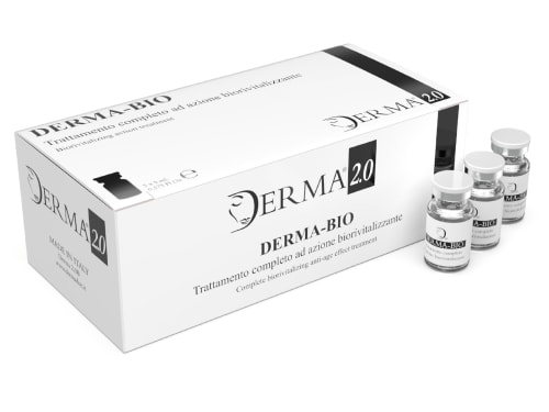
Discover the science behind Derma Bio, a mesotherapy product designed to combat signs of aging and achieve a brighter, smoother, and more youthful appearance.
-

-

Struggling with thinning hair? Discover natural, effective treatments at Creative Touch. We offer iPRF therapy, PRP, and scalp micropigmentation in Rotherham, Sheffield and South Yorkshire.
-
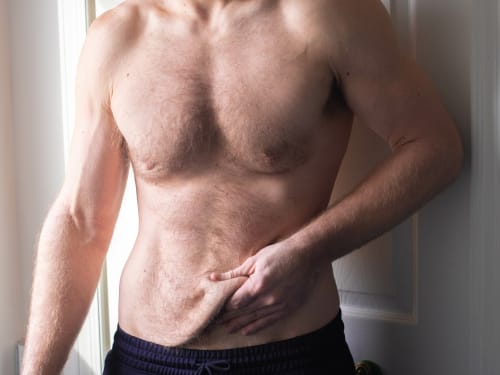
Don’t let loose skin overshadow your weight loss success. Learn why skin sags after significant weight loss and explore our advanced skin tightening and volume restoration treatments.
-

Repair outcomes range from true regeneration to fibrosis, determined by TGFβ isoform balance, macrophage M1→M2 transition, and inflammageing-related suppression of progenitor cells.
-

Discover how to restore and maintain your skin’s natural defences. From the causes of skin barrier damage to advanced treatments. Achieve healthy, radiant skin with our expert advice.