
Corneocyte
The corneocyte is the terminal, anucleate product of keratinocyte differentiation, functioning as the “brick” in the skin’s protective barrier. Encased in a highly cross-linked cornified envelope and filled with a dense keratin- filaggrin matrix, it provides the mechanical strength and UV-shielding capacity of the stratum corneum. Clinical quality is defined by the completeness of the terminal differentiation programme; when inflammatory signals or rapid turnover disrupt maturation, corneocytes arrive at the surface structurally immature and “leaky.” Treatment focuses on restoring the differentiation timeline and the corneodesmosome degradation rate to ensure a robust physical defence and smooth surface texture.
Corneocytes are the terminally differentiated cells that form the stratum corneum, the outermost layer of the skin barrier. They are produced when living keratinocytes complete a tightly regulated programme of self-transformation: the cell dismantles its internal structures, compacts its keratin filament network using filaggrin, and eventually becomes a flattened, protein-dense disc encased in a chemically cross-linked protein shell called the cornified envelope. The result is not simply a dead cell, but a structurally engineered unit whose architecture is specifically suited to its mechanical and biochemical role in barrier function.
Structure and the Cornified Envelope
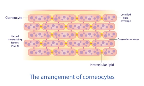
The cornified envelope replaces the cell membrane in a mature corneocyte, forming an insoluble scaffold of proteins – including loricrin, involucrin, and cornifin – that are covalently cross-linked by transglutaminase enzymes. This resistance to physical and chemical disruption is not incidental. It reflects the structural demands placed on cells whose job is to form the outermost surface of a living organism in continuous contact with the environment. Inside, the compacted keratin filament matrix provides the rigidity that gives corneocytes their mechanical strength. The brick-and-mortar analogy commonly used for the stratum corneum captures the corneocyte’s structural role accurately: these cells form the bricks, whilst ceramides, cholesterol, and free fatty acids compose the lipid mortar filling the intercellular spaces around them. But what the analogy can obscure is that the quality of the bricks and the quality of the mortar are not independent variables – the lipid composition of the mortar is determined in part by what the differentiating keratinocyte secretes as it becomes a corneocyte, meaning both elements reflect the same upstream programme.
Anatomy of a Mature Corneocyte
| Feature | Biological Role | Analogy | Clinical Value |
|---|---|---|---|
| Cornified Envelope (CE) | Cross-linked protein “shell.” | The Armour | Chemical and mechanical resistance. |
| Keratin-Filaggrin Matrix | Compacted internal “filling.” | The Concrete | Determines cell shape and density. |
| Lipid Envelope | Lipid layer “glued” to the CE. | The Waterproofing | Essential for lamellar matrix attachment. |
Corneocytes as the Source of Natural Moisturising Factor
The filaggrin that compacted the keratin network during differentiation does not remain intact. In the outer stratum corneum, filaggrin undergoes progressive enzymatic degradation through the action of caspase-14, calpain-1, and bleomycin hydrolase, releasing hygroscopic breakdown products – including pyrrolidone carboxylic acid (PCA), trans-urocanic acid (UCA), and free amino acids – that collectively form natural moisturising factor (NMF). These compounds maintain intracellular hydration within the corneocyte against the osmotic gradient that would otherwise drive water outward. UCA also contributes directly to the acid mantle, supporting the pH 4.5–5.5 environment on which ceramide-processing enzymes depend. Filaggrin deficiency therefore creates a concurrent NMF shortage and acid mantle impairment alongside the structural barrier defect – three failures from one protein’s absence. [7]
Desquamation: The Controlled Exit
Corneocytes are not shed individually by chance; they are released through a regulated enzymatic process called desquamation. Adjacent corneocytes are locked together by corneodesmosomes – specialised protein rivets that provide the stratum corneum with its cohesive strength. As cells migrate toward the surface, serine proteases (specifically kallikreins or KLKs) progressively degrade these rivets.
This process is highly dependent on both hydration and . The enzymes that “clip” the rivets require a water-rich environment and an acidic to function. When the barrier is dehydrated or the acid mantle is compromised, these enzymes fail. The “glue” remains intact, and cells accumulate into visible, disorganized flakes rather than shedding invisibly as single units.
Table 2: The Shedding Cycle (Corneodesmosomes)
| Adhesion State | Condition of “The Glue” | Surface Appearance |
|---|---|---|
| Optimal Adhesion | Regular protease activity. | Smooth, light-reflecting, “glowy” skin. |
| Hyper-Adhesion | Slow degradation (due to dehydration/). | Dull, congested, “ashy” or flaky patches. |
| Premature Shedding | Barrier disruption/Over-exfoliation. | Raw, shiny, “plastic-like” texture, stinging. |
Corneocyte Quality During and After Perimenopause
Corneocyte quality – defined by cornified envelope integrity, internal protein density, and NMF concentration – is meaningfully affected by declining oestrogen during perimenopause and menopause through mechanisms that go deeper than the commonly cited reduction in keratinocyte proliferation. Oestrogen does not simply drive more keratinocytes into differentiation. It has a specific role in the ceramide metabolic pathway within keratinocytes that is independent of its proliferative effects.
Research published in Scientific Reports (Kendall et al., 2022) demonstrated that post-menopausal stratum corneum contains significantly lower ceramide abundance across all major ceramide classes, with ceramides also displaying shorter average carbon chain lengths. This is a structural quality deficit, not just a quantity reduction, since shorter ceramides produce less ordered lamellar packing. Serum oestradiol correlated positively with both ceramide abundance and chain length, and in vitro treatment of primary human keratinocytes with oestradiol directly increased ceramide production, confirming that the hormone acts on ceramide biosynthesis directly rather than through its influence on cell cycling. Women taking HRT did not show these changes. The practical consequence is that the lipid mortar surrounding corneocytes becomes both sparser and less structurally competent as oestrogen declines – a deterioration in the quality of what corneocytes are embedded within, not only in the number being produced.[11]
Epidermal cell turnover also reduces by approximately half between the ages of 30 and 70, meaning that in mature skin, lower-quality corneocytes linger at the surface considerably longer before being shed. [10] Slower turnover compounds the hormonal quality deficit: the barrier is populated by cells that took longer to produce and arrived in a less favourable lipid environment. These two mechanisms – reduced production quality and reduced replacement rate – operate simultaneously at the same life stage. That convergence is what makes perimenopausal barrier change feel more abrupt than the gradual nature of either mechanism alone would suggest.
Clinical Application
The clinical question for corneocytes is not simply “how do we stimulate more of them?” It is more precise than that: what determines whether the corneocytes being produced are structurally competent – with complete cornified envelopes, adequate NMF concentration, and the lipid environment to support them – versus the thinner, less well-organised cells characteristic of photodamaged, hormonally transitioning, or chronically inflamed skin? That distinction is what separates a treatment that transiently resurfaces the stratum corneum from one that genuinely improves the population producing it.
Professional treatments influence this population through four distinct mechanisms: resetting the keratinocyte stem cell pool that feeds the differentiation programme, driving the differentiation programme directly, restoring the lipid synthesis capacity that determines corneocyte quality, and removing the inflammatory suppression that is actively impairing all three. These are not interchangeable. Applying a synthesis-stimulating treatment to skin where inflammation is still suppressing FLG and SPTLC1 expression is stimulating a programme that cannot complete properly. Understanding which mechanism is limiting in a specific client is what makes treatment sequencing rational rather than arbitrary.
Phase 1: Resolving What’s Blocking the Differentiation Programme
For clients presenting with reactive, sensitised, or chronically inflamed skin – the kind of persistent barrier fragility that topical ceramide support hasn’t fully resolved – the differentiation programme itself is compromised upstream. IL-4 and IL-13 suppress not just filaggrin but loricrin and involucrin alongside it, meaning the cornified envelope is being built from a depleted protein toolkit before any structural deficit at the surface level is even considered. Treating the surface whilst this is ongoing is addressing the consequence, not the cause.
Cold atmospheric plasma (CAP) is the treatment most directly positioned at this upstream problem for corneocyte quality. Its RONS-mediated modulation of NF-κB reduces the IL-4 and IL-13 burden in sensitised tissue, removing the cytokine suppression of the cornification programme rather than attempting to stimulate cornification through it. Crucially, at appropriate treatment parameters (short exposures, under 60 seconds) CAP stimulates basal keratinocyte proliferation and migration simultaneously, meaning it is not simply anti-inflammatory but actively pro-regenerative at the epidermal level. For clients where the inflammatory environment has become self-sustaining, CAP is not stimulating corneocyte production directly, but restoring the conditions in which the keratinocyte differentiation programme can complete properly. [1]
Polynucleotides contribute through a complementary route: their adenosine A2A receptor-mediated pathway increases M2 macrophage polarisation and reduces NF-κB activity broadly, quietening the tissue environment in which keratinocyte differentiation is occurring. Where the inflammatory suppression involves both epidermal and dermal compartments – as it often does in reactive perimenopausal skin with concurrent sensitivity and structural thinning – combining CAP and polynucleotides addresses the differentiation block from surface and tissue-depth routes simultaneously. [2]
Phase 2: Driving the Differentiation Programme and Resetting the Cell Population
Once the inflammatory environment is less actively suppressive, the question becomes whether the keratinocyte population producing corneocytes is itself capable of generating higher-quality cells or whether photodamage, hormonal transition, or age-related stem cell depletion has reduced the programme’s capacity.
Microneedling directly activates the cornification and keratinocyte differentiation gene programmes through the wound-healing cascade. This is not simply a collagen signal. Gene expression analysis of microneedled 3D skin models documents upregulation across cornification, keratinocyte differentiation, and epidermis development pathways, alongside the SPTLC3 ceramide synthesis upregulation. This means that the differentiation programme and the lipid production machinery are activated together. Histological follow-up at one year from clinical data shows stratum spinosum thickening and restored rete ridge architecture: structural improvements in how the epidermis is organised, not just surface texture changes. [8] For clients whose primary presentation is dull, unevenly textured skin with impaired moisture retention – the classic picture of accumulated lower-quality corneocytes – microneedling targets the source.
Microneedling combined with topical exosome delivery extends this substantially, and it is worth being precise about why. Exosomes delivered through microneedling channels restore SPTLC1 expression (the rate-limiting ceramide synthesis enzyme) and PPAR-α, the transcription factor that coordinates the full lipid synthesis programme across ceramides, cholesterol, and free fatty acids simultaneously. They also restore filaggrin and involucrin expression in treated keratinocytes. The practical consequence is that exosome delivery through microneedling channels addresses epidermal architecture and the lipid synthesis capacity of newly differentiating keratinocytes at the same time, producing corneocytes that arrive in a better-equipped lipid environment than microneedling alone achieves.[3]
Thulium 1927nm fractional laser takes the approach one step further back in the production chain. Rather than stimulating existing keratinocytes, the MTZ injury triggers re-epithelialisation from hair follicle stem cells that retain COL17A1 expression – the protein governing keratinocyte stem cell self-renewal capacity. The cells migrating to repopulate treated zones carry a younger stem cell signature than the photodamaged cells they replace. Fast responders in clinical studies show markedly greater lipid metabolism gene upregulation, likely because keratinocytes sourced from this follicular reservoir arrive with a more competent cornification and ceramide synthesis programme than the photodamaged population they replace. [4] For clients where the problem is not simply suppressed differentiation but a genuinely depleted or photodamaged keratinocyte pool, thulium addresses the stock of cells producing corneocytes, not just the productivity of existing cells.
iPRF contributes through a growth factor environment that sustains keratinocyte stem cell activity. EGF supports the EGFR–COL17A1 axis relevant to stem cell self-renewal; PDGF and TGF-β stimulate keratinocyte proliferation and migration. For clients whose epidermal renewal has slowed – a more common presentation in their 40s and 50s than it is in younger clients – iPRF creates the growth factor environment in which the differentiation programme operates with more support than the skin can currently generate autonomously. [6]
Phase 3: Supporting Final Maturation and Maintaining What’s Been Built
LED red light (630–660nm) occupies a specific and underappreciated position relative to corneocyte quality. It does not act directly on keratinocyte differentiation. Its mechanism is bottom-up: cytochrome c oxidase activation in fibroblast mitochondria drives TGF-β and AKT signalling, increasing collagen accumulation, which in turn activates integrin signalling in basal keratinocytes. It also directly accelerates lamellar body secretion at the stratum granulosum–stratum corneum interface – the step at which ceramides, cholesterol, and free fatty acids are packaged and released into the intercellular space.[9] This makes red LED particularly well-suited as post-procedure support after thulium or microneedling: the newly differentiating cells arriving at the stratum granulosum complete their lipid secretion step more efficiently, improving the quality of the lamellar matrix the new corneocytes are embedded in. As a completely non-ablative, non-inflammatory modality, it is also appropriate for reactive clients where Phase 1 is still underway.
HA skin boosters, in this context, restore the epidermal hyaluronic acid pool that research confirms is required for the hyperplastic repair response to barrier disruption. This is not simply deep hydration; HA accumulation in the epidermis is a structural precondition for normal regenerative capacity. In clients where ground substance depletion has impaired both the barrier repair response and the epidermal HA environment, skin boosters restore something the differentiation programme needs to function rather than supplementing a cosmetic endpoint.[5]
Putting It Together: Treatment Sequencing for Corneocyte Quality
The sequencing logic follows the mechanism categories directly.
For reactive, sensitive, or inflammatory presentations: Phase 1 first. CAP (and polynucleotides where dermal involvement is present) resolves the IL-4/IL-13 suppression before Phase 2 treatments are introduced. Activating microneedling’s cornification programme into a still-suppressed inflammatory environment produces incomplete results; the differentiation signals are stimulated but the proteins they would generate are still being actively downregulated. Resolve the block, then stimulate.
For photodamaged or post-menopausal presentations where the keratinocyte population itself has declined: thulium provides the population reset, then microneedling with exosomes addresses the differentiation programme and lipid synthesis capacity in the reconstituted population. LED post-procedure supports lamellar body secretion as the new cells mature. iPRF can precede or accompany the differentiation-phase treatments to create a richer growth factor environment throughout.
For age-related decline without active inflammation: microneedling with exosomes, iPRF for growth factor support, and red LED post-procedure form a coherent sequence that addresses the differentiation programme, the lipid synthesis machinery, and the final maturation step without the inflammatory removal phase that more reactive presentations require.
The most complete corneocyte quality improvements – better cornified envelope integrity, higher NMF concentration, more ordered lamellar architecture – come from protocols that address more than one point in the production chain. A treatment that stimulates differentiation but without supporting the lipid environment those cells mature into, produces better bricks but not better mortar. Both need to improve together for barrier competence to follow. [11]
| Goal | Biological Target | Treatment Strategy |
|---|---|---|
| Improve Quality | Ensure complete terminal differentiation. | Exosomes / Microneedling (The “Factory” fix). |
| Support Strength | Enhance cornified envelope cross-linking. | Retinoids (The “Blueprint” fix). |
| Normalize Shedding | Support enzymatic corneodesmosolysis. | PHAs / AHAs (The “Maintenance” fix). |
| Protect Mature Cells | Prevent premature degradation of NMF. | pH-Corrective Homecare (The “Environment” fix). |
References
Hasse S, Duong Tran T, Hahn O, et al. (2016). Induction of proliferation of basal epidermal keratinocytes by cold atmospheric-pressure plasma. Clin Exp Dermatol, 41(2), 202-9 . doi.org/10.1111/ced.12735
Lee KWA, Chan KWL, Lee A, et al. (2024). Polynucleotides in Aesthetic Medicine: A Review of Current Practices and Perceived Effectiveness. Int J Mol Sci, 25(15) . doi.org/10.3390/ijms25158224
Lee YS (2025). Preliminary Histological Evidence of Epidermal and DEJ Remodeling with Microneedling-Assisted Topical Exosome Therapy: A Single-Subject Case Report. Clin Cosmet Investig Dermatol, 18, 2377-2385 . doi.org/10.2147/ccid.s542022
Li X, Qin S, Shi S, et al. (2023). Prospective study of efficacy and safety of non-ablative 1927 nm fractional thulium fiber laser in Asian skin photoaging. Front Surg, 10, 1076848 . doi.org/10.3389/fsurg.2023.1076848
Maytin EV, Chung HH, Seetharaman VM (2004). Hyaluronan participates in the epidermal response to disruption of the permeability barrier in vivo. Am J Pathol, 165(4), 1331-41 . doi.org/10.1016/s0002-9440(10)63391-3
Nanba D, Toki F, Asakawa K, et al. (2021). EGFR-mediated epidermal stem cell motility drives skin regeneration through COL17A1 proteolysis. J Cell Biol, 220(11) . doi.org/10.1083/jcb.202012073
Sandilands A, Sutherland C, Irvine AD, et al. (2009). Filaggrin in the frontline: role in skin barrier function and disease. J Cell Sci, 122(Pt 9), 1285-94 . doi.org/10.1242/jcs.033969
Singh A, Yadav S (2016). Microneedling: Advances and widening horizons. Indian Dermatol Online J, 7(4), 244-54 . doi.org/10.4103/2229-5178.185468
Umino Y, Denda M (2023). Effect of red light on epidermal proliferation and mitochondrial activity. Skin Res Technol, 29(9), e13447 . doi.org/10.1111/srt.13447
Wang Z, Man MQ, Li T, et al. (2020). Aging-associated alterations in epidermal function and their clinical significance. Aging (Albany NY), 12(6), 5551-5565 . doi.org/10.18632/aging.102946
Unknown Author. PMC: PMC9755298. PMC9755298
Also Known As
- anucleate cell
- corneocytes
Anatomical Relationships
Structural Connections
- Requires Ceramides
- Requires Filaggrin
- Thulium Fractional Laser
Referenced in Conditions & Treatments
- this Stimulated by Microneedling
- this Inhibited by Interleukin-13
- this Inhibited by Interleukin-4
- this Preceded by Keratinocyte
- this Part of Stratum corneum PMID: 29262154
Learn More
This topic is discussed in 1 article:
-

Flattened, protein-rich cells that form the ‘bricks’ of the stratum corneum, embedded in a lipid matrix that acts as ‘mortar’