
Adipocyte
Adipocytes are the primary cells of adipose tissue – the cell type responsible for storing energy as triglycerides and releasing it as fatty acids in response to hormonal signals. The familiar framing of fat cells as passive energy reservoirs understates what they actually do: adipocytes are endocrine cells, secreting a range of hormones and cytokines – collectively adipokines – that regulate appetite, insulin sensitivity, immune activity, and inflammatory tone throughout the body. Their dysfunction with age, obesity, or metabolic disease produces consequences that extend well beyond excess fat storage, driving the chronic low-grade inflammation that connects metabolic health to skin quality, hair cycling, and treatment responsiveness. Three functionally distinct adipocyte types exist – white, brown, and beige – each with different metabolic roles and different relevance to aesthetic medicine.
Three Adipocyte Types
Not all adipocytes are metabolically equivalent. The three principal types differ in structure, origin, and function in ways that matter both for understanding body composition and for interpreting treatment mechanisms.
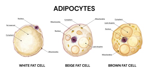
White adipose tissue (WAT) adipocytes are the dominant form in adult human subcutaneous and visceral fat. They are unilocular – a single large lipid droplet occupies the majority of the cell volume, pushing the nucleus and mitochondria to the periphery. Their primary functions are energy storage (as triglycerides) and endocrine secretion. WAT is the source of leptin, adiponectin, resistin, and – in dysfunctional states – pro-inflammatory cytokines including TNF-α and IL-6. [4]
Brown adipose tissue (BAT) adipocytes are multilocular – multiple smaller lipid droplets distributed through a cell rich in mitochondria. BAT is specialised for thermogenesis rather than energy storage: uncoupling protein-1 (UCP-1) in the inner mitochondrial membrane dissipates the proton gradient that would otherwise drive ATP synthesis, releasing energy as heat instead. BAT is most abundant in neonates and persists in adults primarily in the supraclavicular and paraspinal regions. Its metabolic activity is inversely correlated with obesity and declines with age. [6]
Beige (brite) adipocytes are WAT-derived cells capable of adopting a brown-like thermogenic phenotype under cold exposure or adrenergic stimulation – a plasticity mediated by UCP-1 upregulation. They arise within white adipose depots rather than from a separate brown adipocyte lineage. This plasticity is directly relevant to the cryolipolysis secondary mechanism debate: the hypothesis that local cooling activates beige adipocyte thermogenesis as a contributing factor to fat reduction alongside the primary apoptosis pathway. Evidence for this in clinical cryolipolysis is suggestive rather than conclusive, but the beige adipocyte biology makes it mechanistically plausible. [11]
| Type | Structure | Primary function | Key marker | Clinical relevance |
|---|---|---|---|---|
| White (WAT) | Unilocular; lipid-dominant | Energy storage; endocrine secretion | Leptin, adiponectin | Fat reduction targets; adipokine dysregulation in obesity |
| Brown (BAT) | Multilocular; mitochondria-rich | Thermogenesis | UCP-1 | Cold-activated; declines with age and obesity |
| Beige/Brite | Unilocular → multilocular (inducible) | Inducible thermogenesis | UCP-1 (induced) | Potentially activated by cryolipolysis cold stimulus |
Anatomical Distribution – Where Each Type Is Found
WAT and BAT are not uniformly distributed – their anatomical locations reflect their distinct functions, and the distribution of each changes significantly with age, sex, and metabolic status.
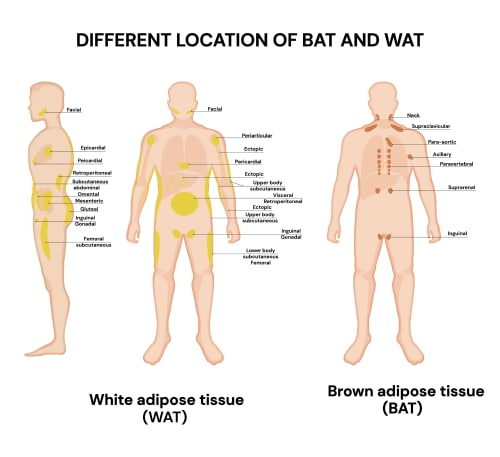
White adipose tissue is distributed across two functionally distinct compartments. Subcutaneous WAT lies beneath the skin in the hypodermis – the abdominal, gluteofemoral, and femoral depots are the major subcutaneous tissue stores – and is the primary target of aesthetic fat reduction treatments. Visceral WAT occupies the abdominal cavity as the omental, mesenteric, and retroperitoneal depots, plus smaller pericardial and epicardial stores. These two compartments are metabolically distinct: visceral adipocytes are more lipolytically active than subcutaneous adipocytes and, in states of dysfunction and expansion, are more strongly associated with insulin resistance, systemic inflammation, and metabolic disease risk. Central adiposity – the accumulation of excess visceral rather than subcutaneous fat – is the metabolically higher-risk pattern, independent of overall fat mass. [14]
Brown adipose tissue in adults is concentrated in a continuous fascial layer in the upper torso – supraclavicular, cervical, and axillary regions collectively account for approximately 67% of total adult BAT volume and activity. Additional depots are present in the paravertebral, para-aortic, and suprarenal regions. BAT is most abundant in neonates and declines progressively with age; by the eighth decade, few individuals have detectable BAT at any site. BAT mass also declines with obesity and is inversely correlated with BMI, creating a feedback pattern in which excess adiposity reduces the thermogenic tissue that would otherwise contribute to energy expenditure. [8]
Adipogenesis – How Adipocytes Form
Adipocytes develop from preadipocytes – uncommitted mesenchymal precursor cells resident in the stromal vascular fraction (SVF) of adipose tissue. The differentiation process, adipogenesis, is governed by a transcriptional cascade in which PPARγ (peroxisome proliferator-activated receptor gamma) acts as the master regulator. PPARγ activation alone is sufficient to drive adipocyte differentiation in preadipocyte cell lines; no other transcription factor has this capacity. C/EBPα (CCAAT/enhancer-binding protein alpha) forms a positive feedback loop with PPARγ – each induces the other’s expression, locking the cell into the adipocyte lineage. C/EBPβ and C/EBPδ act earlier in the cascade, driving the initial PPARγ induction. [12]
The clinical significance of adipogenesis is not simply developmental. Throughout adult life, adipose tissue turns over continuously – new adipocytes differentiate from the SVF preadipocyte pool to replace those that die. This regenerative capacity is directly impaired by SASP from senescent adipocytes: the pro-inflammatory cytokine environment produced by senescent cells suppresses PPARγ expression in neighbouring preadipocytes, blocking their differentiation and progressively depleting the tissue’s replacement capacity. Age-related adipose dysfunction is therefore partly a failure of renewal, not only an accumulation of dysfunctional cells. [7]
The Adipocyte as Endocrine Organ
WAT is the largest endocrine organ in most adults by mass. The adipokines it secretes regulate systemic physiology far beyond the adipose compartment – and their dysregulation in obesity, metabolic syndrome, and ageing contributes directly to the inflammatory baseline relevant to skin quality. [3]
Leptin – the satiety hormone secreted proportionally to fat mass – signals energy sufficiency to the hypothalamus and modulates immune cell activity. In obesity, chronically elevated leptin produces leptin resistance at the hypothalamic level, uncoupling the signal from its intended regulatory effect. Leptin receptors are expressed on sebaceous glands and keratinocytes; elevated leptin in obesity is associated with increased sebum production and altered epidermal differentiation. The full mechanism is covered in the Leptin entity.
Adiponectin – secreted in inverse proportion to fat mass – has insulin-sensitising and anti-inflammatory properties. It activates AMPK in muscle and liver, promoting fatty acid oxidation and improving insulin sensitivity. Adiponectin levels fall significantly in obesity and metabolic syndrome, removing a key anti-inflammatory and metabolic regulatory signal. In skin biology, lower adiponectin levels correlate with increased psoriasis severity, and adiponectin signalling in keratinocytes suppresses NF-κB-mediated inflammation. This makes the adiponectin decline of obesity a contributing factor to inflammatory skin conditions, operating independently of the leptin resistance pathway. [13]
Resistin – a pro-inflammatory adipokine that links adipose tissue dysfunction to insulin resistance and systemic inflammation. It promotes TNF-α and IL-6 secretion from macrophages and impairs insulin signalling in peripheral tissues. Resistin levels are elevated in obesity and inflammatory skin conditions including psoriasis. [13]
TNF-α and IL-6 – under normal conditions, WAT secretes low levels of these cytokines as part of tissue homeostasis. In adipose dysfunction – hypertrophy, hypoxia, macrophage infiltration – their output increases substantially, contributing the chronic low-grade inflammation of inflammageing. These are the same cytokines featured in the SASP and the gut dysbiosis LPS/TLR4 pathway; their adipose-derived contribution represents a third independent upstream driver of the same downstream inflammatory state.
Lipolysis – How Adipocytes Release Fatty Acids
Energy mobilisation from adipocytes proceeds through a coordinated triglyceride hydrolysis cascade involving three lipases acting sequentially. [16]
ATGL (adipose triglyceride lipase) initiates the cascade, cleaving one fatty acid from the triglyceride molecule to produce diacylglycerol (DAG). ATGL is responsible for the majority of triglyceride hydrolysis and is the rate-limiting step for the overall lipolytic rate under basal conditions. [2]
HSL (hormone-sensitive lipase) cleaves the second fatty acid from DAG, producing monoacylglycerol (MAG). HSL is the primary target of hormonal regulation: catecholamines (adrenaline, noradrenaline) bind β-adrenergic receptors, raising cAMP, activating PKA, which phosphorylates and activates HSL. Insulin suppresses this pathway directly, reducing cAMP and promoting fatty acid re-esterification. HSL is thus the molecular switch through which the fasting/fed hormonal state controls fat mobilisation. [1]
MGL (monoglyceride lipase) completes the cascade, cleaving the final fatty acid from MAG to release glycerol and three free fatty acids into circulation.
The Randle Cycle connection is direct: the free fatty acids liberated by HSL-mediated lipolysis are the substrates for CPT-1-dependent mitochondrial β-oxidation. When insulin is low and catecholamines are elevated – the fasted, exercising, or low-carbohydrate-fed state – lipolysis proceeds and fatty acids enter mitochondria for oxidation. When insulin is elevated – the fed, high-carbohydrate state – HSL is suppressed, lipolysis falls, and malonyl-CoA inhibits CPT-1, preventing fatty acid entry into mitochondria. The Randle Cycle entity develops the full implications of this switching for metabolic flexibility and insulin resistance.
Adipocyte Senescence and the SASP
With age, an increasing proportion of adipocytes and adipose-resident cells enter replicative or stress-induced senescence – permanent cell cycle arrest without cell death. Senescent adipocytes secrete the senescence-associated secretory phenotype (SASP): a mixture of pro-inflammatory cytokines (IL-6, IL-8, TNF-α), matrix metalloproteinases (MMP-3, MMP-12), and chemokines that remodel the local tissue environment in ways that amplify dysfunction. [7]
The consequences operate at two levels. Locally: SASP suppresses PPARγ in neighbouring preadipocytes (impairing renewal, as noted above), MMP secretion degrades extracellular matrix in both adipose and overlying dermal tissue, and the cytokine environment promotes further adipocyte death and macrophage recruitment – a self-amplifying cascade. Systemically: the sustained low-level cytokine output of dysfunctional adipose tissue contributes to the chronic sterile inflammation of inflammageing, driving the same NF-κB/MMP/SASP cycle that accelerates fibroblast senescence, degrades dermal collagen, and reduces treatment responsiveness. [9]
The histological marker of this process is the crown-like structure (CLS) – a ring of infiltrating macrophages surrounding a dead or dying adipocyte, formed as the macrophage population responds to lipid and DAG released by the failing cell. CLS density in adipose tissue biopsies correlates with the degree of adipose inflammation, insulin resistance, and systemic SASP burden, making it a validated histological indicator of adipose tissue inflammageing status. [15]
A 2025 Nature study added a further regulatory dimension: nerve-associated macrophages – a resident macrophage population in adipose tissue that maintains homeostasis through sympathetic nerve signalling – decline with age, removing a brake on adipose inflammation. The consequence is that age-related adipose inflammation reflects not only the accumulation of pro-inflammatory signals but the simultaneous loss of the cellular mechanism that would normally restrain them – a dual failure that makes age-related adipose dysfunction self-accelerating in a way that younger tissue’s homeostatic mechanisms would prevent. [5]
Dermal Adipose Tissue and Skin Biology
Beneath the dermis and above the subcutaneous fat layer lies a distinct adipocyte population – dermal white adipose tissue (dWAT) – developmentally and functionally separate from subcutaneous WAT. dWAT is not simply the uppermost layer of subcutaneous fat; it has a distinct developmental origin (dermal fibroblast lineage rather than mesenchymal progenitor), a distinct response to physiological signals, and direct structural and paracrine interactions with the dermis above it. [10]
The hair follicle connection is the most precisely characterised dWAT function. The adipocyte layer surrounding the hair follicle bulb thickens reproducibly during anagen (active growth phase) and thins during telogen (resting phase), tracking the follicle cycle rather than systemic metabolic state. This synchrony is bidirectional: preadipocyte-derived signals including PDGF activate hair follicle stem cells at the onset of anagen; BMP2 secreted by mature intradermal adipocytes regulates follicle cycling; and VEGF from the adipocyte layer supports the vascular supply to the growing follicle. The Hair Follicle entity develops this relationship from the follicle perspective; here the important point is that dWAT is a physiological participant in hair cycling, not merely a structural support layer. [17]
The ageing dermis connection involves a directional transition that is the reverse of what might be expected. With age, dermal fibroblasts in humans acquire an increasingly adipogenic transcriptional profile – they begin to resemble preadipocytes more than mature fibroblasts. This shift reduces the population of matrix-producing fibroblasts available for collagen and elastin maintenance, contributing to dermal thinning, and the resulting dWAT expansion creates the fat infiltration into the dermis described in the Subcutaneous tissue entity – lipid accumulation disrupting the collagen fibre architecture and impairing dermal mechanical properties independently of overall fat volume changes. [10]
Why Adipocytes Are Selectively Targetable
Non-invasive fat reduction depends on biological properties that make adipocytes more vulnerable than surrounding cell types to specific physical and chemical stimuli. These selectivity differentials are what make body contouring treatments possible without damaging overlying dermis, vasculature, or nerve tissue.
- Cold (cryolipolysis): high lipid content causes intracellular crystallisation at −1°C to +5°C – temperatures that do not affect the more aqueous cytoplasm of dermal or epidermal cells. Apoptosis follows, with macrophage-mediated clearance over 8–12 weeks
- Phospholipid membrane disruption ( Aqualyx/deoxycholic acid): dense intracellular lipid load makes adipocytes structurally more susceptible to bile-acid-mediated membrane disruption at concentrations that produce lesser effects on other tissue types
- Acoustic cavitation (fat cavitation/HIFU): lower mechanical resistance of lipid-filled adipocytes to rapid pressure changes; surrounding fibrous septa and vasculature are relatively spared
- RF thermal selectivity: lower thermal conductivity and different dielectric properties cause subcutaneous adipose to reach and retain higher temperatures than dermis or muscle at equivalent energy input
Full mechanism detail for each modality is in the individual treatment entities (Cryolipolysis, Deoxycholic Acid, Radiofrequency Microneedling).
Clinical Application
Adipocytes are relevant at Creative Touch across four practical contexts.
Body contouring treatment selection
Matching treatment modality to clinical objective requires understanding which adipocyte vulnerability each exploits. Cryolipolysis targets cold-sensitivity and produces apoptosis; Aqualyx targets membrane susceptibility and produces adipocytolysis; cavitation targets mechanical fragility; RF targets thermal selectivity with the added benefit of concurrent collagen stimulation. These are not interchangeable mechanisms – treatment selection should be led by the specific tissue change required, not by device availability.
GLP-1 medication context
Rapid WAT volume loss from semaglutide or tirzepatide removes facial compartment structural support faster than the overlying skin can adapt through elastic recoil. The clinical consequence – facial deflation and laxity disproportionate to overall weight loss – is a direct adipocyte volume change, not a drug side effect per se. Clients on GLP-1 medications need consultation addressing compartmental deflation, skin laxity management, and realistic timelines before aesthetic volume treatment is planned.
Metabolic and dietary conversations
The adipokine picture (leptin/adiponectin ratio as a metabolic health marker, adiponectin decline in obesity connecting to inflammatory skin conditions) gives a biologically grounded framing for why metabolic health and skin quality are connected. The lipolysis pathway – HSL activation by catecholamines; HSL suppression by insulin – is the cellular mechanism by which low-carbohydrate dietary patterns mobilise stored fat, and by which chronic hyperinsulinaemia impairs fat mobilisation. The Randle Cycle entity carries the full metabolic flexibility framing.
Senescence and inflammageing
Adipocyte SASP is an upstream driver of the systemic inflammatory baseline that affects treatment responsiveness across collagen-stimulating treatments. Senolytic approaches (quercetin, fisetin) are covered in the Cellular Senescence entity; the adipocyte senescence biology here provides the adipose-specific mechanistic context for why addressing the inflammatory baseline matters independently of treating its downstream skin manifestations.
References
Althaher AR (2022). An Overview of Hormone-Sensitive Lipase (HSL). ScientificWorldJournal, 2022, 1964684 . doi.org/10.1155/2022/1964684
Brejchova K, Radner FPW, Balas L, et al. (2021). Distinct roles of adipose triglyceride lipase and hormone-sensitive lipase in the catabolism of triacylglycerol estolides. Proc Natl Acad Sci U S A, 118(2) . doi.org/10.1073/pnas.2020999118
Clemente-Suárez VJ, Redondo-Flórez L, Beltrán-Velasco AI, et al. (2023). The Role of Adipokines in Health and Disease. Biomedicines, 11(5) . doi.org/10.3390/biomedicines11051290
Giralt M, Villarroya F (2013). White, brown, beige/brite: different adipose cells for different functions? Endocrinology, 154(9), 2992-3000 . doi.org/10.1210/en.2013-1403
Gonzalez-Hurtado E, Leveau C, Li K, et al. (2025). Nerve-associated macrophages control adipose homeostasis across lifespan and restrain age-related inflammation. Nat Aging, 5(9), 1828-1843 . doi.org/10.1038/s43587-025-00952-9
Ikeda K, Maretich P, Kajimura S (2018). The Common and Distinct Features of Brown and Beige Adipocytes. Trends Endocrinol Metab, 29(3), 191-200 . doi.org/10.1016/j.tem.2018.01.001
Ishaq Abbas, Saretzki Gabriele (2022). Senescence and DNA Damage in Adipocytes and Fat Tissues and Its Potential Amelioration through Nutritional Interventions. Recent Progress in Nutrition, 02(03), 1-66 . doi.org/10.21926/rpn.2203016
Leitner BP, Huang S, Brychta RJ, et al. (2017). Mapping of human brown adipose tissue in lean and obese young men. Proc Natl Acad Sci U S A, 114(32), 8649-8654 . doi.org/10.1073/pnas.1705287114
Matacchione G, Perugini J, Di Mercurio E, et al. (2022). Senescent macrophages in the human adipose tissue as a source of inflammaging. Geroscience, 44(4), 1941-1960 . doi.org/10.1007/s11357-022-00536-0
Peng Y, Cheong S, Lu F, et al. (2024). Dermal white adipose tissue: Development and impact on hair follicles, skin defense, and fibrosis. FASEB J, 38(18), e70047 . doi.org/10.1096/fj.202400653r
Pilkington AC, Paz HA, Wankhade UD (2021). Beige Adipose Tissue Identification and Marker Specificity-Overview. Front Endocrinol (Lausanne), 12, 599134 . doi.org/10.3389/fendo.2021.599134
Siersbaek R, Nielsen R, Mandrup S (2010). PPARgamma in adipocyte differentiation and metabolism—novel insights from genome-wide studies. FEBS Lett, 584(15), 3242-9 . doi.org/10.1016/j.febslet.2010.06.010
Słuczanowska-Głabowska S, Staniszewska M, Marchlewicz M, et al. (2023). Adiponectin, Leptin and Resistin in Patients with Psoriasis. J Clin Med, 12(2) . doi.org/10.3390/jcm12020663
Wronska A, Kmiec Z (2012). Structural and biochemical characteristics of various white adipose tissue depots. Acta Physiol (Oxf), 205(2), 194-208 . doi.org/10.1111/j.1748-1716.2012.02409.x
Yamada T, Kamiya M, Higuchi M, et al. (2018). Fat depot-specific differences of macrophage infiltration and cellular senescence in obese bovine adipose tissues. J Vet Med Sci, 80(10), 1495-1503 . doi.org/10.1292/jvms.18-0324
Yang A, Mottillo EP (2020). Adipocyte lipolysis: from molecular mechanisms of regulation to disease and therapeutics. Biochem J, 477(5), 985-1008 . doi.org/10.1042/bcj20190468
Zhang P, Kling RE, Ravuri SK, et al. (2014). A review of adipocyte lineage cells and dermal papilla cells in hair follicle regeneration. J Tissue Eng, 5, 2041731414556850 . doi.org/10.1177/2041731414556850
Also Known As
- adipocytes
- adipose cell
- fat cell
- lipocyte
- lipocytes
Anatomical Relationships
Structural Connections
- Stimulates Interleukin-6 Evidence: Hypertrophic adipocytes upregulate IL-6 secretion via NF-kB. Gao 2025 doi:10.1080/21623945.2025.2485927; Ellulu 2016 doi:10.5114/aoms.2016.58928.
- Stimulates Matrix metalloproteinase Evidence: Senescent adipocytes secrete MMPs (MMP-3, MMP-12) as SASP, degrading ECM in adipose and overlying dermis. Entity text; PMC9616990.
- Stimulates Tumour necrosis factor Evidence: Dysfunctional adipocytes stimulate TNF-alpha; TNF-alpha promotes HSL lipolysis via NF-kB. Itoh 2011; Dicker 2007 doi:10.1194/JLR.M600471-JLR200.
- Inhibits Insulin Evidence: Adipocyte TNF-alpha/IL-6 secretion impairs insulin signalling; adiponectin decline removes insulin-sensitising signal. Gao 2025; Ellulu 2016.
- Produces Free fatty acids Evidence: Lipolysis cascade via ATGL/HSL/MGL releases three free fatty acids per triglyceride from adipocytes. Entity text; PMC7187988.
- Produces Interleukin-6 Evidence: WAT produces IL-6; output increases substantially with adipose dysfunction and hypertrophy. Entity text; Ellulu 2016 doi:10.5114/aoms.2016.58928.
- Produces Leptin Evidence: WAT is the source of leptin, secreted proportionally to fat mass to signal energy sufficiency. Entity text.
- Produces Tumour necrosis factor Evidence: Dysfunctional adipocytes produce TNF-alpha as part of the SASP/obesity-associated inflammatory cascade. Entity text; Gao 2025.
- Affects Cellular senescence Evidence: Adipocytes enter senescence with age; SASP suppresses PPARgamma in preadipocytes, impairing tissue renewal. Entity text; PMC9616990.
- Affects Fibroblast Evidence: dWAT fibroblast adipogenic shift reduces matrix-producing fibroblasts; lipid infiltration disrupts collagen. FASEB doi:10.1096/fj.202400653R.
- Affects Hair follicle Evidence: Adipocyte layer thickens/thins with anagen/telogen; PDGF from preadipocytes activates follicle stem cells. Entity text; PMC4221925.
- Affects Inflammageing Evidence: Dysfunctional adipose tissue contributes sustained low-level cytokine output to inflammageing. Entity text; Ruck 2023 doi:10.1186/s40348-023-00170-6.
- Affects Randle cycle Evidence: Adipocyte lipolysis rate (HSL activation/suppression by insulin) determines fatty acid availability for Randle cycle. Entity text.
- Affects Skin ageing Evidence: SASP drives MMP secretion degrading dermal ECM; dWAT fibroblast shift reduces collagen-producing fibroblasts. Entity text; FASEB doi:10.1096/fj.202400653R.
- Has sub-structure Dermis Evidence: Dermal white adipose tissue (dWAT) is a distinct adipocyte population beneath the dermis. Entity text; FASEB doi:10.1096/fj.202400653R.
- Has sub-structure Subcutaneous tissue Evidence: White adipocytes lie beneath the skin in the hypodermis; subcutaneous depots are major WAT stores. Entity text + PMC22226221.
- Obesity Evidence: Adipocyte dysfunction in obesity drives chronic inflammation, leptin resistance, and metabolic disease. Entity text.
- Senescence-associated secretory phenotype Evidence: Senescent adipocytes secrete the SASP – IL-6, IL-8, TNF-alpha, MMPs – driving local and systemic inflammation. Entity text; PMC9616990.
- Type 2 diabetes Evidence: Adipocyte dysfunction/adipokine dysregulation central to insulin resistance and T2DM. Gao et al. 2025 doi:10.1080/21623945.2025.2485927.
- Cryolipolysis Evidence: Adipocyte cold-sensitivity via intracellular lipid crystallisation is the mechanistic basis of cryolipolysis. Entity text.
- Radiofrequency Microneedling Evidence: RF targets adipocyte thermal selectivity (lower thermal conductivity of lipid-filled cells); concurrent skin tightening via collagen. Entity text.
Referenced in Conditions & Treatments
- this Inhibited by Cryolipolysis Evidence: Controlled cooling triggers adipocyte apoptosis and macrophage-mediated clearance, reducing fat layer thickness. Entity text; Kania 2023 doi:10.1111/jocd.16039.
- this Inhibited by Deoxycholic acid Evidence: DCA physically disrupts adipocyte membrane, causing necrosis and permanent adipocyte destruction. Entity text; Muskat 2022 PMC8988282.
- this Affected by Interleukin-6 Evidence: Elevated IL-6 impairs adipocyte differentiation and adipogenesis via JAK/STAT signalling; contributes to adipose dysfunction. Nicklas et al. 2017 Physiology doi:10.1152/PHYSIOL.00012.2016
- this Affected by Paradoxical adipose hyperplasia Evidence: PAH involves paradoxical preadipocyte/adipocyte proliferation in treated area rather than apoptotic clearance. Entity text; Seaman PMC4678995.
Learn More
This topic is discussed in 1 article:
-

Fat freezing is a revolutionary treatment that helps eliminate stubborn fat cells with minimal downtime. But for optimal results, aftercare is key!