
Hyaluronic acid
Hyaluronic acid is a glycosaminoglycan that forms the hydrating ground substance of the dermis, holding water at up to 1,000 times its own weight and maintaining the mechanical environment fibroblasts require for normal collagen synthesis. Its most clinically significant role is not hydration per se but mechanosensing: the presence of HA in the extracellular matrix creates the tension that keeps fibroblasts biosynthetically active. When HA is lost through chronological ageing, UV exposure, or oestrogen decline, fibroblasts enter a quiescent state and reduce collagen output even when TGF-β signalling remains intact. Injected cross-linked HA restores this mechanical tension and has biopsy-confirmed evidence of procollagen I and III upregulation at 4 and 13 weeks. HA is not a single molecule: its molecular weight determines whether it hydrates the surface, penetrates the epidermis, or signals through CD44 receptors. These distinctions matter for how topical, oral, and injectable forms are understood and positioned. The body contains approximately 15 grams of hyaluronic acid in total, of which roughly one third (around 5 grams) is degraded and resynthesised every day. [5] In the dermis, the half-life of HA is approximately 24 hours, making its dermal pool one of the most rapidly renewed structural components in the body. This turnover rate is the biological foundation for why injectable HA requires maintenance: cross-linking – the process of creating chemical bridges between HA chains to resist enzymatic breakdown – extends the material’s in-tissue lifespan from hours to months, but endogenous hyaluronidase gradually overcomes this resistance, and the visible effect declines accordingly. [3] Maintenance appointments are therefore a property of the biology of the material, not a commercial convention.
Hyaluronic acid (HA) is a glycosaminoglycan – a long, unbranched polysaccharide – found throughout the body’s connective tissues, with the highest concentrations in the dermis, synovial fluid, and vitreous humour of the eye. In the dermis, HA forms the hydrophilic ground substance of the extracellular matrix (ECM), binding water at up to 1,000 times its own weight and maintaining the swollen, turgid environment that gives healthy skin its visible volume and palpable resilience. It is produced by hyaluronan synthase enzymes (HAS1, HAS2, HAS3) in fibroblasts and keratinocytes, and is continuously turned over; the half-life of dermal HA is approximately one to three days, making its synthesis rate a live variable rather than a fixed structural deposit. What makes HA clinically significant is not simply its water-retention capacity: it is the central mediator of fibroblast mechanosensitivity in the dermis, and its loss creates a cascade of consequences that extend well beyond surface dehydration. [7]
Mechanosensing: The Function Hydration Doesn’t Capture
The most important and least understood role of dermal HA is mechanical rather than cosmetic. Fibroblasts in the dermis are mechanosensitive: they detect the tension and stiffness of their surrounding matrix through integrin receptors and adjust their synthetic output accordingly. HA’s water-binding creates the turgor pressure that keeps this matrix mechanically loaded. When HA is present at physiological concentrations, fibroblasts maintain a stretched, biosynthetically active morphology and produce procollagen, elastin precursors, and further HA. When HA is depleted, the matrix loses mechanical tension, fibroblasts retract into a quiescent, non-productive morphology, and collagen synthesis declines even when TGF-β signalling remains intact. Ground substance depletion does not merely reflect dermal ageing, it actively drives it by removing the mechanical stimulus that keeps fibroblasts working. [2]
This mechanosensing relationship is directly relevant to injectable skin boosters. Biopsy-confirmed research has demonstrated that injected cross-linked HA in the mid-dermis produces a mechanically stretched fibroblast morphology and measurably increases procollagen I and III gene expression and protein deposition at 4 and 13 weeks post-injection. Crucially, fibroblasts do not bind the filler directly, the collagen stimulation is not a biochemical interaction with the HA molecule but a mechanical response to restored matrix tension. The implication is significant: skin boosters provide both volumetric and structural signalling benefits that persist for as long as the HA maintains its mechanical integration in the dermis. [8]
How HA Declines
HA loss in the dermis follows three partially independent routes. Chronological ageing reduces HAS enzyme activity over time, gradually lowering the synthesis rate. UV exposure is more aggressive: chronic UVB irradiation produces progressive, time-dependent downregulation of all three HAS isoforms through TGF-β pathway impairment, simultaneously reducing synthesis and increasing hyaluronidase (HYAL) activity. The result is a compounding HA deficit in photoaged skin that contributes directly to fibroblast quiescence and the thinning, reduced regenerative capacity characteristic of chronically sun-exposed tissue. [2] Oestrogen decline is a third route: oestrogen directly stimulates HAS2 expression in dermal fibroblasts, and its withdrawal during perimenopause reduces HA synthesis independently of chronological ageing or UV accumulation. This is why perimenopausal skin often loses volume and resilience faster than the chronological ageing rate alone would predict. [7]
Molecular Weight: Not One Molecule
HA is not a single, fixed molecule, its biological behaviour varies substantially with molecular weight, and this distinction matters for how different delivery routes are understood.
High molecular weight HA (over 1,000 kDa) is the form that dominates healthy dermis. Applied topically, it forms a surface hydration film but does not penetrate beyond the stratum corneum. It is anti-inflammatory at this molecular weight, supporting the barrier environment without provoking immune responses. Low molecular weight HA fragments (under 300 kDa), by contrast, penetrate into the epidermis and dermis following topical application, but carry a pro-inflammatory profile – they are recognised as tissue damage signals via pattern recognition receptors, which is the biological mechanism the body uses to detect matrix degradation. [9]
HA also signals through CD44 receptors on keratinocytes and fibroblasts, with molecular weight determining the nature of the signal. Large HA fragments activate CD44 to promote keratinocyte differentiation and barrier restoration in aged skin; small HA fragments promote keratinocyte proliferation but do not restore barrier function or differentiation through the same pathway. These are not equivalent effects, and the assumption that smaller HA is simply “better” because it penetrates more deeply does not account for the distinct biological signals different fragment sizes carry. [1]
Oral HA: Evidence Worth Knowing
Oral hyaluronic acid supplementation has attracted scepticism on the grounds that ingested HA would be broken down before reaching the skin. The absorption picture is more nuanced. A 2023 double-blind randomised controlled trial of 129 participants demonstrated that oral HA significantly improved skin hydration within 2–8 weeks and increased epidermal thickness at 12 weeks compared to control – effects seen across both younger and older participants and across different skin types. The proposed mechanism is not intact HA reaching the dermis directly, but rather HA oligosaccharides acting on intestinal receptors that stimulate endogenous HA synthesis in skin fibroblasts, alongside a direct fibroblast proliferation effect. [6]
The evidence for oral HA is more developed than for many widely marketed supplements, though the trial pool remains limited. It is a reasonable supplementary strategy for clients prioritising skin hydration, with realistic expectations about magnitude of effect and timeline.
Clinical Application
The treatment context for hyaluronic acid is unusual in that HA itself is both the substance being restored and the active component in several of Creative Touch’s treatments. Understanding what HA does in the dermis, and why its loss matters beyond aesthetics, frames every injectable, topical, and supplementary HA intervention more precisely than the standard “hydration and volume” narrative allows.
Injectable HA: Mechanical Restoration First
The most clinically significant insight from HA research is that injected cross-linked HA restores collagen synthesis not by delivering a biological signal directly, but by restoring the mechanical tension that fibroblasts require to remain biosynthetically active. The volume effect is visible; the fibroblast reactivation is the mechanism that produces durable structural improvement. This distinction matters for how injectable treatments are positioned to clients: skin boosters and dermal fillers are not simply filling spaces but re-establishing the matrix environment that drives ongoing collagen production.
Skin boosters (non-crosslinked or lightly crosslinked HA) target mid-dermal hydration and ground substance restoration. Their primary indication is diffuse quality decline – skin that has lost resilience, suppleness, and the ability to recover quickly – rather than discrete volumetric loss. The biopsy evidence confirming procollagen I and III upregulation following injected HA applies most directly here: in well-hydrated skin with intact ground substance, fibroblasts remain mechanically stimulated and continue contributing to the collagen matrix. For clients with UV-accumulated HA loss or perimenopausal ground substance depletion, skin boosters address the fibroblast quiescence mechanism at its source.
Dermal fillers and Hyaluronic acid lip fillers operate through related but distinct mechanisms, with volumetric restoration as the primary indication alongside mechanical matrix effects. These are covered in their dedicated treatment entities.
Lumi-Pro Lips uses non-crosslinked HA specifically for lip tissue hydration and quality improvement rather than structural augmentation; the relevant mechanism is surface and submucosal hydration restoration rather than deep dermal mechanical signalling.
Protecting What’s There
Before discussing what injectable HA builds, it is worth noting what actively degrades it. Matrix metalloproteinases (MMPs), particularly hyaluronidase activity, continuously turn over dermal HA, and in inflamed, UV-damaged, or hormonally depleted tissue this degradation rate outpaces synthesis. Polynucleotides suppress MMP activity through NF-κB inhibition, reducing the enzymatic burden that degrades both HA and the collagen it scaffolds. For clients with significantly photoaged or inflammation-burdened dermis, polynucleotides as a preparatory or concurrent treatment reduce the rate at which injected HA is broken down, extending the effective window of mechanical stimulation and collagen response. This is the treatment logic applied to ground substance: reduce degradation first, then restore volume and tension.
Topical HA: Calibrated Expectations
Topical HA is effective for surface hydration but does not restore dermal ground substance. High molecular weight HA applied topically forms a hydrating film at the skin surface – genuine and useful for barrier comfort and transient plumping appearance, but not penetrating to the fibroblast layer where mechanosensing occurs. Low molecular weight HA penetrates further but carries a pro-inflammatory profile at the tissue level, making it a less straightforward recommendation for sensitised or barrier-compromised skin.
The realistic positioning for topical HA in a homecare protocol is surface comfort and hydration support between professional treatments, not a substitute for the dermal restoration that injectable delivery achieves. For clients asking whether their topical HA serum is “doing the same thing” as a skin booster, the honest answer is that they are working at different depths with different mechanisms.
Oral HA: A Supplementary Option
For clients interested in nutritional support for skin hydration, oral HA has a more developed evidence base than many widely marketed supplements. A 2023 double-blind RCT of 129 participants showed measurable hydration improvement and epidermal thickness increase at 12 weeks – effects attributed to intestinal HA receptor activation stimulating endogenous fibroblast synthesis rather than intact HA reaching the dermis directly. The trial pool remains limited, and this is supplementary support rather than a treatment-level intervention. For clients already investing in injectable protocols, oral HA is a reasonable adjunct to discuss alongside diet and skin-directed supplementation more broadly.
Treatment Pairings for Dermal HA Restoration
Skin boosters + polynucleotides: Polynucleotides reduce MMP/hyaluronidase activity (Category B) before or alongside HA injection; injectable HA restores matrix tension and fibroblast mechanical signalling (Category A). Degradation slowed + synthesis environment restored.
Skin boosters + iPRF: HA restores the matrix mechanical environment; iPRF delivers growth factors (PDGF, TGF-β1, IGF-1) that directly stimulate fibroblast procollagen synthesis in a now-mechanically activated cell population. The combination addresses ground substance and growth factor signalling concurrently.
Skin boosters + RF microneedling: RF microneedling reverses fibroblast senescence and restores endogenous IGF-1 production; skin boosters restore the mechanosensing environment those reactivated fibroblasts require to translate growth factor signals into structural output. Particularly relevant for significantly aged or hormonally depleted skin where both fibroblast senescence and ground substance depletion are concurrent.
Cross-linking, Longevity, and Maintenance
Injected HA does not persist indefinitely because the body’s hyaluronidase enzyme continues to act on it – more slowly than on native HA, but persistently. Cross-linking is the engineering response to this problem: BDDE (1,4-butanediol diglycidyl ether), the most widely used cross-linking agent in licensed fillers, forms ether bonds between HA chains that physically obstruct hyaluronidase access to the glycosidic bonds it would otherwise cleave. The cross-linked material behaves like a gel rather than a free-floating polysaccharide, degrading from the surface inward rather than all at once.
The degree of cross-linking, alongside manufacturing technology, influences how long this resistance holds. Greater cross-linking density increases resistance to enzymatic breakdown, though the relationship is not strictly linear across all products – manufacturing platform matters as much as cross-linking degree in determining in vitro degradation kinetics. [4] What this means clinically is that different fillers within the HA family are not interchangeable: a lighter cross-linked skin booster restores ground substance differently from a highly cross-linked structural filler, and their respective clinical durations reflect those material differences.
The maintenance rationale follows directly from the turnover biology. Endogenous HA in the dermis is replaced on a cycle of roughly 24 hours; even the most robustly cross-linked injectable formulation will eventually be degraded, with the visible volumetric effect typically declining over six to eighteen months depending on product, placement, and individual metabolism. [3] This is not product failure – it is the biology of HA as a material. Setting expectations accordingly, and framing regular appointments as the normal cadence of working with a biologically active substance, gives clients a more accurate picture than “top-ups required.”
This ground substance depletion that cross-linked HA addresses is one of three converging dermal mechanisms in skin ageing – alongside net collagen loss and elastic fibre fragmentation – which is why volumetric restoration with HA is most effective when understood within that wider structural context rather than as an isolated hydration intervention.
References
Bourguignon LY (2014). Matrix hyaluronan-activated CD44 signaling promotes keratinocyte activities and improves abnormal epidermal functions. Am J Pathol, 184(7), 1912-9 . doi.org/10.1016/j.ajpath.2014.03.010
Dai G, Freudenberger T, Zipper P, et al. (2007). Chronic ultraviolet B irradiation causes loss of hyaluronic acid from mouse dermis because of down-regulation of hyaluronic acid synthases. Am J Pathol, 171(5), 1451-61 . doi.org/10.2353/ajpath.2007.070136
De Boulle K, Glogau R, Kono T, et al. (2013). A review of the metabolism of 1,4-butanediol diglycidyl ether-crosslinked hyaluronic acid dermal fillers. Dermatol Surg, 39(12), 1758-66 . doi.org/10.1111/dsu.12301
Faivre J, Wu K, Gallet M, et al. (2024). Comparison of Hyaluronidase-Mediated Degradation Kinetics of Commercially Available Hyaluronic Acid Fillers In Vitro. Aesthet Surg J, 44(6), NP402-NP410 . doi.org/10.1093/asj/sjae032
Fallacara A, Baldini E, Manfredini S, et al. (2018). Hyaluronic Acid in the Third Millennium. Polymers (Basel), 10(7) . doi.org/10.3390/polym10070701
Gao YR, Wang RP, Zhang L, et al. (2023). Oral administration of hyaluronic acid to improve skin conditions via a randomized double-blind clinical test. Skin Res Technol, 29(11), e13531 . doi.org/10.1111/srt.13531
Papakonstantinou E, Roth M, Karakiulakis G (2012). Hyaluronic acid: A key molecule in skin aging. Dermatoendocrinol, 4(3), 253-8 . doi.org/10.4161/derm.21923
Wang F, Garza LA, Kang S, et al. (2007). In vivo stimulation of de novo collagen production caused by cross-linked hyaluronic acid dermal filler injections in photodamaged human skin. Arch Dermatol, 143(2), 155-63 . doi.org/10.1001/archderm.143.2.155
Widgerow AD, Ziegler ME, Garruto JA, et al. (2022). Designing topical hyaluronic acid technology-Size does matter…. J Cosmet Dermatol, 21(7), 2865-2870 . doi.org/10.1111/jocd.15027
Molecular Structure
- Formula
- C₂₈H₄₄N₂O₂₃
- Weight
- 776.60 g/mol
- IUPAC
- (2S,3S,4S,5R,6R)-6-[(2S,3R,5S,6R)-3-acetamido-2-[(2S,3S,4R,5R,6R)-6-[(2R,3R,5S,6R)-3-acetamido-2,5-dihydroxy-6-(hydroxymethyl)oxan-4-yl]oxy-2-carboxy-4,5-dihydroxyoxan-3-yl]oxy-5-hydroxy-6-(hydroxymethyl)oxan-4-yl]oxy-3,4,5-trihydroxyoxane-2-carboxylic acid
Computational Identifiers
| InChI | InChI=1S/C28H44N2O23/c1-5(33)29-9-18(11(35)7(3-31)47-25(9)46)49-28-17(41)15(39)20(22(53-28)24(44)45)51-26-10(30-6(2)34)19(12(36)8(4-32)48-26)50-27-16(40)13(37)14(38)21(52-27)23(42)43/h7-22,25-28,31-32,35-41,46H,3-4H2,1-2H3,(H,29,33)(H,30,34)(H,42,43)(H,44,45)/t7-,8-,9-,10-,11-,12-,13+,14+,15-,16-,17-,18?,19?,20+,21+,22+,25-,26+,27-,28-/m1/s1 | |
|---|---|---|
| InChIKey | KIUKXJAPPMFGSW-MNSSHETKSA-N | |
| Canonical SMILES | CC(=O)NC1C(C(C(OC1O)CO)O)OC2C(C(C(C(O2)C(=O)O)OC3C(C(C(C(O3)CO)O)OC4C(C(C(C(O4)C(=O)O)O)O)O)NC(=O)C)O)O | |
| Isomeric SMILES | CC(=O)N[C@H]1[C@@H](O[C@@H]([C@H](C1O[C@H]2[C@@H]([C@H]([C@@H]([C@H](O2)C(=O)O)O[C@H]3[C@@H](C([C@@H]([C@H](O3)CO)O)O[C@H]4[C@@H]([C@H]([C@@H]([C@H](O4)C(=O)O)O)O)O)NC(=O)C)O)O)O)CO)O | |
Data sourced from: PubChem (NCBI) ↗ | ||
Also Known As
- HA
- hyaluronan
Biological Relationships
Influenced By
- this Stimulated by Cold Atmospheric Plasma therapy
- this Stimulated by Oestrogen
- this Stimulated by Transforming growth factor beta Evidence: TGF-beta3 upregulates HAS1 and HAS2, driving hyaluronan synthesis; hyaluronan-rich ECM characteristic of regenerative (TGF-beta3-dominant) healing; confirmed in entity full_description (PMC6485243).
- this Inhibited by Matrix metalloproteinase
- this Inhibited by Oestrogen decline Evidence: Oestrogen stimulates glycosaminoglycan (HA) synthesis in dermis; oestrogen withdrawal reduces HA levels, reducing dermal water-binding capacity and turgor. PMC12374573.
- this Produced by Dermis Evidence: Hyaluronic acid content in the dermis declines with age
- this Produced by Fibroblast PMID: 34297930 Evidence: Fibroblasts produce hyaluronic acid and other proteoglycans
- this Affected by Skin ageing Evidence: Ground substance depletion including declining hyaluronic acid is one of the three converging dermal mechanisms of skin ageing. PMC6047276
- this Required by Tissue regeneration Evidence: Hyaluronic acid-rich ECM is characteristic of regenerative (TGF-beta3-dominant) healing; high HA in fetal wounds promotes scarless tissue regeneration; provides cell migration scaffold during the proliferative phase (PMC3663196).
Learn More
This topic is discussed in 11 articles:
-
Humectant well-studied for facial skincare; effectiveness depends on molecular weight with low MW (20-300 kDa) achieving 14-19% penetration versus 2.7-10% for high MW
-

Humectant that draws water into skin, supporting hydration whilst the lipid matrix repairs. Does not replace barrier lipids but supports the aqueous environment.
-
The science behind polynucleotides – a revolutionary treatment that offers a comprehensive approach to skin rejuvenation. How does it compare?
-

Humectant that draws moisture into skin layers. Molecular weight determines penetration depth.
-

Elevate your autumn skincare routine with our expert advice and innovative treatments: achieve a radiant complexion as the seasons change and the leaves fall.
-

-

Getting adequate rest is the best investment you can make into your beauty. We take a look at some of your body’s natural skin rejuvenation processes that occur during sleep.
-

Considering Botox for the first time? Your questions answered in our guide, including essential aftercare tips to optimise your results.
-
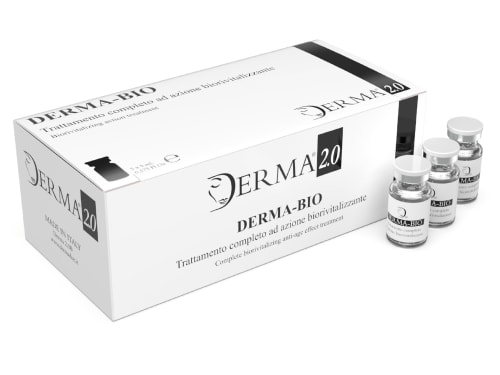
Discover the science behind Derma Bio, a mesotherapy product designed to combat signs of aging and achieve a brighter, smoother, and more youthful appearance.
-

A glycosaminoglycan that forms the water-retaining ground substance of the dermis and maintains the mechanical environment fibroblasts need for collagen synthesis.