
Elastin
Elastin is the dermal protein responsible for skin’s stretch-and-recoil capacity and the most clinically challenging structural protein to work with, because adult skin cannot meaningfully rebuild it once it is damaged. Unlike collagen, which responds to synthesis stimulation with measurable new fibre deposition, elastin recovery is constrained by a biological architecture that is primarily established before birth: functional elastic fibre assembly requires intact fibrillin microfibril scaffolding that UV radiation degrades, and tropoelastin produced by adult fibroblasts without that scaffold cannot form properly functioning fibres. Solar elastosis compounds this; heavily photodamaged skin accumulates degraded, non-functional elastin material that actively impedes new fibre organisation. The strategic priority for elastin is therefore protect-first, degrade-less, then support what synthesis capacity remains – with the clinical goal being improvement in elastic fibre quality and organisation rather than dramatic quantitative restoration.
Elastin is the structural protein responsible for skin’s capacity to stretch and return to shape. Working alongside the collagen scaffold in the dermis, it gives skin its recoil, snap-back, and the subtle responsiveness that distinguishes youthful from aged texture. In young, healthy skin, elastin accounts for approximately 2–4% of dermal dry weight but contributes disproportionately to the mechanical behaviour of the tissue. What makes elastin categorically different from collagen, and what makes the honest clinical conversation about elastin quite different from the one about collagen, is its near-complete inability to regenerate in adult skin once damaged.
Structure and Assembly
Elastin is produced as a soluble monomer called tropoelastin, secreted by fibroblasts and smooth muscle cells. The assembly of tropoelastin into a functional elastic fibre is a hierarchical process that cannot proceed without a pre-existing scaffold. Fibrillin-1 microfibrils, independently assembled proteins in the extracellular matrix, act as the template onto which tropoelastin deposits and organises. Tropoelastin spherules adsorb onto this microfibrillar scaffold, coalesce into larger aggregates through a temperature-dependent process called coacervation, and are then cross-linked by lysyl oxidase (LOX) and its related enzymes (LOXL1–4) into the insoluble, mechanically functional elastin polymer. [7]
The scaffold dependency is clinically significant. Even if adult fibroblasts produce tropoelastin in response to treatment stimuli, the re-formation of structurally functional elastic fibres requires intact fibrillin microfibrils. UV radiation degrades both the elastin itself and the fibrillin scaffold that new tropoelastin would need to deposit onto. This is one of the fundamental reasons elastin repair is genuinely difficult and why claims of dramatic elastin regeneration from topical or professional treatments should be interpreted with considerable caution. [1]
Why Adult Skin Cannot Easily Rebuild Elastin
Elastogenesis is primarily a developmental process: the majority of the body’s elastic fibre network is established during late foetal development and early postnatal life. Adult fibroblasts retain the capacity to produce tropoelastin but at dramatically reduced levels compared to developmental fibroblasts, and the reorganisation of those lower quantities into well-formed elastic fibres is constrained by the fibrillin scaffold availability described above. The current description’s statement that “elastin production essentially stops after puberty” is broadly correct in practical terms, though more precisely: production capability is markedly diminished rather than completely absent, and the assembly pathway becomes increasingly inefficient as the scaffold infrastructure ages and degrades. [7]
This matters for how treatments are framed. Some professional treatments demonstrably stimulate new tropoelastin expression in fibroblasts. Whether that tropoelastin assembles into properly functioning elastic fibres, rather than being degraded intracellularly or deposited inefficiently, depends on the state of the fibrillin microfibril scaffold in the tissue being treated.
Solar Elastosis: Accumulation, Not Just Loss
When people describe elastin declining with age and UV exposure, they typically mean the skin loses its elastic resilience. The full picture is more specific, and understanding it changes how UV damage to elastin is framed clinically. Solar elastosis is not simply a loss of elastin. It is the pathological accumulation of degraded, fragmented, non-functional elastin-like material in the upper dermis, produced when UV irradiation, primarily UVA1, upregulates MMP-12 (metalloelastase). MMP-12 degrades intact elastic fibres, producing fragments that accumulate because they cannot be properly cleared or reassembled. [9]
The result is paradoxical: severely photodamaged skin often contains more total elastin material by mass measurement than healthy aged skin, but virtually none of it is mechanically functional. Clinically this manifests as the characteristic leathery, coarse, yellowish appearance of heavily sun-damaged skin. This distinction matters when interpreting treatment outcomes: a treatment that reduces total measured elastin in photodamaged tissue may be removing solar elastotic material and improving functional elastic fibre quality, rather than simply degrading the barrier further.
The RF Paradox: A Case Study in Honest Evidence
Radiofrequency treatment research produced a finding that clearly illustrates this complexity. A controlled RF facial rejuvenation study showed statistically significant increases in collagen Types I and III alongside a statistically significant decrease in total elastin at 3 months post-treatment. On the surface, this looks negative for elastin. The authors’ explanation is clinically coherent: the decrease in total elastin reflected removal and replacement of abnormal solar elastotic material, with reorientation of elastic fibres and evidence of newly synthesised, properly assembled fibres taking its place. The tissue was, in effect, exchanging non-functional elastin accumulation for a smaller quantity of better-functioning elastic fibre. [3]
Taken honestly, this is a useful marker of how elastin outcomes should not be evaluated purely by quantity. Tissue measurements of total elastin are not a reliable proxy for functional elastin quality, which is why the clinical experience of improved skin elasticity after RF treatment coexists with what looks on paper like an elastin reduction. A combined RF and high-intensity focused electromagnetic stimulation (HIFES) study using biopsy analysis found elastin content increased by 75.9% at one month and 110.8% at two months post-treatment, suggesting that deep tissue heating and mechanical stimulation together can drive more substantive new elastic fibre formation than surface heating alone. [5]
Perimenopause and Oestrogen
Oestrogen receptors are expressed in dermal fibroblasts, and oestrogen directly promotes tropoelastin gene expression alongside its effects on collagen. The perimenopausal decline in oestrogen therefore reduces not only procollagen synthesis but tropoelastin production as part of the same fibroblast downregulation. The fibrillin scaffold also becomes less well-maintained as oestrogen declines, compounding the assembly problem. For perimenopausal clients experiencing skin laxity alongside dryness and texture changes, the elastin dimension of dermal decline is occurring in parallel with collagen loss but through overlapping mechanisms – and protection and synthesis support strategies applied early in hormonal transition are more effective than interventional repair later. [8]
The elastin calcification pathway – prevented by vitamin K2’s MGP carboxylation activity – is also progressive and cumulative, beginning well before clinical laxity is visible. As with glycation-driven collagen rigidity, the most effective intervention is one that begins before significant accumulation has occurred. For perimenopausal clients already addressing oestrogen-related tropoelastin decline, K2 supplementation alongside vitamin D3 (whose calcium absorption-increasing activity makes adequate MGP function more important, not less) is a mechanistically coherent addition to the structural maintenance protocol.
Protecting What Cannot Easily Be Replaced
Given the limited regenerative capacity of adult elastin, protection from primary causes of degradation carries more clinical weight for elastin than it does for collagen. Broad-spectrum sun protection addresses the MMP-12 and ROS-driven elastin degradation pathways simultaneously. Antioxidant-based skincare, particularly vitamin C and vitamin E, reduces the reactive oxygen species load that activates AP-1 and downstream MMP transcription. Retinoids reduce MMP expression broadly and have some evidence for increasing tropoelastin expression in fibroblasts, though the evidence for elastin is considerably less robust than for collagen synthesis. [9]
The strategic hierarchy for elastin is: protect first (UV and oxidative stress prevention), slow the degradation environment (retinoids, antioxidants, MMP modulation), and then support what synthesis capacity remains through professional treatments – with realistic expectations that improvement in elastic fibre quality and organisation, rather than dramatic quantitative restoration, is the achievable goal in most presentations.
Clinical Application
The clinical framework for elastin treatments differs fundamentally from the collagen framework. Where collagen strategy centres on stimulating synthesis and controlling MMP degradation in roughly equal measure, elastin strategy requires a third consideration upfront: the state of the fibrillin microfibril scaffold that newly synthesised tropoelastin must deposit onto. A treatment that successfully stimulates tropoelastin production in tissue where the scaffold is degraded produces limited elastic fibre recovery regardless of how much new protein is expressed.
This means treatment selection for elastin is best structured around three questions: Is there significant solar elastotic material present that is actively interfering with normal elastic fibre organisation? Is the fibrillin scaffold sufficiently intact to support new elastic fibre assembly? And which pathway to fibroblast activation is most appropriate given the inflammatory context of the tissue?
Clearing the Path: Fractional RF and the Solar Elastosis Problem
For photodamaged skin with visible signs of solar elastosis, the priority is not simply stimulating new elastin – it is removing the dysfunctional accumulated material and creating the structural environment in which newly synthesised tropoelastin can assemble correctly. This is where fractional radiofrequency produces its most distinctive elastin benefit.
A biopsy-confirmed clinical study on combined microneedling and sublative fractional RF treatment found statistically significant restoration of normal-appearing elastic fibres alongside a significant reduction in abnormal elastin at three months, with p = 0.0005 for the abnormal elastin reduction. This is the RF paradox resolved in clinical histology: the treatment exchanges degraded, non-functional elastin accumulation for a smaller quantity of properly organised elastic fibres. For clients with heavily photodamaged or significantly aged skin, this outcome represents a genuine structural improvement even though total elastin content may not increase dramatically. [4]
RF energy’s thermal mechanism is particularly relevant for elastin because the fibrillin scaffold itself responds to controlled heat exposure. Thermal injury in the papillary and reticular dermis triggers a fibroblast activation cascade that includes fibrillin-1 expression alongside tropoelastin production, partially restoring the assembly infrastructure that makes new elastic fibre formation possible. In acne scar tissue, where the dermal architecture is significantly disrupted, fractional RF has demonstrated histological evidence of elastic fibre regeneration alongside reduction of dermal fibrosis – confirming that the treatment can remodel the fibrillar environment rather than simply stimulating synthesis into unchanged tissue. scirp
RF Microneedling: Two Depths, Two Mechanisms
RF microneedling contributes to elastin recovery through both its needling and radiofrequency components, which operate at different tissue depths and on different timelines.
The needling-driven wound healing cascade stimulates fibroblast activation across the treated zone, with TGF-β signalling supporting tropoelastin transcription alongside collagen synthesis as part of the general repair response. The radiofrequency thermal component, delivered into the mid-to-deep dermis via the needle tips, reaches depths where the elastic fibre network is more likely to be structurally intact, creating the thermal remodelling signal at the level where new fibre assembly can be supported by surviving fibrillin infrastructure. The combination produces both the abnormal elastin clearing effect associated with surface fractional RF and the deeper fibroblast activation associated with needling. For clients with mixed presentations – some solar elastosis alongside general laxity from age-related elastin decline – this combined mechanism addresses more of the clinical picture than either component alone.
iPRF: Growth Factor Support for a Slow Process
iPRF contributes to elastin recovery through growth factor delivery, though the evidence base here is more appropriately described as mechanistically plausible than fully characterised for elastin specifically. The TGF-β, PDGF, and IGF-1 released from platelet concentrates all have established fibroblast activation roles, and the significantly elevated TGF-β and fibronectin mRNA expression demonstrated with iPRF versus PRP supports a more complete fibroblast activation environment. Fibronectin is specifically relevant to elastin assembly: it supports tropoelastin deposition onto the fibrillin scaffold during early elastic fibre formation, making it a useful co-factor in the assembly pathway rather than just a general matrix protein. [10]
The honest framing for iPRF and elastin is: it creates a growth factor environment highly conducive to whatever elastin synthesis capacity remains in the tissue, and it supports the matrix protein production that makes assembly possible – but it does not directly address solar elastotic material or degraded fibrillin scaffold. For maximum elastin benefit, iPRF is most rationally paired with RF or RF microneedling rather than used in isolation for elastin-specific concerns.
Polynucleotides: Elasticity Via the Macrophage Axis
The in vivo polynucleotide research that characterised the macrophage PCK1 mechanism for collagen also measured skin elasticity as an outcome, finding significant improvement in skin elasticity over the treatment period alongside collagen density increases. The mechanism extends naturally to elastin: M2 macrophage polarisation and IL-10/TGF-β secretion drive fibroblast activation broadly, and the NF-κB suppression that reduces MMP-driven collagen degradation simultaneously reduces the MMP elastase activity that degrades elastic fibres and their fibrillin scaffold. Polynucleotides do not specifically target tropoelastin transcription, but the combined effect of activated fibroblasts, reduced elastase activity, and a calmer inflammatory environment creates conditions in which whatever residual elastin synthesis capacity exists can function more effectively. [2]
For clients with senescent fibroblasts – the perimenopausal and post-menopausal presentation, or significantly photo-aged skin – polynucleotides’ route through macrophage reprogramming to reach senescent fibroblasts is particularly relevant, because direct fibroblast stimulation approaches become progressively less effective as fibroblast responsiveness declines with age. [2]
PLLA: A Modest but Real Elastin Contribution
PLLA’s primary clinical value in this context is collagen neogenesis rather than elastin-specific intervention, and it should be framed as such. However, its direct PI3K/AKT fibroblast activation upregulates tropoelastin production alongside Type I and III collagen synthesis as part of the broader fibroblast capacity restoration it drives. [6] For clients undergoing a PLLA course primarily for structural collagen decline, this elastin contribution is a genuine secondary benefit rather than an incidental one – particularly in the post-menopausal presentation where the fibroblast senescence that limits collagen output is simultaneously limiting whatever residual tropoelastin synthesis capacity remains. PLLA does not address solar elastotic material, does not remodel the fibrillin scaffold, and does not replace the clearing and remodelling role of RF microneedling for elastin-specific presentations. Where it adds value is in the senescent fibroblast environment, restoring the cellular capacity that makes any elastin synthesis at all possible – as the endpoint of a protocol rather than its starting point.
Treatment Pairings for Elastin
Because elastin recovery requires clearing degraded material, supporting the scaffold, and stimulating synthesis in that logical sequence, combination approaches outperform any single modality.
For photoaged/solar elastosis presentations: Fractional RF or RF microneedling as the primary treatment (clearing abnormal elastin, thermal scaffold remodelling), combined with iPRF in the same session (growth factor support for new fibre assembly), with polynucleotides as a separate preparatory or interval treatment to reduce the MMP-elastase burden before the RF session. The sequencing matters: resolving the inflammatory elastase environment before thermal treatment allows the wound-healing response to operate into a less destructive extracellular environment.
For age/hormonal-related laxity without significant solar elastosis: RF microneedling primary (fibroblast activation at appropriate depth, TGF-β signalling for tropoelastin and fibrillin), combined with polynucleotides (macrophage axis fibroblast activation, NF-κB/MMP suppression), with iPRF as an enhancing co-treatment. This combination targets the fibroblast activation signal from three independent pathways simultaneously – wound cascade, macrophage-fibroblast axis, and growth factor delivery – giving the limited elastin synthesis capacity of adult fibroblasts the strongest possible stimulus from multiple angles. For clients where fibroblast senescence is prominent – post-menopausal presentations, or where previous stimulation treatments have underdelivered – PLLA can be incorporated as a longer-horizon course running alongside or between RF microneedling sessions, partially restoring the fibroblast capacity that determines how effectively both the elastin and collagen synthesis signals are acted upon. [6]
The homecare layer that applies to both: No professional treatment for elastin achieves its potential without consistent broad-spectrum UV protection, which prevents new MMP-12 activity from degrading both the treated tissue and the fibrillin scaffold between sessions. Topical antioxidants, particularly vitamin C and vitamin E, reduce the ROS-driven AP-1 signalling that upregulates MMP transcription. Retinoids reduce MMP expression broadly.
Vitamin K2 – specifically MK-4 and MK-7 – adds a third homecare mechanism that is distinct from both UV protection and antioxidant MMP suppression. Through γ-glutamyl carboxylase (GGCX)-dependent carboxylation of matrix Gla protein (MGP), K2 prevents the deposition of calcium into elastic fibres; a progressive mineralisation process that reduces elastin’s stretch-and-recoil capacity through stiffening rather than degradation. Where glycation drives collagen rigidity through AGE cross-linking and UV-driven MMP-12 degrades the elastic fibre network, elastin calcification represents a third parallel stiffening mechanism operating independently of both. Adequate K2 status does not reverse established calcification – carboxylated MGP prevents further deposition rather than dissolving existing mineral deposits – making early and sustained supplementation more protective than remedial. For clients managing elastin decline comprehensively, MK-7 at 100–200 µg daily addresses this calcification pathway alongside the UV protection and antioxidant protocols addressing the other two.
For elastin specifically, this homecare layer is not supplementary to professional treatment, it is the maintenance environment that determines whether the improvement achieved in clinic is preserved or progressively lost between sessions. [9]
References
Baumann L, Bernstein EF, Weiss AS, et al. (2021). Clinical Relevance of Elastin in the Structure and Function of Skin. Aesthet Surg J Open Forum, 3(3), ojab019 . doi.org/10.1093/asjof/ojab019
Byun KA, Park HJ, Oh S, et al. (2025). Polynucleotides Enhance Collagen Synthesis via Modulating Phosphoenolpyruvate Carboxykinase 1 in Senescent Macrophages: Experimental Evidence. Int J Mol Sci, 26(17) . doi.org/10.3390/ijms26178720
el-Domyati M, el-Ammawi TS, Medhat W, et al. (2011). Radiofrequency facial rejuvenation: evidence-based effect. J Am Acad Dermatol, 64(3), 524-35 . doi.org/10.1016/j.jaad.2010.06.045
El-Domyati M, Moawad O, Abdel-Wahab H, et al. (2025). A New Approach with Combined Microneedle and Sublative Fractional Radiofrequency for Photoaging Management: A Clinical, Histometric, and Immunohistochemical Study. Aesthetic Plast Surg, 49(5), 1435-1443 . doi.org/10.1007/s00266-024-04416-0
Kent DE, Fritz K, Salavastru C, et al. (2024). First Evidence of Cutaneous Remodelling Induced by Synchronized Radiofrequency Aided by High-Intensity Facial Muscle Stimulation: Porcine Animal Model. Dermatol Surg, 50(2), 178-181 . doi.org/10.1097/dss.0000000000004028
Oh S, Lee JH, Kim HM, et al. (2023). Poly-L-Lactic Acid Fillers Improved Dermal Collagen Synthesis by Modulating M2 Macrophage Polarization in Aged Animal Skin. Cells, 12(9) . doi.org/10.3390/cells12091320
Ozsvar J, Yang C, Cain SA, et al. (2021). Tropoelastin and Elastin Assembly. Front Bioeng Biotechnol, 9, 643110 . doi.org/10.3389/fbioe.2021.643110
Remoué N, Molinari J, Andres E, et al. (2013). Development of an in vitro model of menopause using primary human dermal fibroblasts. Int J Cosmet Sci, 35(6), 546-54 . doi.org/10.1111/ics.12075
Shin JW, Kwon SH, Choi JY, et al. (2019). Molecular Mechanisms of Dermal Aging and Antiaging Approaches. Int J Mol Sci, 20(9) . doi.org/10.3390/ijms20092126
Wang X, Yang Y, Zhang Y, et al. (2019). Fluid platelet-rich fibrin stimulates greater dermal skin fibroblast cell migration, proliferation, and collagen synthesis when compared to platelet-rich plasma. J Cosmet Dermatol, 18(6), 2004-2010 . doi.org/10.1111/jocd.12955
Also Known As
- ELN
Biological Relationships
Biological Interactions
- Associated disease Perimenopausal skin changes Evidence: Oestrogen-driven tropoelastin decline causes perimenopausal skin laxity; elastin decline occurs in parallel with collagen loss. Entity text; Thornton MJ 2013 PMC3772914; Lephart 2022 PMC9397534.
- Affects Skin ageing Evidence: Elastin degradation and solar elastosis are defining drivers of skin ageing phenotype; loss of elastic recoil is primary visible sign. Entity text; PMC6540032.
Influenced By
- this Stimulated by Cold Atmospheric Plasma therapy
- this Stimulated by Microneedling Evidence: Microneedling wound healing cascade supports tropoelastin transcription via TGF-beta signalling as part of general repair response. PMC11965193.
- this Stimulated by Oestrogen Evidence: Academic: 17β-estradiol stimulates tropoelastin and elastic fibre proteins in fibroblasts; biorxiv.org/lookup/doi/10.1101/728865
- this Stimulated by Peptides
- this Stimulated by Platelet-Rich Plasma Evidence: PRP/iPRF TGF-beta, PDGF and IGF-1 from platelet concentrates support fibroblast activation for tropoelastin synthesis. Entity text; jocd.12955 Wiley.
- this Stimulated by Poly-L-Lactic Acid Evidence: PLLA PI3K/AKT fibroblast activation upregulates tropoelastin alongside Type I and III collagen synthesis. PMC10177436.
- this Stimulated by Polynucleotides Evidence: Polynucleotides drive M2 macrophage-fibroblast axis activation supporting elastin synthesis; in vivo study showed significant skin elasticity improvement. PMC12429772.
- this Stimulated by Radiofrequency Microneedling Evidence: RF microneedling drives elastic fibre restoration: biopsy shows 75.9% elastin increase at 1 month, 110.8% at 2 months. PMC10833192; PMC11965193.
- this Stimulated by Retinoid Evidence: Retinoids increase tropoelastin expression in fibroblasts and reduce MMP-12 elastase activity via AP-1 antagonism. Entity text; PMC6540032; PMC10095221.
- this Stimulated by Transforming growth factor beta Evidence: TGF-beta drives ECM deposition including elastic fibre components; Smad pathway activates tropoelastin and fibrillin gene expression in fibroblasts during wound healing and remodelling (PMC3857353; entity full_description).
- this Inhibited by Matrix metalloproteinase
- this Inhibited by Oestrogen decline Evidence: Oestrogen decline reduces tropoelastin gene expression in dermal fibroblasts and impairs fibrillin scaffold maintenance. Entity text; PMC3772914 Thornton; PMC9397534 Lephart.
- this Produced by Dermis Evidence: Responsible for synthesising… elastin
- this Produced by Fibroblast PMID: 34297930 Evidence: Tropoelastin is assembled onto a fibrillin microfibril scaffold to form functional elastic fibres
- this Affected by Hallmarks of ageing Evidence: Epigenetic alterations and cellular senescence reduce elastic fibre synthesis; SASP-derived MMPs degrade elastin; cross-linked elastin accumulation is a hallmarks-driven dermal ageing feature (PMC10676801).
- this Affected by Inflammageing Evidence: SASP-derived MMPs (MMP1, MMP3, MMP10, MMP12) degrade elastin in the dermis; chronic inflammation drives elastin fragmentation, producing loss of skin elasticity (PMC10178737 – Inflammaging and Immunosenescence as Part of Skin Aging).
- this Affected by Skin ageing Evidence: Elastic fibre fragmentation is a core skin ageing mechanism via MMP-12 and solar elastosis; elastin decreases in intrinsically aged skin. PMC6540032
Learn More
This topic is discussed in 11 articles:
-
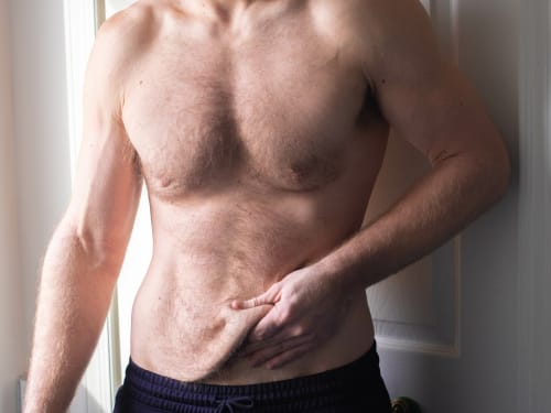
Don’t let loose skin overshadow your weight loss success. Learn why skin sags after significant weight loss and explore our advanced skin tightening and volume restoration treatments.
-
The science behind polynucleotides – a revolutionary treatment that offers a comprehensive approach to skin rejuvenation. How does it compare?
-

Discover the detailed science behind PRP and iPRF therapies. Learn how growth factors, platelets, and cellular mechanisms drive hair restoration and skin rejuvenation at a molecular level.
-

Elevate your autumn skincare routine with our expert advice and innovative treatments: achieve a radiant complexion as the seasons change and the leaves fall.
-

Protein providing skin elasticity and resilience
-

Your 40s bring real hormonal shifts that affect your skin. Discover what’s actually happening and the skincare, nutrition, and treatments that support your skin through perimenopause.
-

Discover the surprising ways air pollution can damage your skin. Learn how to protect your complexion from environmental pollutants and achieve a healthier, more radiant glow.
-

Getting adequate rest is the best investment you can make into your beauty. We take a look at some of your body’s natural skin rejuvenation processes that occur during sleep.
-

Fine lines and wrinkles? Say hello to your skin’s new BFFs: microneedling and RF microneedling! Discover the difference and find your perfect treatment at Creative Touch.
-

Discover how plasma treatments offer a safe, effective, and minimally invasive way to achieve tighter skin, reduce wrinkles, improve acne, and remove skin tags.