
Lip
Lip ageing operates across four structural layers – skeletal resorption, orbicularis atrophy, fat compartment deflation, and vermilion tissue changes – plus a fifth, often overlooked dimension: colour loss. Reduced submucosal vascular density and declining vermilion melanocyte activity progressively desaturate lip colour, making lips appear dull and less distinct from surrounding skin independently of structural change. Cutaneous philtrum lengthening compounds structural ageing by rotating the vermilion inward. Volume restoration addresses only one of these five concurrent processes. Accurate pre-treatment assessment of architecture, proportion, tissue quality, and colour saturation together – not volume alone – is the clinical prerequisite for proportionate, natural, and complete lip rejuvenation outcomes.
The lip complex spans three structural zones: the cutaneous lip (the skin between the vermilion border and the nose superiorly, and the mental crease inferiorly), the vermilion (the keratinised mucosa visible as the red or pink lip body), and the labial mucosa (the wet inner surface). Each zone ages differently and is subject to different treatment considerations. The visible aesthetic unit that clients and practitioners focus on – the volume, definition, and proportion of the vermilion and its borders – is the product of all three zones working in architectural concert, and treating any one layer without understanding its relationship to the others is how lip treatment outcomes become disproportionate or unnatural.
Youthful Lip Architecture
The youthful lip has a set of measurable architectural characteristics that serve as reference points for aesthetic assessment:
Upper-to-lower lip ratio: The youthful ideal in the Caucasian female population is approximately 1:1.6, consistent with the golden ratio – the upper lip vermilion height being approximately 60% of the lower. This ratio is broadly accepted across assessor groups, though variation exists across ethnicities and generations – a 2024 observer study found Baby Boomers preferred the classical 1:1.6 ratio more strongly than younger age groups. The ratio refers specifically to vermilion height, not total lip height including the cutaneous portion.
Cupid’s bow: The double-peaked curvature of the upper lip vermilion border, defined by the two peaks of the bow at the philtral columns and the central dip at the tubercle midpoint. Cupid’s bow definition – the crispness and three-dimensionality of this architecture – is a primary visual marker of lip youth and is among the first structural features to efface with age.
Philtral columns: The two ridges running from the base of the columella to the peaks of the Cupid’s bow. Their prominence creates shadow and three-dimensionality in the upper lip and is architecturally dependent on the underlying orbicularis oris tone and subcutaneous tissue volume below them.
Lip projection: The anterior protrusion of the vermilion relative to the cutaneous lip and the subnasal plane – determined by the underlying bone (anterior maxilla and pyriform aperture), orbicularis oris muscle bulk, and the volume of the labial fat compartments.
Vermilion show: The visible height of the vermilion in frontal view – determined by the eversion of the lip margin and the position of the vermilion border relative to the cutaneous lip.
The Ageing Lip: A Multi-Layer Process
Lip ageing is not a single-tissue event – it is a simultaneous deterioration across four structural layers, each contributing to the composite visual change. [12]
1. Skeletal resorption
Maxillary bone resorption at the pyriform aperture and anterior maxilla withdraws the bony support platform from which the upper lip projects anteriorly. As the skeletal foundation retreats posteriorly, the overlying soft tissue follows – the upper lip effectively migrates backward, reducing both lip projection and vermilion show. Alveolar ridge resorption, particularly in the context of tooth loss, compounds this effect by reducing the anterior lip support from the dental arch. The mandible undergoes parallel resorption with increasing mandibular angle and loss of ramus height, contributing to lower lip and chin ptosis in the lower third. [13]
2. Orbicularis oris muscle atrophy and contracture
The orbicularis oris demonstrates two concurrent age-related changes that work in opposite directions but produce a combined unfavourable outcome. The marginal fibres atrophy – with histological evidence of reduced myosin heavy chain MYH-2 and MYH-7 expression, muscle bundle shrinkage, and increased epimysial connective tissue replacing contractile mass. [12] Simultaneously, the peripheral fibres develop progressive contracture and elevated resting tone, generating radial compression forces around the perioral zone that produce the concentric perioral rhytides characteristic of aged lips. The combined effect converts the youthful “J”-shaped cross-sectional muscle profile – which produces anterior eversion of the lip margin – to an “I”-shaped atrophied profile that allows the lip to invert. [15]
3. Soft tissue and fat compartment deflation
The superficial labial fat compartments – which underlie the dermis of the cutaneous lip and contribute to the fullness of the Cupid’s bow architecture – atrophy with age in the same pattern as facial fat compartments elsewhere. This deflation reduces the three-dimensional fullness of the lip surface and effaces the Cupid’s bow peaks that depend on subcutaneous volume beneath the philtral columns. [4]
4. Vermilion and cutaneous tissue changes
The vermilion itself undergoes intrinsic ageing changes: hyaluronan content decreases through simultaneous reduction in hyaluronan synthase-1 (HAS1) expression and upregulation of CEMIP (cell migration-inducing hyaluronidase), reducing the tissue’s water-binding capacity and producing a drier, thinner appearance. Collagen and elastin degradation at the dermis-vermilion junction reduces turgor and increases fine-line formation. [10] Cupid’s bow architecture flattens and lip width increases as the vermilion border loses structural support – a finding confirmed by stereophotogrammetric morphometric analysis showing significant increases in lip width and cutaneous lip height with age, alongside reduction in upper vermilion height. [2]
The Philtrum Lengthening Phenomenon
The cutaneous philtrum – the distance between the base of the columella and the Cupid’s bow – lengthens progressively with age through soft tissue descent and weakening of perioral support structures. This lengthening has a cascading effect on lip appearance: as the cutaneous philtrum elongates, the vermilion rotates inward and downward, reducing visible vermilion show in frontal view, obscuring the Cupid’s bow architecture, and producing the characteristic aged perioral appearance of a long, flat upper lip with little visible pink tissue. The philtrum-to-lip proportion therefore functions as an ageing clock – a lengthening philtrum relative to vermilion height is one of the earlier and more diagnostically reliable indicators of perioral ageing, and is the anatomical target of surgical lip lift procedures.
Colour and Pigmentation Changes
Lip colour in youth derives primarily from the submucosal capillary plexus – the rich vascular network beneath the thin translucent vermilion epithelium – supplemented by a modest melanin contribution from vermilion melanocytes. Both components diminish with age. Vascular density in the submucosal plexus reduces as part of the general age-related decline in cutaneous microvasculature, and the vermilion epithelium undergoes subtle thickening that increases the optical distance between the surface and the underlying vessels. The combined effect is progressive desaturation of lip colour – a greyish, muted, or washed-out tone that loses the vivid pink-red saturation characteristic of younger lips, independent of any structural change.
Melanocyte density and activity at the vermilion decline in parallel with the general reduction in epidermal melanocyte number seen in intrinsically aged skin. This reduces the depth and warmth of the lip’s natural colour tone and diminishes the chromatic contrast between the vermilion and the surrounding cutaneous lip – making the lip appear less defined even when the Cupid’s bow architecture remains structurally intact. Perioral hyperpigmentation from cumulative UV exposure and chronic irritation compounds this asymmetrically, creating an uneven colour environment around the lip border that further undermines definition.
The clinical significance is that colour desaturation is a distinct ageing dimension from structural change – and one that neither volume restoration nor hydration treatments address. A client who has received technically excellent lip filler and Lumi Pro Lips may still present with a visually aged lip if the colour saturation deficit remains uncorrected. Lip blush – cosmetic tattooing that deposits pigment in the superficial dermis of the vermilion – restores colour saturation and border definition by replacing what vascular and melanocyte decline has removed, addressing the chromatic ageing dimension directly. It is complementary to structural and hydration treatments rather than competing with them, and frequently produces the most visible improvement in clients whose primary complaint is that their lips have “disappeared” or lost their colour rather than their volume.
Lip Tissue Damage
Lip tissue is structurally more vulnerable to acquired damage than facial skin: the stratum corneum’s 3–5 cell layer depth provides limited resilience, the absence of sebaceous glands removes the endogenous lipid replenishment that would otherwise support barrier recovery, and the vermilion’s high TEWL baseline means any additional barrier disruption produces disproportionate consequences. The principal modes of acquired damage most relevant to clinical lip care are covered below. For barrier failure mechanisms, contact cheilitis, angular cheilitis, and the full cheilitis classification framework, see the Chapped Lips page.
The Saliva-Driven Dehydration Cycle
Lip-licking temporarily resolves the sensation of dryness – which is why the behaviour is reinforced – but deposits salivary enzymes onto tissue poorly equipped to manage them. Amylase and lipase degrade residual surface lipids; sodium chloride creates a hyperosmotic surface stress that draws moisture out of epithelial cells; and the subsequent rapid evaporation leaves lips net drier than before contact. Damaged keratinocytes release IL-1α and TNF-α, heightening neural sensitivity and reinforcing the urge to lick – converting a behavioural habit into a physiological feedback loop. [5] The characteristic perioral ring of erythema in established lip-lick cheilitis extends to the boundary of tongue reach rather than the vermilion margin – a distribution that distinguishes it from allergic contact cheilitis and helps guide management decisions. [11]
Irritant “Plumping” Products
Products formulated to induce temporary lip swelling – typically containing capsaicin, cinnamaldehyde, or menthol – act on TRP ion channels in the vermilion epithelium, triggering neurogenic inflammation, vasodilation, and tissue oedema. The swelling is real but transient, and the mechanism producing it is the same one that, with repeated use, degrades the tissue it is acting on. The receptor pharmacology, tolerance profiles, and structural consequences of each agent are covered in the Irritant Lip Plumping Agents page.
The clinical picture worth naming here is the dependency cycle. With habitual capsaicin-containing products, progressive TRPV1 tachyphylaxis reduces the perceived tingling whilst barrier disruption continues – users escalate application frequency in response to diminishing sensation, accelerating damage without the sensory feedback that would otherwise signal it. Cinnamaldehyde does not desensitise in the same way, meaning the inflammatory response reproduces at consistent magnitude on every application with no natural ceiling on cumulative exposure. Recovery following cessation is achievable; barrier function typically restores within four to six weeks with consistent barrier-supportive care.
Clinical Pearl Clients who describe their lips as feeling “addicted” to a specific balm – worse without it, needing constant reapplication, with a characteristic tingle – are usually describing TRP-mediated sensitisation rather than product efficacy. The product is maintaining a low-grade inflammatory state that it is simultaneously marketed as resolving.
When Topical Care Is Not Sufficient
Persistent lip changes that do not respond within two weeks of a well-formulated topical routine warrant professional assessment. Angular cheilitis (commissural fissuring indicating Candida or bacterial infection), actinic cheilitis (persistent lower lip whitish plaque or ulceration – a premalignant condition), sudden reactivity to previously tolerated products, and chronic refractory dryness with possible systemic cause (Sjögren’s syndrome, coeliac disease, retinoid use, B-vitamin or iron deficiency) are all outside the scope of topical lip care and are covered in detail in the Chapped Lips page.
Vascular Anatomy and Filler Safety
The lips are among the highest-risk injection sites in aesthetic medicine, not because of their structural complexity per se, but because of the calibre and course of the arteries that supply them. Understanding this vascular anatomy is not supplementary knowledge for lip practitioners – it is foundational to safe practice.
The Superior and Inferior Labial Arteries
The primary blood supply to the lips is delivered by the superior labial artery (SLA) and the inferior labial artery (ILA), both arising from the facial artery at or near the angle of the mouth. The facial artery, a branch of the external carotid, winds anteriorly from the submandibular region and crosses the mandibular border before ascending toward the lips. The SLA and ILA branch from it in the pericommissural region and run medially toward the midline, supplying each lip through a rich submucosal and intramuscular network.
Their depth relative to the skin surface is the critical variable for injection safety. Cadaveric studies report the SLA at approximately 4.8–5.6 mm from the skin surface and the ILA at approximately 4.1–5.4 mm, with both arteries running generally from a deeper position at the commissure to a more superficial position toward the midline. [6] A 3D CT analysis of 52 cadaveric heads reported a mean SLA depth of 5.68 mm at its commissural origin, reducing across the medial course; lumen diameter at the branch point averages approximately 1.36 mm – a calibre large enough to admit a standard 27-gauge needle tip. [16]
Within the lip, the arteries typically run within or immediately adjacent to the orbicularis oris – most commonly in the intramuscular plane or just deep to the muscle. However, a superficial course anterior to the muscle is well documented, and neither artery reliably follows a predictable plane throughout its entire medial course. This is not a minor anatomical footnote: studies confirm that the SLA alone displays multiple distinct positional patterns relative to the vermilion and orbicularis, and its level of origin from the facial artery varies considerably between individuals and between left and right sides of the same face. [9]
Anatomical Variation and Its Clinical Significance
The variability is the point. A literature review synthesising cadaveric dissection data found that the labial arteries display great variability with respect to path, presence, and location – including cases where one or both arteries are absent or hypoplastic on one side, replaced by a contralateral dominant supply or by collateral contributions from the mental artery or submental artery. [9] An anatomical study of 102 facial arterial specimens identified 35 distinct branching pattern combinations and marked left-right asymmetry within individuals – meaning a practitioner cannot assume symmetrical vascular anatomy bilaterally. [7]
A specific anatomical risk zone – described in cadaveric dissection work – is the fibro-muscular compartment above the philtrum, where the SLA passes and gives off the columellar and septal branches. In this region, rapid bolus injection or filler migration can compress the SLA against the fibromuscular tissue, producing ischaemia through extrinsic compression rather than direct intravascular penetration. [6]
Occlusion Risk and Mechanism
Vascular complications in lip filler procedures arise through two principal mechanisms: direct intravascular injection (needle or cannula tip enters the arterial lumen and filler is deposited intra-arterially) and extravascular compression (the volume of filler compresses the arterial wall sufficiently to impede or arrest flow). The ischaemic consequences – pallor, pain, delayed capillary refill, and ultimately cutaneous necrosis if untreated – are produced by the same physiological endpoint regardless of mechanism: cessation of arterial supply to the downstream capillary bed.
The anastomotic connections between the SLA and ILA at the midline, and between the labial arterial system and adjacent vessels (including the columellar branches and septal arteries), mean that occlusion at any point can redistribute to produce ischaemia in tissue that appears anatomically distant from the injection site. The SLA’s anastomotic continuity with the nasal septal artery and dorsal nasal arterial branches is of particular relevance – this pathway can transmit an embolic filler bolus toward the ophthalmic artery system, the mechanism implicated in the rare but catastrophic complication of filler-related visual loss. [1] [14]
For these reasons, technical principles – aspiration before injection where clinically indicated, slow retrograde deposition, small aliquots, the use of cannulas where appropriate, and familiarity with hyaluronidase reversal protocols – are not stylistic preferences but mechanistically grounded responses to the vascular anatomy described here. [8] [3]
Clinical Application
The pre-treatment lip assessment in aesthetics practice should evaluate the following in combination rather than treating volume alone as the target:
- Vermilion height (upper and lower) and upper-to-lower ratio
- Cupid’s bow definition and philtral column prominence
- Philtrum length relative to vermilion show
- Lip projection relative to subnasale and pogonion
- Vermilion border definition and blurring pattern
- Lip symmetry – including commissure position and any asymmetric volume loss
- Perioral rhytide pattern and depth (indicating orbicularis contracture severity)
- Prior filler burden assessment – palpation and patient history
- Lip colour saturation and tone – degree of desaturation relative to surrounding skin; greying or washed-out appearance; whether colour loss precedes or accompanies structural change
- Perioral pigmentation pattern – UV-related hyperpigmentation at the vermilion border; uneven colour framing; relevance to lip blush candidacy
The clinical implication of the multi-layer ageing model is that volume restoration alone – the default approach of simply adding cross-linked HA to the body of the lip – addresses only one of four concurrent structural changes. A client with significant philtrum lengthening and Cupid’s bow effacement may benefit more from targeted philtral column definition and vermilion border reinforcement than from bulk volume addition, which can produce disproportionate results if the structural architecture has not been addressed first. Clients with significant skeletal resorption-driven lip recession present a different challenge than those with primarily soft tissue deflation, and the treatment approach – volume placement depth, product selection, and whether adjunct treatments such as deep-plane biostimulators for soft tissue support or surgical referral for skeletal correction are appropriate – differs accordingly.
References
Beleznay K, Carruthers JD, Humphrey S, et al. (2015). Avoiding and Treating Blindness From Fillers: A Review of the World Literature. Dermatol Surg, 41(10), 1097-117 . doi.org/10.1097/dss.0000000000000486
Chong Y, Dong R, Liu X, et al. (2021). Stereophotogrammetry to reveal age-related changes of labial morphology among Chinese women aging from 20 to 60. Skin Res Technol, 27(1), 41-48 . doi.org/10.1111/srt.12906
Cohen JL, Biesman BS, Dayan SH, et al. (2015). Treatment of Hyaluronic Acid Filler-Induced Impending Necrosis With Hyaluronidase: Consensus Recommendations. Aesthet Surg J, 35(7), 844-9 . doi.org/10.1093/asj/sjv018
Coleman SR, Grover R (2006). The anatomy of the aging face: volume loss and changes in 3-dimensional topography. Aesthet Surg J, 26(1S), S4-9 . doi.org/10.1016/j.asj.2005.09.012
Fonseca A, Jacob SE, Sindle A (2020). Art of prevention: Practical interventions in lip-licking dermatitis. Int J Womens Dermatol, 6(5), 377-380 . doi.org/10.1016/j.ijwd.2020.06.001
Lupu F, Iliuta CP, Enyedi M, et al. (2022). The Assessment of the Anatomical Risk in the Perioral Region. Maedica (Bucur), 17(4), 820-825 . doi.org/10.26574/maedica.2022.17.4.820
Nguyen VH, Cheng-Kuan L, Nguyen TA, et al. (2024). The branching patterns and termination points of the facial artery: a cadaveric anatomical study. Arch Craniofac Surg, 25(2), 77-84 . doi.org/10.7181/acfs.2024.00038
Nikolis A, Cohen JL, Enright KM, et al. (2024). Deliberations of the Safety Task Force: Risk factors and treatment of adverse events associated with aesthetic injectables. J Cosmet Dermatol, 23(11), 3551-3564 . doi.org/10.1111/jocd.16476
Samizadeh S, Pirayesh A, Bertossi D (2019). Anatomical Variations in the Course of Labial Arteries: A Literature Review. Aesthet Surg J, 39(11), 1225-1235 . doi.org/10.1093/asj/sjy235
Shang J, Feng X, Chen Y, et al. (2024). Human lip vermilion: Physiology and age-related changes. J Cosmet Dermatol, 23(8), 2676-2680 . doi.org/10.1111/jocd.16317
Stone RJ, Labert GM, Norman RA (2024). Lip-Lick Cheilitis and Its Connection to the Brain. Cureus, 16(7), e64312 . doi.org/10.7759/cureus.64312
Sun F, Liu Y, Zhang T (2025). Aging of the Human Lip: Current Knowledge and Clinical Implications. J Cosmet Dermatol, 24(8), e70310 . doi.org/10.1111/jocd.70310
Swift A, Liew S, Weinkle S, et al. (2021). The Facial Aging Process From the “Inside Out”. Aesthet Surg J, 41(10), 1107-1119 . doi.org/10.1093/asj/sjaa339
Thanasarnaksorn W, Cotofana S, Rudolph C, et al. (2018). Severe vision loss caused by cosmetic filler augmentation: Case series with review of cause and therapy. J Cosmet Dermatol, 17(5), 712-718 . doi.org/10.1111/jocd.12705
Yi KH, Wan J (2025). The Aging Process of Facial Muscles. J Cosmet Dermatol, 24(12), e70590 . doi.org/10.1111/jocd.70590
Zhu GS, Liao ZF, Chen CL, et al. (2024). Three-Dimensional Computed Tomography Scanning Study of the Superior Labial Artery in Chinese Individuals for Assessing Filler Injection Safety. Aesthetic Plast Surg, 48(19), 3962-3970 . doi.org/10.1007/s00266-024-04187-8
Also Known As
- lips
Learn More
This topic is discussed in 4 articles:
-
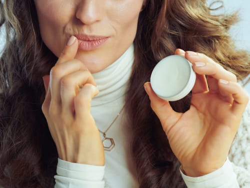
The fleshy, movable muscular folds surrounding the opening of the oral cavity. They are composed of skin, the orbicularis oris muscle, and mucous membrane, serving as the entry to the digestive tract and playing key roles in speech, consumption, and tactile sensory reception.
-

If a treatment it isn’t safe, or won’t naturally enhance your looks, we will say no to you. Find out why.
-
Celebrate the achievements of Zoe Bayliss and her team at Creative Touch, an award-winning salon in South Yorkshire.
-
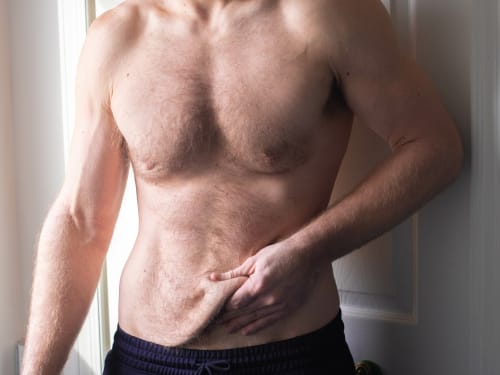
Don’t let loose skin overshadow your weight loss success. Learn why skin sags after significant weight loss and explore our advanced skin tightening and volume restoration treatments.