
Tumour necrosis factor
Tumour necrosis factor alpha (TNF-α) is a pro-inflammatory cytokine produced by macrophages, adipocytes, and T-cells that acts through NF-κB to amplify and sustain inflammatory signalling. In skin, it operates across three axes: it suppresses filaggrin and loricrin expression via the FRA1:c-JUN:HDAC1 complex and JNK activation, compromising barrier integrity; it drives type I collagen degradation through MMP-1/ MMP-3 upregulation, accelerating dermal ageing; and it induces serine phosphorylation of IRS-1, blocking insulin receptor signalling and creating peripheral insulin resistance in obesity. In psoriasis it is a primary pathogenic driver and the target of anti-TNF biologic therapies. TNF-α is not directly targeted in aesthetic practice but is a consistently relevant upstream driver of barrier failure, collagen loss, and skin quality decline.
TNF-α is released in response to cellular stress, infection, injury, or sustained metabolic dysfunction. It signals through two cell-surface receptors – TNFR1 (ubiquitously expressed) and TNFR2 (primarily immune cells and endothelium) – activating the NF-κB transcription factor pathway as its primary downstream signal. NF-κB activation drives a pro-inflammatory gene expression programme: upregulation of IL-1β, IL-6, IL-8, COX-2, and additional TNF-α itself, creating an amplification loop that sustains inflammation beyond the initial trigger. In skin, this downstream cascade operates across three clinically relevant axes. nature.com
Barrier Protein Suppression
TNF-α directly suppresses the expression of filaggrin (FLG) and loricrin (LOR) – the two principal structural proteins of the stratum corneum. The mechanism involves TNF-α (synergistically with IFN-γ) promoting formation of the FRA1:c-JUN:HDAC1 transcriptional repressor complex, which binds the filaggrin gene promoter and inhibits its transcription. [1] TNF-α additionally downregulates filaggrin and loricrin through c-Jun N-terminal kinase (JNK) activation – a Smad-independent pathway that reduces barrier protein synthesis independently of the FRA1 complex. [5] The functional consequence is a thinner, protein-depleted stratum corneum with elevated TEWL – the same pattern observed in both psoriasis and atopic dermatitis, where TNF-α is chronically elevated. This connects TNF-α directly to the barrier disruption cycle: reduced filaggrin → elevated TEWL → increased allergen and irritant penetration → further immune activation → further TNF-α production.
Collagen Degradation
TNF-α drives type I collagen degradation in human skin through a two-step MMP activation mechanism. It upregulates MMP-1 secretion and induces MMP-3 production, which acts as an endogenous activator of MMP-1’s collagenolytic activity – the combination producing significantly accelerated type I collagen degradation compared to either agent alone. [7] TNF-α also activates pro-MMP-2 through NF-κB-dependent induction of MT1-MMP (membrane type 1 MMP) in fibroblasts embedded in collagen matrix, providing a parallel pathway for ECM degradation. [2] The net effect is accelerated collagen turnover weighted toward degradation over synthesis – the same imbalance that characterises photoaged skin and chronically inflamed skin conditions, and that explains why sustained systemic TNF-α elevation from adipose inflammation contributes to accelerated dermal ageing independent of UV exposure.
Insulin Resistance and the Metabolic-Skin Axis
Adipose tissue is not merely a passive energy store – it is an active endocrine organ, and hypertrophied adipocytes in obesity secrete substantially elevated TNF-α into the systemic circulation. TNF-α induces serine phosphorylation of insulin receptor substrate-1 (IRS-1) in adipocytes, muscle, and liver cells, converting IRS-1 from a positive mediator of insulin receptor signalling into an inhibitor – blocking the tyrosine kinase activity of the insulin receptor and preventing downstream glucose uptake signalling. [3] This is the primary molecular mechanism by which adipose inflammation in obesity creates peripheral insulin resistance – establishing TNF-α as the biochemical link between excess adiposity and type 2 diabetes risk. For skin, the consequence is a dual pathway of damage: direct MMP-driven and filaggrin-suppressive effects on the dermis and epidermis, compounded by the glycation, AGE accumulation, and IGF-1 dysregulation that accompany insulin resistance and elevated blood glucose.
TNF-α in Psoriasis
Psoriatic lesions are characterised by substantially elevated TNF-α produced by keratinocytes, dermal macrophages, and T-lymphocytes. TNF-α drives the keratinocyte hyperproliferation and defective differentiation of psoriatic plaques through NF-κB activation, and synergises with IL-17A – the other primary psoriatic cytokine – to amplify chemokine production, neutrophil recruitment, and the self-sustaining inflammatory loop of the psoriatic plaque. [6] The clinical validation of TNF-α’s central role in psoriasis pathogenesis comes from the therapeutic success of anti-TNF biologics – etanercept, adalimumab, infliximab – which produce substantial plaque clearance by neutralising TNF-α bioavailability, dismantling the TNF-α/IL-17A synergy that sustains plaque inflammation before T-cell numbers or keratinocyte differentiation patterns visibly normalise. [4]
References
Ahn SS, Yeo H, Jung E, et al. (2022). FRA1:c-JUN:HDAC1 complex down-regulates filaggrin expression upon TNFα and IFNγ stimulation in keratinocytes. Proc Natl Acad Sci U S A, 119(37), e2123451119 . doi.org/10.1073/pnas.2123451119
Han YP, Tuan TL, Wu H, et al. (2001). TNF-alpha stimulates activation of pro-MMP2 in human skin through NF-(kappa)B mediated induction of MT1-MMP. J Cell Sci, 114(Pt 1), 131-139 . doi.org/10.1242/jcs.114.1.131
Hotamisligil GS, Peraldi P, Budavari A, et al. (1996). IRS-1-mediated inhibition of insulin receptor tyrosine kinase activity in TNF-alpha- and obesity-induced insulin resistance. Science, 271(5249), 665-8 . doi.org/10.1126/science.271.5249.665
Johnston A, Guzman AM, Swindell WR, et al. (2014). Early tissue responses in psoriasis to the antitumour necrosis factor-α biologic etanercept suggest reduced interleukin-17 receptor expression and signalling. Br J Dermatol, 171(1), 97-107 . doi.org/10.1111/bjd.12937
Kim BE, Howell MD, Guttman-Yassky E, et al. (2011). TNF-α downregulates filaggrin and loricrin through c-Jun N-terminal kinase: role for TNF-α antagonists to improve skin barrier. J Invest Dermatol, 131(6), 1272-9 . doi.org/10.1038/jid.2011.24
Victor FC, Gottlieb AB (2002). TNF-alpha and apoptosis: implications for the pathogenesis and treatment of psoriasis. J Drugs Dermatol, 1(3), 264-75 . pubmed.ncbi.nlm.nih.gov/12851985
Ågren MS, Schnabel R, Christensen LH, et al. (2015). Tumor necrosis factor-α-accelerated degradation of type I collagen in human skin is associated with elevated matrix metalloproteinase (MMP)-1 and MMP-3 ex vivo. Eur J Cell Biol, 94(1), 12-21 . doi.org/10.1016/j.ejcb.2014.10.001
Also Known As
- TNF
- TNF-alpha
- TNF-α
Biological Relationships
Biological Interactions
- Stimulates Cellular senescence Evidence: Chronic TNF-α exposure drives fibroblast entry into senescence via NF-kB-mediated stress signalling. Yu et al. 2023 Aging Cell doi:10.1111/acel.14054
- Stimulates Inflammageing Evidence: TNF-alpha is a primary SASP cytokine driving inflammageing; elevated serum TNF-alpha is a defining biomarker; TNF-alpha-senescence creates a positive feedback loop sustaining the inflammatory state (doi:10.18632/aging.101328; PMC10178737).
- Stimulates Interleukin-6 Evidence: TNF-α activates NF-kB, driving upregulation of IL-6 as part of the pro-inflammatory gene expression programme. doi:10.1038/s41419-022-04523-3
- Stimulates Keratinocyte Evidence: TNF-α activates NF-κB in keratinocytes driving IL-6 IL-8 CXCL1 production; TNF receptor signalling triggers IL-24-dependent psoriasis-like inflammation. Kumari et al. Immunity 2013 doi:10.1016/j.immuni.2013.10.009
- Stimulates Matrix metalloproteinase Evidence: TNF-alpha activates NF-kB and AP-1 in dermal fibroblasts driving MMP-1/3/9 upregulation. Entity text; Radtke et al. 2019 PMC6829232.
- Stimulates Senescence-associated secretory phenotype Evidence: TNF-α is a core SASP component and positive regulator of SASP expression in senescent fibroblasts. Campisi et al. 2013 JCI doi:10.1172/JCI64098
- Interacts with Interleukin-6 Evidence: TNF-α and IL-6 form positive amplification loop; TNF induces IL-6, both converge on NF-kB-driven inflammation. doi:10.1038/s41419-022-04523-3
- Inhibits Collagen Evidence: TNF-α drives type I collagen degradation via MMP-1/MMP-3 upregulation; net collagen turnover weighted toward degradation. PMID:25457675
- Inhibits Fibroblast Evidence: TNF-α suppresses collagen synthesis in dermal fibroblasts and increases MMP-1/collagen I ratio. Huuskonen et al. Microorganisms 2023 doi:10.3390/microorganisms11061465
- Inhibits Filaggrin Evidence: TNF-α promotes FRA1:c-JUN:HDAC1 repressor complex binding filaggrin gene promoter, suppressing FLG transcription. PNAS doi:10.1073/pnas.2123451119
- Inhibits Loricrin Evidence: TNF-α suppresses loricrin via JNK activation and reduced epidermal differentiation complex gene expression. PMID:21346775
- Associated disease Dermatitis Evidence: TNF-α chronically elevated in dermatitis lesional skin driving barrier dysfunction and inflammation. Kim et al. 2023 Sci Rep doi:10.1038/s41598-023-41831-w
- Associated disease Obesity Evidence: Hypertrophied adipocytes in obesity secrete elevated TNF-α; TNF drives insulin resistance via IRS-1 serine phosphorylation. PMID:8571133
- Associated disease Psoriasis Evidence: TNF-α is a primary pathogenic driver of psoriasis; anti-TNF biologics (etanercept, adalimumab) validate its central role. PMC4115021
- Affects Dermis Evidence: TNF-α stimulates MMP expression in dermal fibroblasts and upregulates collagen degradation. Huuskonen et al. Microorganisms 2023 doi:10.3390/microorganisms11061465
- Affects Rosacea Evidence: TNF-alpha is among the macrophage-derived mediators contributing to rosacea pathology alongside MMPs and interferon-gamma.
- Affects Skin ageing Evidence: TNF-α-driven collagen degradation and barrier protein suppression contribute to accelerated skin ageing independent of UV. PMID:25457675
Influenced By
- this Stimulated by Adipocyte Evidence: Dysfunctional adipocytes stimulate TNF-alpha; TNF-alpha promotes HSL lipolysis via NF-kB. Itoh 2011; Dicker 2007 doi:10.1194/JLR.M600471-JLR200.
- this Stimulated by Deoxycholic acid Evidence: PDC/DCA induces TNF-alpha release; post-necrotic inflammatory cascade drives TNF-alpha. Muskat 2022 PMC8988282; Frontiersin doi:10.3389/fendo.2022.841889.
- this Interacts with Interleukin-13 Evidence: IL-13 and TNF-α co-drive barrier dysfunction; TNF amplifies IL-13-induced keratinocyte changes in AD. Bay-Jensen et al. 2025 Sci Rep doi:10.1038/s41598-024-84151-3
- this Interacts with Interleukin-4 Evidence: IL-4 and TNF-α co-regulate keratinocyte gene expression; TNF counteracts some IL-4-mediated differentiation effects. Kanelleas et al. 2022 JCM doi:10.3390/jcm11195633
- this Produced by Adipocyte Evidence: Dysfunctional adipocytes produce TNF-alpha as part of the SASP/obesity-associated inflammatory cascade. Entity text; Gao 2025.
- this Affected by Dermatitis Evidence: Irritant contact dermatitis mechanism: damaged keratinocytes release pro-inflammatory cytokines including TNF-alpha, IL-1alpha, and IL-8 to recruit neutrophils and macrophages (NBK562228).
- this Affected by Psoriasis Evidence: TNF-alpha is a core pathogenic cytokine overproduced in psoriatic lesions by multiple cell types; it amplifies inflammatory signalling and suppresses filaggrin via c-Jun pathway (PMC8657643; PMC8609659).
- this Affected by Subcutaneous tissue Evidence: SASP from senescent subcutaneous adipocytes explicitly includes TNF-alpha; entity text cites PMC10409694.
Learn More
This topic is discussed in 4 articles:
-

While many patients experience positive weight loss results from Mounjaro injections, here’s what you should know about tiredness as a potential side effect of Mounjaro.
-

TNF is a pro-inflammatory cytokine produced by macrophages, adipocytes, and T-cells that acts through NF-κB to amplify and sustain inflammatory signalling.
-
TNF is a pro-inflammatory cytokine produced by macrophages, adipocytes, and T-cells that acts through NF-κB to amplify and sustain inflammatory signalling.
-

Pro-inflammatory cytokine reduced by ketogenic and low-carbohydrate dietary approaches
-
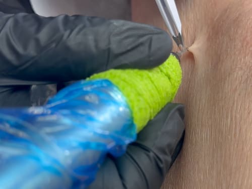
Not liking those pesky skin tags? Learn about causes, blemish removal options at Creative Touch, and prevention tips for smoother, bump-free skin.