
Subcutaneous tissue
Subcutaneous tissue – the hypodermis – is the deepest of the three skin layers, composed predominantly of white adipose tissue organised into adipocyte lobules separated by fibrous septa. It provides structural support, thermal insulation, energy storage, and endocrine signalling, and contributes directly to the surface architecture of skin through its volume distribution in facial and body fat compartments. With ageing, subcutaneous tissue undergoes three distinct processes that affect skin appearance: compartmental volume loss removing structural support, fat infiltration upward into the dermis impairing elasticity, and adipocyte senescence driving a local and systemic pro-inflammatory state through SASP cytokine secretion. Multiple non-invasive body contouring modalities – cryolipolysis, ultrasonic cavitation, HIEMT, radiofrequency, and deoxycholic acid injection – act on subcutaneous adipocytes through distinct mechanisms that exploit different aspects of adipocyte biology. Subcutaneous tissue is also the primary route for drug delivery of GLP-1 agonists, insulin, and biological therapies.
Subcutaneous tissue (the hypodermis) is the third and deepest layer of the skin complex, situated below the dermis and above the deep fascia or periosteum depending on anatomical location. It is not merely a passive cushioning layer – it is a metabolically active endocrine organ, a structural determinant of surface skin architecture, and a site of significant biological change with age. Its composition, distribution, and cellular behaviour are directly relevant to a wide range of aesthetic treatments and to the visible ageing changes that many of those treatments address.
Anatomy and Composition
Subcutaneous tissue is organised into lobules of adipocytes – lipid-storing cells that constitute the majority of tissue volume – separated by fibrous septa of connective tissue that anchor the layer to the overlying dermis and the underlying fascia. These septa are not uniformly distributed: in areas prone to cellulite (primarily female thighs and buttocks), the septa run perpendicularly between dermis and fascia, creating the tethered surface dimpling characteristic of that condition; in other areas they run at oblique angles, producing a smoother surface.
The vasculature, lymphatics, and sensory nerve supply of the skin all pass through subcutaneous tissue, making it anatomically relevant to any treatment that affects tissue circulation or lymphatic drainage. The lymphatic network within subcutaneous tissue is a key clearance route for the cellular debris and triglycerides produced by fat-reduction treatments – a dependency that has direct implications for post-treatment protocols and expected outcome timelines.
Adipose tissue types. Not all subcutaneous fat is metabolically identical. White adipose tissue (WAT) is the predominant form – unilocular cells storing energy as triglycerides, with endocrine function including leptin and adiponectin secretion. Brown adipose tissue (BAT) contains multilocular cells rich in mitochondria and specialised for thermogenesis through uncoupling protein-1 (UCP-1); it is most abundant in neonates but persists in adults around the supraclavicular and paraspinal regions. Beige (brite) adipocytes are white adipocytes capable of adopting a brown-like thermogenic phenotype under cold or adrenergic stimulation – a plasticity relevant to the thermogenesis debate in cryolipolysis. nature.com The functional distinction between WAT and BAT matters for treatment response: modalities that induce adrenergic stimulation (HIEMT, cold exposure) may activate beige adipocyte thermogenesis as a secondary mechanism alongside their primary fat-reduction pathway.
The stromal vascular fraction. Subcutaneous tissue is not composed solely of adipocytes. The stromal vascular fraction (SVF) – the non-adipocyte cellular component – includes preadipocytes, fibroblasts, endothelial cells, pericytes, resident macrophages, and adipose-derived stem cells (ADSCs). This cellular diversity gives subcutaneous tissue its regenerative and immune capacity; ADSCs in particular have attracted significant research interest for their ability to differentiate into multiple cell lineages and their paracrine secretion of growth factors. The SVF is also the compartment in which the inflammatory changes of ageing are initiated, through macrophage accumulation and adipocyte senescence.
Facial fat compartments. In the face, subcutaneous fat is organised into discrete anatomical compartments – superficial and deep – whose boundaries are maintained by fibrous septae and retaining ligaments. The distinct compartments of the midface (nasolabial, medial cheek, lateral cheek, infraorbital), periorbital region, forehead, and temporal fossa each age independently rather than uniformly, which is why facial volume loss produces characteristic patterns of deflation rather than global flattening. This compartmental architecture is the anatomical basis for precise aesthetic volume restoration – filling compartments in a sequence that respects their structural interdependence – and is covered in more detail in the Facial Fat Compartments entity.
Why Adipocytes Are Selectively Targetable
The ability to reduce subcutaneous fat non-invasively without damaging overlying dermis, muscle, or neurovascular structures depends on biological properties that make adipocytes specifically more vulnerable than surrounding cell types to certain physical and chemical stimuli. Understanding these selectivity mechanisms explains why non-invasive fat reduction is possible at all – and why each modality is suited to specific clinical scenarios.
Cold sensitivity differential. Adipocytes are more sensitive to cold-induced injury than dermal or epidermal cells. The high lipid content of adipocytes means their intracellular contents begin to crystallise at temperatures between approximately −1°C and +5°C – temperatures that do not initiate crystallisation in the more aqueous cytoplasm of other cell types. Cryolipolysis exploits this differential: controlled cooling to temperatures within this range triggers adipocyte crystallisation and subsequent apoptosis whilst leaving overlying dermis and epidermis physiologically intact. [7]
Phospholipid membrane as chemical target. The adipocyte plasma membrane, like all cell membranes, is a phospholipid bilayer – but the large volume of liquid lipid it contains makes it structurally more susceptible to membrane-disrupting agents than cells without this internal lipid load. Deoxycholic acid, a bile salt, inserts into and disrupts phospholipid bilayers, causing cellular lysis; adipocytes in the subcutaneous compartment are more densely packed with lipid and therefore more susceptible to this disruption at concentrations that produce less effect on other tissue types.
Acoustic mechanical fragility. Adipocytes have lower mechanical resistance to rapid pressure changes than the denser surrounding connective tissue. Focused ultrasound at therapeutic frequencies generates acoustic cavitation – rapidly alternating compression and rarefaction pressure waves that form and implode microbubbles – producing localised mechanical shear forces that rupture adipocyte membranes whilst leaving the fibrous septa and vasculature relatively intact. [1]
Electromagnetic thermal susceptibility. Radiofrequency energy heats tissue through dielectric or resistive mechanisms depending on frequency. Subcutaneous adipose tissue has different dielectric properties and lower thermal conductivity than dermis or muscle, meaning it reaches higher temperatures at equivalent RF energy input and retains heat longer. This selective thermal loading allows RF to achieve temperatures that trigger adipocyte apoptosis (approximately 43–45°C sustained) whilst the better-vascularised and more thermally conductive dermis is simultaneously stimulated for collagen remodelling rather than damaged.
Subcutaneous Tissue and Skin Ageing
The relationship between subcutaneous tissue and skin ageing involves three distinct mechanisms operating simultaneously and in different directions. Standard content addresses only the first – volume loss – leaving two important and less intuitive processes largely unexplained.
Compartmental Volume Loss
The most visible aspect of subcutaneous ageing is the loss of fat volume from facial compartments, producing the hollowing, ptosis, and loss of convex contour that characterise an aged face. As described in the Lip, Facial Fat Compartments, and Ageing Face entities, compartmental deflation removes the structural support from which overlying skin projects anteriorly, allows retaining ligaments to become relatively lax relative to reduced tissue bulk, and produces the characteristic deepening of the nasolabial folds, hollowing of the tear trough, and flattening of the midface convexity. The deflation is compartment-specific rather than global, and its sequence – typically the periorbital and medial cheek compartments earliest, the lateral cheek later – determines the progression pattern of facial ageing in most individuals. [18]
Fat Infiltration into the Dermis
A less intuitive but mechanistically important ageing process is the directional movement of lipid material from subcutaneous adipocytes upward into the dermis – a process sometimes called ectopic fat deposition or dermal fat infiltration. With age, the boundary between dermis and subcutaneous tissue becomes less well-defined; lipid droplets accumulate within the dermal layer, disrupting the collagen and elastin fibre architecture and impairing the mechanical properties that maintain skin elasticity and resistance to surface deformation. A 2023 PMC study confirmed that subcutaneous fat infiltration into the dermal layer produces a measurable increase in wrinkle formation and skin laxity independent of overall fat volume changes – the fat is not simply redistributing downward through gravity but actively infiltrating the layer above it. [4] This mechanism is structurally distinct from volumetric deflation and is not addressed by volume restoration treatments – it requires interventions that address the dermal architecture directly.
Adipocyte Senescence and Inflammageing
The most biologically significant – and most underappreciated – mechanism connecting subcutaneous tissue to skin ageing is adipocyte cellular senescence and its downstream inflammatory consequences. With age, an increasing proportion of adipocytes enter a state of permanent cell cycle arrest (senescence) without dying, and begin secreting the senescence-associated secretory phenotype (SASP) – a pro-inflammatory mixture of cytokines including IL-6, IL-8, TNF-α, and MMP-3. [10]
SASP secretion from senescent adipocytes operates at both local and systemic levels. Locally, the pro-inflammatory cytokine environment suppresses preadipocyte differentiation – impairing the tissue’s ability to replace lost adipocytes – whilst simultaneously degrading extracellular matrix through MMP secretion, contributing to the structural breakdown of both subcutaneous and dermal architecture. [15] Systemically, the sustained low-level cytokine output of senescent adipose tissue contributes to the chronic sterile inflammation of ageing – inflammageing – through the same IL-6 and TNF-α pathways implicated in cardiovascular risk, metabolic dysfunction, and the suppression of barrier protein synthesis covered in the Filaggrin, Loricrin, and Ceramides entities.
The visible marker of this process in histological sections of aged subcutaneous tissue is the crown-like structure (CLS) – a ring of infiltrating macrophages surrounding a dead or dying adipocyte. CLS density correlates with the degree of adipose tissue inflammation, insulin resistance, and systemic SASP burden, making it a histological indicator of adipose inflammageing status. [8]
A 2025 Nature study added a further dimension: nerve-associated macrophages – a resident macrophage population in adipose tissue that normally maintains homeostasis and restrains inflammatory activity through sympathetic nerve signalling – decline with age, removing a key regulatory brake on adipose inflammation. [6] The loss of this homeostatic population means age-related adipose inflammation is not simply the accumulation of pro-inflammatory signals but the simultaneous loss of the cellular mechanism that would normally restrain them.
Clinical Pearl The GLP-1 medication context is directly relevant here. Rapid subcutaneous volume loss from GLP-1 agonists (semaglutide, tirzepatide) removes facial compartment volume faster than the overlying skin can adapt through elastic recoil – a phenomenon informally termed “Ozempic face.” The mechanism is not the drug itself but the rate of subcutaneous volume loss relative to skin laxity. Clients on GLP-1 medications asking about facial aesthetics treatment need a consultation that addresses compartmental volume loss, skin laxity management, and realistic timeline expectations – not simply a reflexive offer of filler.
Treatment Mechanisms Acting on Subcutaneous Tissue
The five principal non-invasive body contouring modalities available in aesthetics each exploit a different aspect of adipocyte biology – the selectivity mechanisms described above. This section covers the mechanism, evidence basis, and clinical positioning of each. Individual treatment entities contain full protocol detail, session planning, and client experience information; this section focuses on what each modality does at the tissue level and why.
Cryolipolysis
Cryolipolysis delivers controlled, sustained cooling to subcutaneous tissue via applicator panels held against the skin surface, reducing local tissue temperature to between −1°C and +5°C for a treatment duration typically of 35–60 minutes. At this temperature range, adipocyte lipid contents begin to crystallise, initiating a sequence of cellular stress responses culminating in apoptosis – programmed cell death – rather than necrosis. The apoptosis pathway is clinically significant: necrotic cell death releases intracellular contents abruptly, producing an acute inflammatory response; apoptotic death triggers an orderly macrophage-mediated clearance process that is slower, more controlled, and produces less collateral tissue damage. [9]
The clearance timeline is important for managing client expectations. Following cryolipolysis, an acute inflammatory phase peaks at approximately 14 days as macrophages infiltrate and begin phagocytosing apoptotic adipocytes. This phase resolves progressively over 30–90 days, during which the cleared adipocyte volume is replaced by fibrous tissue contraction. Clinical results – measurable fat layer reduction – are typically detectable at 4 weeks and reach their maximum at 8–12 weeks. A 2024 systematic review found a mean subcutaneous fat layer reduction of 14–28% per treatment cycle, with patient satisfaction rates consistently above 70% in well-selected candidates. [7]
A contested secondary mechanism is cold-induced thermogenesis: the hypothesis that local cooling activates beige adipocyte UCP-1 expression, driving metabolic fat oxidation as a contribution to the overall reduction. The evidence for this pathway in cryolipolysis specifically is suggestive rather than conclusive – animal models support cold-induced beige adipocyte activation, and some human studies show elevated thermogenic markers post-treatment – but it has not been definitively separated from the apoptosis-clearance contribution. Presenting this as an established mechanism overstates the evidence; omitting it ignores a plausible contributing pathway.
Paradoxical adipose hyperplasia (PAH) is a rare but real complication in which the treated area develops a visible increase in adipose volume rather than reduction, typically appearing 2–6 months post-treatment. Its mechanism is not fully established; current evidence suggests the cold stimulus may paradoxically stimulate preadipocyte proliferation in susceptible individuals rather than triggering apoptosis. Its incidence is estimated at approximately 0.025–0.39% of treatment cycles, with higher rates in male patients and certain anatomical sites. PAH does not resolve spontaneously and typically requires surgical liposuction for correction. Honest pre-treatment disclosure of this risk is a clinical and ethical requirement.
Fat Cavitation (Ultrasonic)
Ultrasonic cavitation delivers focused low-frequency ultrasound (typically 20–80 kHz) to the subcutaneous layer, generating rapidly alternating pressure cycles within the tissue. During the rarefaction (negative pressure) phase, dissolved gases in the interstitial fluid nucleate into microbubbles; during the compression phase, these bubbles implode violently – a process called inertial cavitation. The implosion generates highly localised shear forces, temperature spikes, and hydroxyl radical production that mechanically rupture adipocyte membranes. [1]
The released triglycerides and cellular debris enter the interstitial fluid and are cleared predominantly via the lymphatic system to the liver, where triglycerides are processed through normal hepatic lipid metabolism. This lymphatic dependency has a direct clinical implication: clients with compromised lymphatic function – lymphoedema, post-surgical lymphatic disruption – are not suitable candidates, and post-treatment lymphatic drainage support is mechanistically rational rather than simply a commercial add-on.
Histological evidence confirms adipocyte size reduction and membrane disruption following ultrasonic cavitation treatment. Clinical volume reduction evidence is moderate rather than strong – studies demonstrate measurable circumference reduction and improved body composition markers, but results are more variable than cryolipolysis data, likely reflecting the greater sensitivity of cavitation outcomes to device parameters, operator technique, and treatment depth calibration. [2] Cavitation is often combined with radiofrequency in the same treatment session – cavitation reducing fat volume whilst RF addresses concurrent skin laxity – a combination with mechanistic rationale given their independent and complementary tissue targets.
HIEMT
High-intensity electromagnetic muscle training (HIEMT) delivers focused high-intensity focused electromagnetic energy to motor neurons in the target muscle group, generating supramaximal muscle contractions – contractions at an intensity that voluntary effort cannot produce because the neuromuscular system’s normal fatigue and inhibition mechanisms prevent it.
The supramaximal contraction stimulus produces two distinct tissue responses. In muscle, the extreme metabolic demand triggers adaptive hypertrophy – muscle fibre thickening and hyperplasia – through the same mTOR-mediated protein synthesis pathway as conventional resistance exercise, but at a stimulus intensity that produces more rapid adaptation. In subcutaneous tissue, the intense adrenergic stress of supramaximal contraction triggers catecholamine release that drives free fatty acid mobilisation from adjacent adipocytes via hormone-sensitive lipase – a lipolytic effect secondary to the muscle stimulus rather than a direct physical action on fat.
The clinical positioning of HIEMT is importantly distinct from the other fat-reduction modalities: it is primarily a muscle-building treatment whose secondary consequence is localised fat reduction, not primarily a fat-reduction treatment. Clients seeking fat reduction without interest in muscle development may find other modalities more appropriate; clients seeking body composition change – simultaneous fat reduction and muscle development – are the target population for HIEMT. This distinction should be explicit in consultation rather than collapsed into generic “body contouring” language.
Aqualyx (Deoxycholic Acid Injection Lipolysis)
Aqualyx is a CE-marked injectable formulation containing deoxycholic acid – a secondary bile acid produced endogenously by intestinal bacteria from primary bile acids secreted by the liver – complexed with a plant-derived galactose polymer matrix. The endogenous context is relevant: deoxycholic acid is the same molecule the digestive system uses to emulsify dietary fat for absorption. Applied to subcutaneous adipose tissue, it disrupts the phospholipid bilayer of adipocyte cell membranes, causing adipocytolysis – physical destruction of the adipocyte. [16]
The galactose polymer matrix in Aqualyx serves two functions: it slows the diffusion of deoxycholic acid from the injection site, reducing the risk of membrane disruption affecting non-target tissue (nerve sheaths, vascular endothelium, overlying dermis), and it allows the injection to be administered with a degree of spatial control that pure deoxycholic acid solution does not provide. Following adipocytolysis, the released lipid content and cellular debris are cleared by macrophages and the lymphatic system over a period of weeks.
The clinical significance of injection lipolysis is that adipocyte destruction is permanent – adipocytes cannot regenerate, and destroyed cells are replaced by fibrous connective tissue rather than new fat cells. This makes the fat reduction from Aqualyx durable in a way that modalities reducing adipocyte size (rather than number) are not, provided the caloric environment does not cause hypertrophy of remaining adipocytes. A 2019 systematic review of injection lipolysis found consistent evidence of measurable fat reduction in treated areas with an acceptable safety profile when administered correctly, noting that adverse effects – bruising, swelling, firmness, and rarely nodule or abscess formation – are predominantly technique-dependent. [16] Nodule formation, whilst uncommon, is documented and requires clinical management; clients should be counselled on this risk explicitly before treatment.
Radiofrequency (Body)
Radiofrequency body treatments heat subcutaneous tissue through dielectric energy deposition, achieving temperatures that trigger adipocyte apoptosis in the fat layer whilst simultaneously inducing controlled thermal injury in the dermis that stimulates collagen remodelling and skin tightening. This dual action – fat reduction and skin tightening in a single modality – addresses the clinical problem that fat reduction treatments alone can leave behind redundant skin, particularly in areas with pre-existing laxity or following significant volume reduction. Full mechanism detail – including heat shock protein activation, fibroblast stimulation pathways, and collagen fibre contraction – is covered in the Radiofrequency entity. [3]
Subcutaneous Injection
The subcutaneous route is one of the principal parenteral drug delivery routes in clinical medicine, used for insulin, GLP-1 receptor agonists (semaglutide, tirzepatide, liraglutide), low-molecular-weight heparins, and a growing range of biological therapies. Its anatomical rationale is the rich vascularity of subcutaneous tissue combined with the absence of direct muscle mass – factors that produce slower, more consistent absorption than intramuscular injection and avoid the sharp peak-and-trough pharmacokinetics associated with IM administration of drugs designed for sustained release.
Absorption from subcutaneous injection sites depends on local blood flow, injection depth, injection site (abdominal subcutaneous tissue absorbs insulin more rapidly and predictably than thigh or buttock sites), and the physicochemical properties of the formulation. GLP-1 agonists are typically injected into abdominal, thigh, or upper arm subcutaneous tissue; the once-weekly dosing of semaglutide and tirzepatide reflects formulations engineered for slow subcutaneous release rather than rapid systemic uptake.
Lipohypertrophy
Lipohypertrophy is a clinically important complication of repeated subcutaneous injection at the same site. Repeated local trauma and insulin’s lipogenic signalling causes localised adipocyte hypertrophy, producing a firm, rubbery subcutaneous mass that absorbs drug erratically – unpredictably fast or slow depending on the degree of tissue alteration. Lipohypertrophy is common in people with diabetes using insulin and is strongly associated with failure to rotate injection sites. It does not spontaneously resolve once established and can persist for months after site rotation is implemented. Clients using GLP-1 medications or insulin should be advised on injection site rotation as standard.
Facial Fat Compartments and Aesthetic Anatomy
Compartmental Architecture
The subcutaneous fat of the face is not a continuous layer that thins uniformly with age. Cadaveric dye-injection studies by [13] established the foundational model: the facial subcutaneous fat is organised into discrete, anatomically bounded compartments, each contained by septal barriers and supplied by an independent vascular network. Thirty hemifacial dissections confirmed that what had previously been described as a unified malar fat mass is in fact composed of three separate superficial compartments – the medial, middle, and lateral temporal-cheek fat. The forehead, orbital zone, and jowl are similarly partitioned. Septal boundaries between abutting compartments correspond to what clinicians recognise as retaining ligaments; the architecture of the compartments and the architecture of the ligamentous system are, in part, the same structure.
This compartmental model has direct clinical implications. Because each compartment has its own boundaries and vascular supply, an injection placed within one compartment does not redistribute freely into adjacent compartments. Volume placed in the superficial medial cheek fat, for example, remains confined to that territory. This independence is also why facial ageing does not produce a uniform, concentric deflation: different compartments lose volume at different rates and by different mechanisms, generating the characteristic asymmetries and irregularities of the aged face.
The Deep Facial Fat System
Beneath the superficial compartments lies a distinct deep fat system that is of greater clinical importance than its superficial counterpart in most cases of moderate-to-advanced facial ageing. The key components identified by [14] are:
Deep medial cheek fat (medial and lateral compartments) – lying on the maxilla, directly deep to the malar fat. The medial component abuts the pyriform membrane; the lateral component overlies the maxilla. Loss of volume here produces pseudoptosis of the overlying superficial fat, generating the illusion of a more prominent nasolabial fold without any true descent of the malar fat pad having occurred.
Sub-orbicularis oculi fat (SOOF) – situated deep to the orbicularis oculi muscle along the orbital rim. [11] confirmed two distinct SOOF regions: a medial component extending from the medial limbus to the lateral canthus, and a lateral component extending to the temporal fat pad. The medial SOOF is continuous with the deep cheek fat system across the face of the maxilla, creating a unified deep fat platform beneath the orbital rim and the midface.
Sub-orbicularis oris fat and submentalis fat – deep fat compartments relevant to perioral volume loss and the labiomental hollow respectively, characterised by [12].
The conceptual architecture that emerges from these cadaveric studies is of the face as a bilaminar system: a deep fat layer that provides the structural platform and projection, and a superficial fat layer that provides surface contour and volume transitions. Ageing affects these layers unequally.
Differential Atrophy and the Mechanism of Heterogeneous Ageing
The characteristic heterogeneity of the aged face – hollowing in some zones, heaviness in others – reflects two distinct ageing processes operating on the two fat layers at different rates.
[5] used computed tomography of twelve cadaver heads across two age groups (54–75 years and 75–104 years) to quantify midfacial fat compartment changes with age. Key findings relevant to clinical practice:
- The deep medial cheek fat shows the most pronounced compartment-specific volume loss. This is primarily an atrophic process: the tissue is lost rather than displaced.
- The superficial compartments – medial, middle, and lateral temporal-cheek fat – predominantly descend rather than atrophy. CT measurements showed that the distance between the cephalad border of these compartments and the infraorbital rim increased significantly with age, indicating inferior migration. Concurrently, volume shifted from the upper third to the lower third of each compartment, consistent with gravitational redistribution within a compartment whose ligamentous boundaries have relaxed.
- The buccal extension of the buccal fat pad behaves as an independent compartment, and its relative prominence in older subjects reflects the atrophy of the surrounding deep compartments rather than hypertrophy of the buccal fat itself.
The net clinical consequence is that the aged midface presents as the product of two concurrent changes: deep fat atrophy creating hollowing and loss of projection, combined with superficial fat descent creating heaviness in the nasolabial and jowl zones. These are mechanistically distinct, require different treatment approaches, and are often present simultaneously in the same patient.
The Malar Fat Pad: Descent Mechanism and Clinical Significance
The malar fat pad – encompassing the medial, middle, and lateral temporal-cheek superficial compartments – is the structural determinant of the midface highlight, the triangular zone of convexity that characterises youthful midface morphology. Its descent, which proceeds inferiorly and medially with age, is among the most consequential soft-tissue events in midface ageing.
The mechanism of descent is composite. [19] describe how reduced skeletal projection from bony resorption reduces the structural foundation upon which overlying soft tissues rest, whilst progressive laxity of the facial retaining ligaments releases the upper boundary of the compartments, allowing gravity-driven inferior displacement. Crucially, the descent of the superficial malar fat does not occur in isolation: the deep medial cheek fat atrophies simultaneously, removing the internal support platform upon which the malar compartments previously rested. The superficial fat descends into a space that has been volumetrically vacated by deep fat loss beneath it.
The clinical consequences of this composite mechanism are well recognised:
- Nasolabial fold deepening – The descended malar fat accentuates the nasolabial fold by increasing the soft-tissue volume in the nasolabial zone. The fold itself is not created by fat; it is a fixed anatomical boundary. But the density of fat on its medial side increases as the malar compartment descends.
- Loss of the malar highlight – Superior malar projection is lost as the malar fat descends away from the zygoma, flattening the transition from the infraorbital zone to the cheek.
- Infraorbital hollowing – The descent of the malar fat, combined with SOOF volume loss, uncovers the inferior orbital rim, creating the skeletonised appearance associated with the tear trough and nasojugal groove.
Periorbital Compartments: The Tear Trough
The tear trough (nasojugal groove) and the infraorbital hollow are anatomically distinct regions that are often grouped clinically. From a compartmental perspective, the tear trough marks the inferior boundary of the lower eyelid preseptal fat and the transition to the SOOF. SOOF atrophy – particularly of the medial component, which underlies this transition zone – is the primary structural event behind infraorbital hollowing. As the medial SOOF thins, the inferior orbital rim becomes visible through the overlying thin skin, creating the characteristic shadowing of the tear trough. The position of the orbital rim does not change; the fat cushion anterior to it does.
This distinction matters clinically: a prominent tear trough in a younger patient typically reflects orbital fat prolapse from the fat pads anterior to the orbit (causing positive volume beneath the rim), whilst in an older patient it more commonly reflects medial SOOF atrophy and loss of the soft-tissue transition zone (causing a deficit beneath the rim). Filler placement must be calibrated to the underlying anatomy rather than to the surface appearance alone.
Perioral Compartments
The perioral zone contains both superficial and deep fat compartments that age independently of one another. [12] identified fat deep to the orbicularis oris and mentalis muscles – compartments distinct from the superficial lip fat. Volume loss in the sub-orbicularis oris fat is associated with loss of lip eversion and convexity of the cutaneous lip, whilst submentalis fat atrophy contributes to the labiomental hollow. The superficial cutaneous lip fat, when lost, produces vertical lip rhytides and deflation of the red lip. These are three distinct anatomical targets for distinct clinical presentations.
Commissure descent and deepening of the marionette lines reflect both inferior displacement of the jowl fat compartment (the most inferior of the superficial compartments in Rohrich & Pessa’s original map) and loss of support from the mandibular retaining ligament. Volume deficit and ligamentous laxity operate concurrently here, as they do throughout the lower face.
The Fat Compartment Framework as the Basis for Structural Filler Placement
The anatomical model described above has direct implications for filler technique. A practitioner who places hyaluronic acid filler into the superficial medial cheek fat of a patient with dominant deep medial cheek fat atrophy will restore surface volume but will not restore the structural platform that underlies it. The malar highlight will not be recreated; the facial morphology will remain consistent with ageing because the deep deficit has not been addressed. The filler has been correctly placed within a compartment, but within the wrong compartment.
The principle of structural restoration – deep before superficial, scaffold before surface – follows directly from the compartmental anatomy. [17] characterise this as a multilayering approach: volumisation of deep compartments to restore projection and structural support, followed by superficial compartment work to refine contour transitions. The sequence is not merely technical preference; it reflects the anatomical relationship between the layers, in which the superficial fat sits on the deep fat as a mobile drape over a structural framework.
For the midface, this translates clinically as follows:
- Deep medial cheek fat augmentation restores anterior projection, reduces pseudoptosis of the overlying malar fat, and diminishes the apparent nasolabial fold without redraping tissue superiorly.
- SOOF augmentation restores the deep fat platform beneath the orbital rim, reducing infraorbital hollowing and recreating the structural support for the lower eyelid-cheek junction.
- Superficial malar compartment augmentation, when performed after deep fat restoration, refines cheek convexity and restores the malar highlight position without adding disproportionate volume to the nasolabial region.
Vascular Architecture and Injection Safety
Each facial fat compartment has an independent perforating blood supply from named facial and deep arterial branches. Because filler placed within a compartment remains anatomically confined, the bolus volume for a given compartment is limited by the compartment’s dimensions. Overfilling a compartment risks distorting the surrounding tissue architecture, and – in the case of intravascular injection – an embolic bolus originating within a compartment will be distributed within that compartment’s vascular territory. Understanding the compartment boundaries therefore informs both the maximum safe bolus size and the zone of vascular risk for a given injection site.
Clinical Application
Body contouring is not weight loss. This is the single most important expectation-management point for all subcutaneous fat treatments. Non-invasive body contouring modalities reduce local adipocyte volume or number within the treated area; they do not reduce total body adiposity, alter systemic metabolism, or produce weight scale changes that correlate meaningfully with their local effects. A client who gains 3 kg of adipose tissue in the six months following cryolipolysis will not retain the treatment result regardless of the local adipocyte reduction achieved. Clients seeking weight management require a different clinical pathway; clients seeking localised contour refinement at a stable body weight are the appropriate target population.
Lymphatic clearance as a shared dependency. Cryolipolysis, cavitation, and Aqualyx all rely on macrophage recruitment and lymphatic transport to clear adipocyte debris and released triglycerides from the treatment zone. Treatment outcomes are therefore not immediate and are partly dependent on the efficiency of the client’s lymphatic system. Post-treatment manual lymphatic drainage, hydration, and avoidance of factors that impair lymphatic function (alcohol, prolonged sedentary behaviour, compression of drainage pathways) in the days following treatment have mechanistic rationale – the clearance rate of treated tissue directly affects the speed and completeness of visible results.
Treatment selection by presenting concern:
- Isolated subcutaneous fat deposits, stable weight, no skin laxity: cryolipolysis or cavitation; cavitation preferred where applicator geometry makes cryolipolysis impractical
- Fat deposits with concurrent skin laxity: RF body or cavitation + RF combination; laxity treatment concurrent with volume reduction avoids the redundant skin problem
- Body composition change (fat reduction + muscle development): HIEMT as primary, possibly combined with RF or cavitation for additional fat clearance
- Small, anatomically specific deposits (submental, jowl, localised fat pockets): Aqualyx for permanent localised reduction in areas too small for applicator-based treatments
- Facial volume loss from GLP-1 medication or age-related compartmental deflation: volume restoration rather than further fat reduction – this is a distinctly different clinical problem that fat-reduction treatments worsen rather than resolve
The GLP-1 facial ageing consideration is increasingly relevant in aesthetics consultations as semaglutide and tirzepatide prescribing has increased substantially. Rapid subcutaneous volume loss from these medications removes facial compartment support faster than the overlying skin’s elastic recoil can compensate, producing or accelerating the compartmental deflation pattern of facial ageing. This is not a contraindication to GLP-1 medication – the metabolic benefits of these drugs are well-established and clinically significant – but it is a predictable aesthetic consequence that merits proactive discussion. Clients should understand that maintaining skin quality and facial volume through appropriate aesthetics interventions during GLP-1-associated weight loss is a legitimate and evidence-consistent clinical strategy, not an attempt to offset a healthy intervention.
Pre-Treatment Facial Fat Compartment Assessment
Assessment of the facial fat compartments precedes filler placement as a cognitive map, not merely a clinical observation. The practitioner’s objective before injection is to determine, for each facial zone, the relative contribution of atrophy versus descent to the presenting contour change – these require different corrections at different tissue depths.
Malar zone: Palpation of malar projection whilst asking the patient to look slightly downward (relaxing the orbicularis) differentiates between deep fat deficit (reduced anterior projection on palpation; the cheek feels thin over the maxilla) and superficial fat descent (adequate palpable deep tissue but inferior displacement of the cheek bulk, with compensatory heaviness in the nasolabial zone). The malar highlight position on upright frontal view confirms which mechanism dominates: a descended, anteriorly flattened highlight indicates a mixed mechanism; an absent highlight in the setting of visible maxillary surface indicates a predominantly deep fat deficit.
Infraorbital zone: The depth and character of the tear trough distinguishes structural from positional aetiology. A tear trough with a firm, bony floor palpable beneath thin skin at the orbital rim indicates SOOF atrophy with rim visibility; augmentation targets the SOOF or deep orbital rim position. A softer, more variable groove that shifts with facial movement is more consistent with ligamentous laxity at the lid-cheek junction; the treatment geometry differs accordingly.
Perioral zone: Lip deflation assessed in relation to the vermilion border, lip body, and cutaneous lip separately permits identification of which anatomical compartment has predominantly atrophied, guiding placement to the correct layer.
Treatment sequencing principle: The practical implication of the compartmental anatomy is that deep restoration should precede superficial refinement. Placing filler deep to the malar fat first – in the deep medial cheek fat – restores the internal support platform and corrects the pseudoptosis mechanism. Subsequent superficial work, if required, operates on a tissue arrangement that now more closely approximates the youthful baseline, reducing the risk of over-correction in the nasolabial zone.
Injection safety context: Each compartment has independent vascular perforators. Familiarity with compartment boundaries permits more precise estimation of maximum safe bolus volumes for a given anatomical location, and supports accurate anatomical reasoning in the event of a vascular complication – knowing which compartment was targeted narrows the field of concern for vascular territory.
References
Bani D, Quattrini Li A, Freschi G, et al. (2013). Histological and Ultrastructural Effects of Ultrasound-induced Cavitation on Human Skin Adipose Tissue. Plast Reconstr Surg Glob Open, 1(6), e41 . doi.org/10.1097/gox.0b013e3182a7f222
da Silva RMV, Santos JC, de Carvalho WLM, et al. (2022). Effects of Ultracavitation and Radiofrequency on Abdominal Adiposity. J Clin Aesthet Dermatol, 15(1), E66-E71 . PMC8903231
el-Domyati M, el-Ammawi TS, Medhat W, et al. (2011). Radiofrequency facial rejuvenation: evidence-based effect. J Am Acad Dermatol, 64(3), 524-35 . doi.org/10.1016/j.jaad.2010.06.045
Ezure T (2023). Subcutaneous fat infiltration into the dermal layer induces wrinkle formation. Skin Res Technol, 29(3), e13296 . doi.org/10.1111/srt.13296
Gierloff M, Stöhring C, Buder T, et al. (2012). Aging changes of the midfacial fat compartments: a computed tomographic study. Plast Reconstr Surg, 129(1), 263-273 . doi.org/10.1097/prs.0b013e3182362b96
Gonzalez-Hurtado E, Leveau C, Li K, et al. (2025). Nerve-associated macrophages control adipose homeostasis across lifespan and restrain age-related inflammation. Nat Aging, 5(9), 1828-1843 . doi.org/10.1038/s43587-025-00952-9
Lopes-Martins RAB, Barbosa LV, Sousa MMB, et al. (2024). The Effects of Body Cold Exposure (Cryolipolysis) on Fat Mass and Plasma Cholesterol. Life (Basel), 14(9) . doi.org/10.3390/life14091082
Lu B, Huang L, Cao J, et al. (2021). Adipose tissue macrophages in aging-associated adipose tissue function. J Physiol Sci, 71(1), 38 . doi.org/10.1186/s12576-021-00820-2
Meyer PF, da Silva RM, Oliveira G, et al. (2016). Effects of Cryolipolysis on Abdominal Adiposity. Case Rep Dermatol Med, 2016, 6052194 . doi.org/10.1155/2016/6052194
Nerstedt A, Smith U (2023). The impact of cellular senescence in human adipose tissue. J Cell Commun Signal, 17(3), 563-573 . doi.org/10.1007/s12079-023-00769-4
Rohrich RJ, Arbique GM, Wong C, et al. (2009). The anatomy of suborbicularis fat: implications for periorbital rejuvenation. Plast Reconstr Surg, 124(3), 946-951 . doi.org/10.1097/prs.0b013e3181b17b76
Rohrich RJ, Pessa JE (2009). The anatomy and clinical implications of perioral submuscular fat. Plast Reconstr Surg, 124(1), 266-271 . doi.org/10.1097/prs.0b013e3181811e2e
Rohrich RJ, Pessa JE (2007). The fat compartments of the face: anatomy and clinical implications for cosmetic surgery. Plast Reconstr Surg, 119(7), 2219-2227 . doi.org/10.1097/01.prs.0000265403.66886.54
Rohrich RJ, Pessa JE, Ristow B (2008). The youthful cheek and the deep medial fat compartment. Plast Reconstr Surg, 121(6), 2107-2112 . doi.org/10.1097/prs.0b013e31817123c6
Sawada D, Kato H, Kaneko H, et al. (2023). Senescence-associated inflammation and inhibition of adipogenesis in subcutaneous fat in Werner syndrome. Aging (Albany NY), 15(19), 9948-9964 . doi.org/10.18632/aging.205078
Thomas MK, D’Silva JA, Borole AJ (2018). Injection Lipolysis: A Systematic Review of Literature and Our Experience with a Combination of Phosphatidylcholine and Deoxycholate over a Period of 14 Years in 1269 Patients of Indian and South East Asian Origin. J Cutan Aesthet Surg, 11(4), 222-228 . doi.org/10.4103/jcas.jcas_117_18
Trévidic P, Trévidic T, Imanilov A, et al. (2022). Midface Multilayering Filler Injection Technique: Understanding of the Dynamic Facial Anatomy Through a “Smiling Cadavers” Anatomical Study. Plast Reconstr Surg, 149(6), 1326-1336 . doi.org/10.1097/prs.0000000000009158
Wollina U, Wetzker R, Abdel-Naser MB, et al. (2017). Role of adipose tissue in facial aging. Clin Interv Aging, 12, 2069-2076 . doi.org/10.2147/cia.s151599
Wong CH, Mendelson B (2015). Newer Understanding of Specific Anatomic Targets in the Aging Face as Applied to Injectables: Aging Changes in the Craniofacial Skeleton and Facial Ligaments. Plast Reconstr Surg, 136(5 Suppl), 44S-48S . doi.org/10.1097/prs.0000000000001752
Also Known As
- hypoderm
- hypodermis
- Subcutaneous fat
- subcutis
- superficial fascia
Anatomical Relationships
Structural Connections
- Produces Leptin Evidence: White adipose tissue endocrine function includes leptin and adiponectin secretion; explicitly stated in entity full_description.
- Affects Inflammageing Evidence: Subcutaneous SASP secretion (IL-6, TNF-alpha) contributes to the chronic sterile inflammageing state; PMC10409694 confirms adipose senescence drives inflammageing.
- Affects Interleukin-6 Evidence: SASP from senescent subcutaneous adipocytes explicitly includes IL-6; entity text cites PMC10409694.
- Affects Skin ageing Evidence: Three distinct mechanisms – compartmental volume loss, fat infiltration into dermis, and adipocyte senescence/SASP – all directly drive skin ageing (executive_summary).
- Affects Tumour necrosis factor Evidence: SASP from senescent subcutaneous adipocytes explicitly includes TNF-alpha; entity text cites PMC10409694.
- Part of system Integumentary system Evidence: Described as the third and deepest layer of the skin complex; SAT is the deepest component of the three-layered cutaneous integument (PMC11657049).
- Obesity Evidence: Subcutaneous adipose tissue is the structural site of obesity-related body composition change; clinical context explicitly distinguishes weight management from local contour treatment.
- Psoriasis Evidence: SAT harbours immune/stromal cells impacting inflammatory skin diseases including psoriasis (PMC11657049: subcutaneous adipose tissue in dermatological diseases).
- Senescence-associated secretory phenotype Evidence: Adipocyte senescence driving SASP cytokine secretion is described as a central ageing mechanism of subcutaneous tissue (PMC10409694).
- Cryolipolysis Evidence: Subcutaneous adipose tissue is the direct target tissue for cryolipolysis; treatment acts via cooling of subcutaneous fat. Entity text.
Referenced in Conditions & Treatments
- this Affected by Cryolipolysis Evidence: Cryolipolysis acts directly on subcutaneous adipose tissue via controlled cooling to -5 to -6 degrees C. Entity text; Andrade 2023 doi:10.1111/jocd.16002.
- this Affected by Deoxycholic acid Evidence: Correct placement in subcutaneous compartment is the selectivity mechanism; protein-poor environment sustains DCA activity. Entity text; PMC8988282.
- this Required by Cryolipolysis Evidence: Sufficient subcutaneous fat for vacuum applicator tissue draw is mechanistically required; low body fat is a contraindication. Entity text.
- this Connected to Dermis Evidence: Lying beneath the dermo-epidermal junction and extending down to the subcutaneous tissue
- this Part of Adipocyte Evidence: White adipocytes lie beneath the skin in the hypodermis; subcutaneous depots are major WAT stores. Entity text + PMC22226221.
- this Part of Skin Evidence: Skin third layer is subcutaneous tissue; provides mechanical protection and thermal insulation.
- this Associated condition Paradoxical adipose hyperplasia Evidence: PAH manifests as subcutaneous adipose tissue volume increase – firm, well-demarcated mass in treated area. Jalian 2014 PMC4171727.
Learn More
This topic is discussed in 4 articles:
-
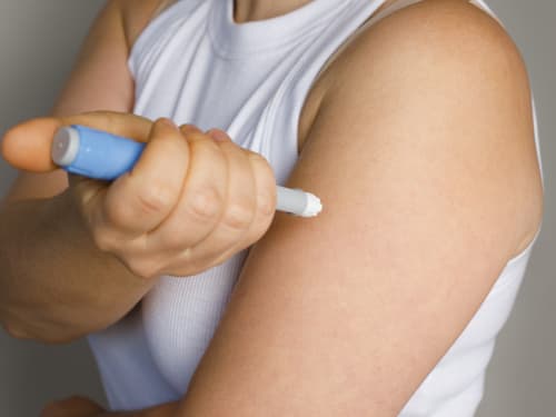
The layer of fat tissue beneath the skin where subcutaneous injections should be delivered. Provides consistent, slow drug absorption.
-
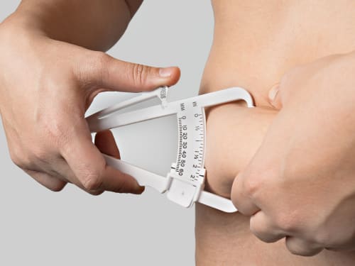
Understand what body composition really measures, why BMI is limited, and how waist‑to‑height ratio, body fat %, ABSI and BRI relate to health risk in UK adults.
-
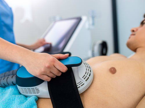
A body sculpting treatment that can build muscle and reduce fat at the same time – too good to be true? We look into some of the science behind it.
-
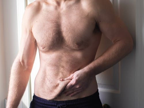
Don’t let loose skin overshadow your weight loss success. Learn why skin sags after significant weight loss and explore our advanced skin tightening and volume restoration treatments.