
Facial skin
Facial skin is the most structurally complex region of the integument, characterised by regional thickness ranging from 0.75mm at the upper eyelid to over 1.2mm at the nasal dorsum, high sebaceous gland density concentrated in the central T-zone, and a dermis mechanically connected to the superficial musculoaponeurotic system (SMAS) and approximately 20 mimetic muscles. These anatomical characteristics mean all three mechanisms of dermal ageing – net collagen loss, elastic fibre fragmentation, and ground substance depletion – manifest earlier and more visibly here than elsewhere on the body. Effective treatment requires regional anatomical awareness matched to the dominant ageing mechanism in each zone, and a sequenced approach that resolves inflammatory suppression before stimulating synthesis.
Facial skin is the body’s most expression-active integument, distinguished by its regional anatomical complexity, continuous dynamic loading from mimetic muscle activity, and disproportionate susceptibility to photoageing and hormonal change. Its thickness varies considerably across its surface – from approximately 0.75mm at the upper eyelid to over 1.2mm at the nasal dorsum – and its dermis is physically connected to the superficial musculoaponeurotic system (SMAS), linking every facial expression directly to the structural collagen and elastin network beneath. [9][26] These anatomical characteristics mean that the three core mechanisms of dermal ageing operate at different intensities in different facial regions, producing the distinct patterns of change that clinical assessment needs to distinguish before treatment is selected.
Regional Thickness and What It Means for Treatment
The face’s thickness variation is not merely an anatomical footnote: it is one of the most practically significant facts in facial treatment planning. A depth, energy level, or injection volume calibrated for the mid-cheek can be inappropriate – or contraindicated – at the periorbital level. The figures below are drawn from high-frequency ultrasound measurements in a study of 118 participants across eight facial sites, with total thickness calculated as the combined epidermis and dermis measurement. Sex differences were statistically significant at most sites; age effects were detectable primarily in the zygoma and submandibular regions in women aged 45 and over. [9]
| Facial Region | Approx. Total Thickness | Notes for Treatment Planning |
|---|---|---|
| Upper eyelid | 0.75–0.85 mm | Extremely thin – requires the gentlest approach for any modality |
| Periorbital area | 0.8–1.0 mm | Early volume loss and fine lines common; conservative depth essential |
| Forehead / Glabella | 1.0–1.4 mm | Adequate depth for collagen-stimulating treatments; notably thinner in women |
| Cheeks / Zygoma | 1.1–1.3 mm | Balanced for volume restoration and stimulation protocols |
| Nasal dorsum | 1.1–1.2 mm | Mid-range; nasal tip and ala likely thicker due to larger sebaceous glands but this site is not well-characterised in HF ultrasound literature |
| Neck | ~1.2 mm | Comparable to mid-face at surface measurement; more prone to laxity due to less underlying structural support |
Men carry measurably thicker facial skin than women at most sites, and in women, post-menopausal thinning becomes detectable at the zygoma and submandibular region – consistent with the 1.13% per-year reduction in skin thickness documented in post-menopausal populations. [9]
Clinical Pearl The upper eyelid is the thinnest site on the face with a total thickness of approximately 0.75–0.85mm in the HF ultrasound dataset. A treatment calibrated for the cheeks can be heavy-handed there. Conservative depth settings in the periorbital zone aren’t caution for its own sake – they reflect genuine anatomical constraint.
The SMAS and Dynamic Mechanical Loading
The dermis of the face is not a passive structural layer. It is anchored to the superficial musculoaponeurotic system (SMAS), a continuous fibromuscular sheet originally described by Mitz and Peyronie in 1976, which connects the mimetic muscles of facial expression to the overlying skin and subcutaneous tissue. [26] The anatomical function of the SMAS is to transmit, distribute, and amplify the activity of the facial muscles directly into the dermis above – meaning every contraction cycles mechanical force through the collagen and elastin network with each expression, many times daily. [12]
The face contains approximately 20 named mimetic muscles, including the orbicularis oculi, frontalis, zygomaticus major, and orbicularis oris. [26] This repeated mechanical loading along predictable expression vectors creates the characteristic lines of facial ageing: nasolabial folds, glabellar lines, crow’s feet, and perioral lines all reflect the intersection of structural dermal decline with repetitive movement patterns. This is categorically different from body skin, where dynamic muscle loading on the dermis is minimal.
The SMAS also explains why facial assessment requires three-dimensional thinking. Volume changes in the fat compartments lying beneath the SMAS alter the mechanical environment experienced by the overlying dermis. As fat compartments shift and deflate with age, the dermis above progressively loses the structural support that maintained both its position and the fibroblast tension environment that sustained collagen synthesis. Addressing the face as a flat, two-dimensional surface misses this volumetric component entirely.
Sebaceous Gland Distribution
The face contains the body’s highest concentration of sebaceous glands, with density in the central T-zone (forehead, nose, and chin) reaching approximately 400–900 glands per cm² – significantly higher than the lateral cheeks or periorbital area. [pmc.ncbi.nlm.nih.gov(https://pmc.ncbi.nlm.nih.gov/articles/PMC6068117/) This regional distribution drives the characteristic oiliness gradient between central and lateral zones and explains why inflammatory acne, sebaceous hyperplasia, and enlarged pore appearance concentrate centrally.
One anatomical nuance worth noting: the nose shows high visible sebum output not because sebaceous gland count per cm² is highest there, but because nasal sebaceous glands are anatomically larger than those elsewhere on the face. [10] This distinction matters when interpreting the sebum literature, as gland density and sebum output are not directly interchangeable.
Sebaceous gland activity is androgen-regulated, making it particularly sensitive to hormonal shifts. In the perimenopausal transition, declining oestrogen with relative androgen persistence can alter both sebum volume and composition, creating conditions in which the sebaceous follicle environment simultaneously promotes low-grade inflammation and impairs the barrier lipid architecture of the surrounding skin.
The Dermo-Epidermal Junction
The dermo-epidermal junction (DEJ) on the face is subject to the same rete ridge flattening that occurs throughout ageing skin, but the combination of chronic UV exposure and constant dynamic loading accelerates this process in photoexposed facial zones. Flattening of the rete ridges reduces the surface area of contact between epidermis and dermis, impairing the exchange of nutrients and differentiation signals across the junction, and weakening the mechanical anchorage of the epidermis to the structures beneath it.
Collagen Type VII, the anchoring fibril protein that connects the basement membrane to the papillary dermis, is particularly vulnerable to UV-driven MMP activity. [https://pubmed.ncbi.nlm.nih.gov/38007090/] Its degradation is one of the earliest structural changes in photoaged facial skin and contributes directly to the thin, fragile quality of chronically sun-exposed areas – visible as crepiness, particularly at the periorbital level where the starting thickness provides the least structural reserve.
How Facial Skin Ages
All three mechanisms of the dermal ageing model operate across the face, but their relative dominance and visible expression vary by region.
Net collagen loss – reduced fibroblast synthesis combined with increased MMP-1 and MMP-3 activity – is most visible as progressive loss of structural firmness and the replacement of well-organised collagen bundles with fragmented material. UV exposure compounds the chronological decline, making photoexposed zones show collagen loss earlier and more severely than UV-protected areas. Post-menopausal skin carries the additional compounding factor that increased MMP activity degrades collagen faster than the already-impaired maturation pathway can replace it. [14]
Elastic fibre fragmentation – driven by MMP-12 degradation and solar elastosis accumulation – underlies the loss of skin recoil and the development of persistent rather than transient expression lines. When functional elastin fibres fragment and non-functional elastotic material accumulates, the skin can no longer spring back between expressions. Solar elastosis concentrates on chronically sun-exposed areas: the lower cheeks, nose, and perioral region show this accumulation most prominently.
Ground substance depletion – the decline in hyaluronic acid (HA), decorin, and other proteoglycans – reduces the mechanical tension environment that fibroblasts require for sustained collagen synthesis. In the midface, declining HA concentration contributes to loss of subtle volume and progressive deflation of the overlying soft tissue; at the periorbital level, it accelerates crepiness and hollowing. Decorin loss compounds this further: without decorin regulating collagen fibril spacing, the collagen that remains becomes progressively disorganised even before quantitative loss is significant.
Rete ridge flattening at the DEJ accelerates as both ground substance and dermal thickness decline, compounding structural fragility in the thinnest facial regions and reducing the signalling exchange between dermis and epidermis that healthy keratinocyte differentiation depends on.
The Three Facial Thirds: Structural Drivers of Surface Change
Aesthetic medicine divides the face into three assessment zones – upper, middle, and lower thirds – as the standard pre-treatment framework for reading surface change in structural terms before selecting interventions. The anatomical boundaries are: the upper third from hairline to glabella, encompassing the forehead, brow, and temporal region; the middle third from glabella to the base of the nose, including the cheeks, periorbital zone, and midface; and the lower third from the base of the nose to the chin, including the lips, perioral region, jaw, and the neck transition. This division is well-established in the operative and aesthetic literature as an organising principle for regional assessment – used consistently across surgical planning, toxin treatment, and filler protocols. [20]
What the thirds framework adds to the regional skin biology content above is a vertical layer of organisation: the surface changes visible in each third are the endpoint of a cascade that originates in the deeper structural layers – bone, then fat, then muscle – and propagates upward to the skin surface. Understanding that cascade by zone allows the practitioner to identify the primary driver of each patient’s surface change before selecting treatment depth and modality.
Upper Third: Forehead, Brow, and Temporal Region
The surface changes characteristic of the upper third – horizontal forehead lines, glabellar rhytides, brow position change, and temporal concavity – reflect both dynamic loading from the mimetic musculature and progressive structural loss in the layers beneath.
The frontalis is the sole brow elevator; its opposing depressors – corrugator supercilii and procerus – draw the brow medially and inferiorly. Brow resting position at any given age is the product of the balance between these muscle groups acting on tissue that has progressively lost the collagen density and elastic recoil to resist their pull. The SMAS–dermis mechanical connection means that every frontalis contraction and corrugator contraction directly stresses the dermal matrix, driving rhytid formation in skin that has already begun to lose its structural resilience. Horizontal forehead lines are the physical record of frontalis action on progressively atrophied tissue; glabellar frown lines are the record of corrugator and procerus action on the same tissue.
The bony substrate of the upper third also changes with age. Measurements of the glabellar angle – the angular prominence of the glabella relative to the nasofrontal suture – show significant reduction with increasing age, indicating recession of the glabellar bony landmark in both men and women. [19] A longitudinal CT study of 96 individuals scanned approximately eleven years apart confirmed a significant decrease in glabellar angle over time, part of a broader pattern of facial skeletal remodelling that proceeds across all three thirds simultaneously. [20] Reduction in the bony support structure of the upper orbital rim contributes to brow descent by reducing the rigid platform against which soft tissues sit; supraorbital and orbital rim resorption is identifiable with increasing age in both sexes. [8]
The temporal hollowing characteristic of upper third ageing represents atrophy of the fat compartments occupying the temporal fossa. The temporal region contains distinct superficial and deep fat compartments, including the deep temporal fat pad – a well-defined anatomical space between the layers of the deep temporal fascia. [17] Volume loss in these compartments is clinically visible as a progressive concavity that frames the lateral brow and lateral eye, contributing to the characteristic hollowed, skeletonised appearance of the upper face in older individuals. This change is compounded by the same skin biology mechanisms described for the face generally – reduced collagen density and ground substance loss – but the primary driver of visible temporal hollowing is volumetric, not dermal.
Middle Third: Cheeks, Midface, and Periorbital Zone
The surface presentation of the ageing middle third – malar flattening, nasolabial fold deepening, infraorbital hollowing, tear trough formation, and loss of the convex cheek – is the endpoint of a cascade whose primary drivers are skeletal and then adipose, with skin biology as the amplifying factor.
The subcutaneous fat of the midface is not a continuous mass. Cadaveric methylene blue injection studies have established that what was previously described as “malar fat” is in fact composed of three discrete superficial compartments – medial, middle, and lateral temporal-cheek – each with distinct anatomical boundaries and each ageing independently. [17] As the ligamentous boundaries that define these compartments relax with age, the compartments descend, producing the characteristic inferior displacement of the malar fat mass that deepens the nasolabial fold and shifts fullness away from the zygomatic arch.
Beneath the superficial compartments, the deep medial cheek fat – a distinct compartment lying directly on the maxilla – provides the anterior projection platform that gives the youthful midface its convexity. Loss of volume in the deep medial cheek fat leads to pseudoptosis: the overlying skin envelope becomes relatively excessive for its diminished supporting volume, producing the illusion of descent and a more prominent nasolabial fold. [18] This mechanism explains why the nasolabial fold can deepen significantly without the superficial fat compartments themselves descending substantially – the fold is in part a volume deficit, not only a descent problem.
The skeletal platform on which these soft tissues sit also diminishes. The maxillary angle – measuring anterior skeletal projection – shows significant reduction with age in both sexes, consistent with progressive resorption of the maxilla. [19] [20] The superomedial and inferolateral orbital rim undergoes selective resorption with age, reducing the bony scaffolding that supports the periorbital soft tissues and exposing progressively more of the orbital rim. [8] These skeletal changes compound the adipose changes: maxillary resorption reduces the skeletal base from which the deep fat compartments project, so that even if fat volumes were preserved, the forward projection they provide would diminish because the platform beneath them recedes.
The sub-orbicularis oculi fat (SOOF) – a distinct anatomical compartment with medial and lateral components extending along the orbital rim – is a third structural contributor. Its atrophy produces the infraorbital hollowing and tear trough appearance characteristic of the ageing periorbital zone, and its position in anatomical continuity with the deep cheek fat means that changes across the deep fat system of the midface occur as an interconnected volumetric unit. [15]
The skin surface in the middle third – including the periorbital skin, which is the structurally most vulnerable zone of the face – is the visible endpoint of this bone-fat-skin cascade. Collagen loss, elastic fibre fragmentation, and ground substance depletion operate here as they do throughout the face, but the degree of surface change cannot be explained by skin biology alone. A patient with maintained dermal collagen density but significant maxillary resorption and deep fat atrophy will still present with malar flattening that reflects the structural loss beneath.
Lower Third: Perioral, Jaw, Chin, and Neck Transition
The ageing lower third presents with perioral lines (both dynamic and static), commissure descent, marionette lines, jowling, jawline definition loss, labiomental groove deepening, and early platysmal banding in the neck. As in the middle third, this surface presentation reflects a structural cascade operating below the skin.
The mandibular skeleton undergoes progressive resorption with age, with the prejowl region of the mandible being a characteristically observed site of resorption in both sexes. [8] Longitudinal CT data confirm that the mandibular angle increases with age – indicating loss of mandibular height and retrognathic rotation of the lower border – consistent with a shrinking bony platform beneath the lower third soft tissues. [20] As the bony base retreats, the overlying soft tissue drapes over a smaller skeletal scaffold, producing excess soft-tissue envelope relative to skeletal support – the fundamental condition that creates jowling.
The fat anatomy of the lower third is similarly compartmental. Jowl fat comprises at least two distinct subcutaneous compartments – a superior and an inferior – positioned above the mandibular border, with a further compartment descending below it. These compartments are separated by the mandibular septum, a structure adherent to the mandibular body; as this septal support weakens with age, jowl fat descends below the mandibular border, obliterating the jawline contour. [13] Buccal fat is anatomically independent from jowl fat and plays a distinct role in lower face shape; its relevance to jowling is indirect.
Sub-orbicularis oris fat and the cutaneous lip fat are discrete compartments with distinct anatomical boundaries. The sub-orbicularis oris fat is a contributor to lip shape and eversion; its atrophy is associated with the characteristic perioral volume loss – reduced lip eversion, shallowing of the Cupid’s bow, and deepening of the labiomental hollow – seen with advancing age. [16]
The platysma occupies a structurally distinctive position in the lower third: it is the only facial muscle without bony attachment, originating in the subcutaneous fascia of the upper chest and inserting into the lower face skin and SMAS. As the SMAS and skin support of the lower face weakens – and as the bony platform recedes – the platysma’s downward and lateral traction is thought to become progressively unresisted, contributing to commissure descent and the deepening of marionette lines. Platysmal banding in the neck becomes visible as the muscle’s midline fibres separate from their previous state of tension.
Perioral skin is thinner on average than mid-cheek skin and is subject to both orbicularis oris action (dynamic loading) and progressive collagen loss (intrinsic ageing). Vertical perioral lines accumulate as the orbicularis repeatedly compresses skin that has lost its elastic recoil – the same SMAS–dermis mechanical coupling that drives rhytid formation in the upper third operates here through the orbicularis, which has direct dermal insertions across the perioral zone.
Why the Thirds Framework Matters for Treatment Sequencing
The clinical value of the thirds framework is that it provides a structural layer of organisation that sits over the regional skin biology. Each third shares the dermal changes described elsewhere in this entity – collagen loss, elastic fibre fragmentation, ground substance depletion, and the inflammatory–synthetic imbalance of ageing skin – but the structural drivers that amplify those changes differ zone by zone and patient by patient.
A practitioner reading the upper third for brow descent will be asking whether the primary driver is frontalis tone (favouring toxin), temporal volume loss (favouring structural filler at the temporal hollow), or skin atrophy (favouring collagen stimulation modalities). A practitioner reading the middle third for malar flattening will be asking whether the primary driver is deep fat atrophy (favouring deep-plane filler), superficial compartment descent (favouring mid-depth filler or lifting approach), skeletal resorption (favouring periosteal volumising), or skin atrophy (favouring resurfacing or collagen stimulation). A practitioner reading the lower third for jowling will be asking whether the primary driver is mandibular resorption (favouring skeletal-level filler), jowl fat descent (favouring contour-restoration filler and possibly platysma toxin), or skin laxity (favouring collagen stimulation and tightening modalities).
Structural changes in adjacent thirds interact across zone boundaries. Midface volume loss augments the apparent heaviness of the lower third by displacing the midface landmark inferiorly, making the lower third appear proportionally larger than it is. Temporal volume loss in the upper third changes the visual framing of the midface lateral cheek. These cross-zone interactions mean that assessment of each third is informed by what is happening in its neighbours, and that single-zone correction without considering the adjacent structural context can produce results that are locally correct but globally inconsistent with the patient’s anatomy.
Clinical Application
The face is where all three dermal ageing mechanisms tend to compound most visibly, and where the dramatic variation in skin thickness from one zone to the next most significantly shapes what treatment can and should do. The periorbital skin at 0.75–0.85mm total thickness is genuinely different tissue from the mid-cheek – not just thinner, but less structurally forgiving. A depth, energy level, or injection volume that works well on the cheeks can look heavy or unnatural at the eye, and vice versa. So before any treatment conversation starts, we’re always reading the face regionally: where is the skin thinnest, where is the inflammatory burden highest, where has ground substance loss created deflation rather than simply surface change? [9]
What shapes our sequencing logic on the face is a consistent observation: treatments that stimulate collagen synthesis, restore ground substance, or activate new fibre production work more durably when the inflammatory environment they’re working into has been quietened first. UV-driven MMP activity, sebaceous-related low-grade inflammation, and the cytokine-mediated suppression of filaggrin and ceramide synthesis can all be active simultaneously in a photoaged or perimenopausal facial presentation. Stimulating collagen production into an environment where MMP degradation is still running at full pace is like rebuilding whilst the structure is still under strain – the results are slower, less complete, and less lasting. So for many of our clients, we start by removing that suppression before introducing the treatments designed to build.
Restoring What the Dermis Has Lost: HA Skin Boosters
For clients where the most visible change is subtle deflation – a quiet loss of plumpness in the midface, or a slight hollowing at the temples that no amount of topical hydration seems to reach – the underlying mechanism is usually ground substance depletion rather than collagen loss alone. Hyaluronic acid (HA) skin boosters address this directly. Injected cross-linked HA restores the mechanical tension that the dermis needs: within one week of treatment, fibroblasts stretch and reorganise in response to the structural support, and this physical signal activates TGF-β through mechanoreception rather than any external growth factor delivery. Measurable new procollagen I synthesis follows at four weeks, sustained for up to twelve months. [22] This matters particularly on the face because UV-damaged fibroblasts often have reduced responsiveness to direct TGF-β stimulation – the mechanical route reaches them when the biochemical route is less reliable.
Regionally, we work with skin boosters in the mid-to-deep dermis of the cheeks, temples, and perioral area. In the periorbital zone, we use preparations specifically formulated for that thinner tissue – standard mid-face volumes are not appropriate there, and the anatomy makes that a non-negotiable rather than a matter of preference.
The Longer-Horizon Option: PLLA
For clients who are thinking further ahead – or who are seeing the kind of gradual structural change that speaks to years of collagen loss rather than a recent shift – poly-L-lactic acid (PLLA) offers a different kind of conversation. Where HA skin boosters restore mechanical tension and deliver relatively prompt results, PLLA works on a longer timeline through a different mechanism entirely: its microspheres trigger a controlled foreign body response in the tissue, recruiting macrophages that polarise toward a repair-oriented state and begin secreting TGF-β. This activates fibroblasts via TGF-β/SMAD2/3 signalling, driving new Type I and Type III collagen production whilst simultaneously upregulating TIMP-1 and TIMP-2 – the tissue inhibitors that slow MMP-mediated degradation. [11] [28]
What makes PLLA particularly relevant for the perimenopausal and post-menopausal presentations we see most often is a second, independent mechanism: PLLA directly activates fibroblast PI3K/AKT signalling and, in doing so, measurably reduces senescence markers in aged fibroblasts. It is not simply stimulating cells that are already functional, it appears to partially restore activity in cells that have entered the senescent state that is characteristic of post-menopausal skin. [11] A 2025 multicenter RCT of 331 subjects found PLLA achieved 90.57% improvement in midfacial volume at 12 months, outperforming HA fillers in that comparison. [27]
The honest expectation-setting for PLLA is that results are gradual and require patience. Most clients need three sessions approximately four to six weeks apart; first visible improvement typically begins at six to eight weeks as the collagen cascade accumulates, and the full result continues developing well beyond the final session. Longevity is 24 to 36 months, considerably longer than HA-based approaches. This makes PLLA most appropriate where a client is willing to invest in a slower build for a more structurally durable outcome, rather than seeking prompt visible change.
Collagen Synthesis and Solar Elastosis: iPRF and RF Microneedling
For the most common facial presentations we see – photoageing, perimenopausal skin, or a combination of both – the challenge is rarely that collagen synthesis is completely inactive. More often, degradation is outpacing what the skin can repair and replace. That’s why iPRF is our most mechanistically complete collagen treatment for these clients: it activates TGF-β/SMAD signalling to drive new procollagen production whilst simultaneously suppressing MMP-1 expression, addressing both sides of the net collagen balance. [6] [23] Treating synthesis alone without reducing the degradation running alongside it is addressing only half the problem.
RF microneedling brings in the elastin dimension. The thermal energy at the needle tips creates controlled denaturation in the mid-to-deep dermis, clearing the accumulated solar elastotic material that blocks new fibre assembly. The apparent short-term reduction in total elastin content after treatment is therapeutic – it reflects clearance of non-functional material rather than further loss – with functional fibre formation following at the three-to-six month mark. [3] On the face, depth calibration across zones is essential: the reticular dermis at the periorbital level may sit within 1.0mm of the surface, which is why we don’t simply apply cheek settings to that area. [9]
Calming the Inflammatory Environment First: Polynucleotides and CAP
For clients with reactive or sensitised skin, sebaceous-driven congestion, or the perimenopausal skin sensitivity that so often arrives alongside hormonal change, our first step is usually to quieten the inflammatory signals that are actively working against repair – before introducing treatments that ask the skin to produce and synthesise more.
Polynucleotides do this at the dermal level: they suppress the MMP activity that is degrading collagen and elastin, reprogramme macrophages toward a repair-oriented state, and restore the TGF-β and IL-10 signals that fibroblasts need to resume normal collagen production. [2] For clients in the perimenopausal years whose fibroblasts have become less responsive to direct stimulation, this indirect route through the tissue’s own macrophage population is often more effective than targeting fibroblasts head-on.
Cold atmospheric plasma (CAP) addresses the epidermal side of the same problem. Its reactive oxygen and nitrogen species reduce IL-4, IL-13, and IL-31 – the cytokines that directly suppress both filaggrin expression and the elongase enzymes responsible for ceramide synthesis. [1] For clients whose skin has become chronically reactive, where topical barrier support has helped but plateaued, CAP targets the upstream signal driving that reactivity rather than managing its consequences at the surface.
Surface Renewal: Thulium Laser and LED
Where the main concern is epidermal quality – uneven texture, lingering pigmentation, or the dull, slightly tired appearance that reflects a slowed keratinocyte renewal cycle – thulium 1927nm fractional laser provides a direct reset. It activates Wnt/β-catenin signalling and preserves COL17A1 expression in the follicular reservoir, with keratinocyte migration creating a new epidermal bridge across treated zones within 24 hours of treatment, meaning surface recovery is faster than many clients expect. [25] [24]
Post-procedure red LED (630–660nm) then supports and extends the benefit. Red light activates TGF-β and AKT signalling in dermal fibroblasts, driving collagen I production via SMAD2/3/4 nuclear translocation whilst simultaneously reducing MMP expression through the NRF2/HO-1 pathway. The accumulated collagen I then activates integrin signalling in basal keratinocytes, increasing their proliferation and differentiation rate-— a dermis-first mechanism that renews the epidermis through strengthened support from below. [4]
Treatment Pairings for Facial Skin
Polynucleotides + iPRF: We resolve the MMP-driven inflammatory suppression first, then introduce iPRF to activate collagen synthesis into a quieter, more receptive environment. For photoaged and perimenopausal presentations where both degradation and synthesis are impaired, this sequencing consistently produces more complete and durable results than either treatment alone.
PLLA + polynucleotides (structural decline with senescent fibroblast environment): Polynucleotides resolve the inflammatory and MMP-driven suppression via macrophage reprogramming; PLLA then builds into a quieter tissue environment, activating both the macrophage-mediated TGF-β cascade and the direct PI3K/AKT fibroblast route simultaneously. For clients where structural collagen decline is significant and the fibroblast population is showing age-related reduced responsiveness, this pairing addresses both the suppression environment and the senescence component that limits how well other collagen stimulators perform.
RF microneedling + HA skin boosters: RF thermal remodelling addresses elastic fibre fragmentation and structural depth loss in the reticular dermis; HA skin boosters simultaneously restore ground substance and activate mechanical fibroblast signalling. Together they reach all three dermal ageing mechanisms within a single combined session.
Thulium + post-procedure red LED: A direct epidermal renewal stimulus followed by a dermis-up accelerator for the fibroblast-keratinocyte cycle. Most appropriate where epidermal quality, texture, and pigmentation are the primary concerns alongside superficial collagen support. [25] [4]
CAP + microneedling-assisted exosomes (reactive or barrier-compromised presentation): CAP removes the IL-4/IL-13 suppression of filaggrin and ceramide synthesis; exosome delivery via microneedling channels then supports collagen I production and improvements in skin texture and quality at the recovering tissue surface. [1] [21] For clients with persistent barrier reactivity that hasn’t fully resolved with topical support alone, this combination addresses the inflammatory driver and the structural repair pathway from complementary directions.
The Thirds as a Pre-Treatment Checklist
The three-zone framework translates directly into a consultation structure: before selecting any treatment modality or injection plane, a practitioner can systematically assess each third for the balance of structural versus skin-level change.
Upper third assessment: Is brow descent present, and if so, is it driven primarily by excess frontalis tone relative to gravity (neurotoxin to the frontalis and corrugator complex will rebalance), or by temporal hollowing withdrawing lateral brow support (volumising the temporal hollow addresses the underlying structural loss), or by skin atrophy reducing the tissue’s resistance to dynamic loading (collagen stimulation is appropriate here but will not address structural volume deficit)? These are not mutually exclusive drivers – most patients have all three – but identifying the primary driver guides the sequencing.
Middle third assessment: For malar flattening and infraorbital hollowing, assess the relative contribution of skeletal recession (best addressed at deep-plane periosteal level), deep fat atrophy (best addressed at sub-muscular depth), superficial compartment descent (mid-depth filler or lifting approach), and skin quality (surface-level collagen stimulation). The critical integration point with the skin biology content in this entity is that treating skin quality without addressing structural volume deficit produces incomplete correction of surface change in the middle third more than in any other zone – the skin’s surface reflects the bony and adipose scaffold beneath it.
Lower third assessment: Jowling and commissure descent require assessment of mandibular skeletal support, jowl fat compartment position, and platysmal tension. Platysma toxin addresses the traction component; filler at the mandibular border restores the skeletal scaffold; but neither of these substitutes for assessing whether the overlying skin has the quality to re-drape appropriately after volumising. This is the integration point with the inflammatory sequencing logic described elsewhere in this entity: a lower third with active inflammation (perioral rosacea, barrier reactivity) will not respond as predictably to volumising as one where skin quality has been optimised first.
Cross-zone considerations: The thirds framework is most powerful when applied across all three zones simultaneously rather than in isolation. Midface volume loss creating apparent lower-third heaviness, or temporal hollowing changing the upper-third frame, are findings that require multi-zone thinking to address accurately. Correcting the lower third without addressing midface descent that is contributing to it produces a result that looks locally treated rather than naturally restored. The structural cascade model – where bone is the primary driver, fat compartments the secondary distributor, and skin the visible endpoint – applies in each third but must be read as a whole-face system.
References
Bai F, Ran Y, Zhai S, et al. (2023). Cold Atmospheric Plasma: A Promising and Safe Therapeutic Strategy for Atopic Dermatitis. Int Arch Allergy Immunol, 184(12), 1184-1197 . doi.org/10.1159/000531967
Byun KA, Park HJ, Oh S, et al. (2025). Polynucleotides Enhance Collagen Synthesis via Modulating Phosphoenolpyruvate Carboxykinase 1 in Senescent Macrophages: Experimental Evidence. Int J Mol Sci, 26(17) . doi.org/10.3390/ijms26178720
Chandra S, Mysore V, Shah S, et al. (2024). Physics of fractional microneedle radiofrequency – A review. J Cutan Aesthet Surg, 17(3), 177-183 . doi.org/10.25259/jcas_98_23
Chang H, Shen Q, Tan Y, et al. (2025). Red light promotes dermis-epidermis remodeling via TGFβ and AKT-mediated collagen dynamics in naturally aging mice. Zool Res, 46(5), 967-982 . doi.org/10.24272/j.issn.2095-8137.2024.405
Conradt G, Hausser I, Nyström A (2024). Epidermal or Dermal Collagen VII Is Sufficient for Skin Integrity: Insights to Anchoring Fibril Homeostasis. J Invest Dermatol, 144(6), 1301-1310.e7 . doi.org/10.1016/j.jid.2023.11.003
Li Y, Song P, He J, et al. (2022). Comparison Between Injectable Platelet-rich Fibrin and Platelet-rich Plasma in Ameliorating UVA-induced Photoaging in Human Dermal Fibroblasts via the Activation of TGF-β/Smad Signaling Pathway. Photochem Photobiol, 98(6), 1395-1401 . doi.org/10.1111/php.13628
Ludovici M, Kozul N, Materazzi S, et al. (2018). Influence of the sebaceous gland density on the stratum corneum lipidome. Sci Rep, 8(1), 11500 . doi.org/10.1038/s41598-018-29742-7
Mendelson B, Wong CH (2012). Changes in the facial skeleton with aging: implications and clinical applications in facial rejuvenation. Aesthetic Plast Surg, 36(4), 753-60 . doi.org/10.1007/s00266-012-9904-3
Meng Y, Feng L, Shan J, et al. (2022). Application of high-frequency ultrasound to assess facial skin thickness in association with gender, age, and BMI in healthy adults. BMC Med Imaging, 22(1), 113 . doi.org/10.1186/s12880-022-00839-w
Mosca S, Ottaviani M, Briganti S, et al. (2025). The Sebaceous Gland: A Key Player in the Balance Between Homeostasis and Inflammatory Skin Diseases. Cells, 14(10) . doi.org/10.3390/cells14100747
Oh S, Lee JH, Kim HM, et al. (2023). Poly-L-Lactic Acid Fillers Improved Dermal Collagen Synthesis by Modulating M2 Macrophage Polarization in Aged Animal Skin. Cells, 12(9) . doi.org/10.3390/cells12091320
Park HJ, Hur MS (2024). Anatomical Study of the Superficial Musculoaponeurotic System in Relation to the Zygomaticus Major. Diagnostics (Basel), 14(18) . doi.org/10.3390/diagnostics14182066
Reece EM, Pessa JE, Rohrich RJ (2008). The mandibular septum: anatomical observations of the jowls in aging-implications for facial rejuvenation. Plast Reconstr Surg, 121(4), 1414-1420 . doi.org/10.1097/01.prs.0000302462.61624.26
Remoué N, Molinari J, Andres E, et al. (2013). Development of an in vitro model of menopause using primary human dermal fibroblasts. Int J Cosmet Sci, 35(6), 546-54 . doi.org/10.1111/ics.12075
Rohrich RJ, Arbique GM, Wong C, et al. (2009). The anatomy of suborbicularis fat: implications for periorbital rejuvenation. Plast Reconstr Surg, 124(3), 946-951 . doi.org/10.1097/prs.0b013e3181b17b76
Rohrich RJ, Pessa JE (2009). The anatomy and clinical implications of perioral submuscular fat. Plast Reconstr Surg, 124(1), 266-271 . doi.org/10.1097/prs.0b013e3181811e2e
Rohrich RJ, Pessa JE (2007). The fat compartments of the face: anatomy and clinical implications for cosmetic surgery. Plast Reconstr Surg, 119(7), 2219-2227 . doi.org/10.1097/01.prs.0000265403.66886.54
Rohrich RJ, Pessa JE, Ristow B (2008). The youthful cheek and the deep medial fat compartment. Plast Reconstr Surg, 121(6), 2107-2112 . doi.org/10.1097/prs.0b013e31817123c6
Shaw RB Jr, Kahn DM (2007). Aging of the midface bony elements: a three-dimensional computed tomographic study. Plast Reconstr Surg, 119(2), 675-81; discussion 682-3 . doi.org/10.1097/01.prs.0000246596.79795.a8
Sifil MK, Kahn DM (2026). Three-Dimensional Computed Tomography-Based Longitudinal Analysis of Facial Skeletal Aging. Aesthet Surg J, 46(4), 366-371 . doi.org/10.1093/asj/sjaf138
Wan J, Yoon SE, Yi KH (2025). The Efficacy of Combined Exosome (Exodew) and Microneedling Treatment for Facial Pore Reduction and Skin Texture Improvement. Plast Reconstr Surg Glob Open, 13(6), e6849 . doi.org/10.1097/gox.0000000000006849
Wang F, Do TT, Smith N, et al. (2024). Implications for cumulative and prolonged clinical improvement induced by cross-linked hyaluronic acid: An in vivo biochemical/microscopic study in humans. Exp Dermatol, 33(1), e14998 . doi.org/10.1111/exd.14998
Wang X, Yang Y, Zhang Y, et al. (2019). Fluid platelet-rich fibrin stimulates greater dermal skin fibroblast cell migration, proliferation, and collagen synthesis when compared to platelet-rich plasma. J Cosmet Dermatol, 18(6), 2004-2010 . doi.org/10.1111/jocd.12955
Wardhani PH, Rahmatika A, Thendria T, et al. (2025). Application of Thulium 1927-nm Laser for Androgenic Alopecia Treatment in Indonesian Patients. J Lasers Med Sci, 16, e42 . doi.org/10.34172/jlms.2025.42
Wenande E, Jacobsen K, Grove GL, et al. (2025). Imaging 1927 nm Fractional Thulium Laser-Tissue Interactions: A Spectrum of Nonablative to Ablative Effects. J Cosmet Dermatol, 24(7), e70304 . doi.org/10.1111/jocd.70304
Whitney ZB, Jain M, Zito PM (2026). Anatomy, Skin, Superficial Musculoaponeurotic System (SMAS) Fascia. StatPearls Publishing. ncbi.nlm.nih.gov/books/NBK519014
Zhang Y, Zhang X, Gao X, et al. (2025). Efficacy and Safety of Poly-l-Lactic Acid for Correction of Midfacial Volume Loss and Contour Defects: A Prospective, Multicenter, Randomized, Parallel-Controlled, Evaluator-Blinded, Superiority Trial. J Cosmet Dermatol, 24(7), e70230 . doi.org/10.1111/jocd.70230
Zhu W, Dong C (2023). Poly-L-Lactic acid increases collagen gene expression and synthesis in cultured dermal fibroblast (Hs68) through the TGF-β/Smad pathway. J Cosmet Dermatol, 22(4), 1213-1219 . doi.org/10.1111/jocd.15571
Anatomical Relationships
Referenced in Conditions & Treatments
- this Associated condition Rosacea Evidence: Rosacea is a chronic, relapsing inflammatory condition of the central facial skin – primarily the cheeks, nose, chin, and forehead.
Learn More
This topic is discussed in 4 articles:
-

Searching for the best lip balm for dry lips? Discover which ingredients have real evidence behind them, why your lips get dry, and how to choose products that actually work.
-

Discover how to restore and maintain your skin’s natural defences. From the causes of skin barrier damage to advanced treatments. Achieve healthy, radiant skin with our expert advice.
-
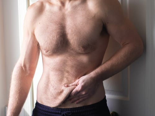
Don’t let loose skin overshadow your weight loss success. Learn why skin sags after significant weight loss and explore our advanced skin tightening and volume restoration treatments.
-
