
Sebaceous gland
Sebaceous glands are holocrine secretory organs embedded in the dermis, producing sebum through complete cellular rupture and release. The highest gland density is on the face and scalp – up to 900 glands per cm² at the nose and forehead – and their complete absence from the vermilion border, palms, and soles creates the structural context for some of the most clinically important body-site differences in skin biology. Sebum is not simply a surface lubricant. It generates the antimicrobial sapienic acid pathway unique to human skin, provides squalene as an antioxidant, and directly shapes the microbial ecology of the body sites it reaches. Androgen-driven dysregulation, age-related composition changes, and the sebaceous gland’s role as an ecological niche for Malassezia and Demodex all have specific, actionable relevance for aesthetics practice.
Sebaceous glands are holocrine secretory glands embedded in the dermis, found across most of the body surface with the highest density on the face and scalp – up to 400–900 glands per cm² on the forehead and nose – and absent entirely from the palms, soles, and the vermilion border of the lips. They are almost always associated with a hair follicle, opening into the upper portion of the follicular canal to form the pilosebaceous unit; the small number of sebaceous glands not associated with a follicle – found on the eyelid margins, nipples, and oral mucosa – are termed free or ectopic sebaceous glands. The gland itself is a cluster of lobed acini surrounding a central duct, composed entirely of sebocytes at progressive stages of differentiation. [9]
Structure and Secretion
Sebaceous gland function depends on a continuous cycle of sebocyte differentiation that is unique among skin cell types. Undifferentiated basal progenitor cells at the periphery of each acinus proliferate and move centrally, progressively accumulating lipid droplets as they differentiate. As maturation completes, the sebocyte has expanded substantially in volume, its nucleus has condensed, and it ruptures entirely, releasing its lipid contents into the sebaceous duct. This mode of secretion, holocrine secretion, means the secretory product includes the entire cellular contents rather than selected molecules; the cell itself becomes the secretion. The resulting sebum travels up the follicular canal and onto the skin surface, where lipases from cutaneous microbiota – primarily Cutibacterium acnes – hydrolyse triglycerides to free fatty acids, completing the surface lipid film. [8]
Sebum Composition and Function
Human sebum is a complex mixture whose composition is distinctive among mammals. In its glandular form it consists primarily of triglycerides (approximately 57%), wax esters (26%), squalene (12%), and free fatty acids generated progressively by surface hydrolysis. The fatty acid profile is dominated by sapienic acid – a C16:1 Δ6 fatty acid produced exclusively by human sebaceous FADS2 activity and absent from all animal and plant fats – alongside its elongation product sebaleic acid. This uniquely human fatty acid pathway produces sebum with antimicrobial properties against gram-positive pathogens and contributes to the chemically distinctive surface environment of human skin. Squalene, present at approximately 12% of sebum, is a potent antioxidant that protects the skin surface from UV-induced lipid peroxidation, but is itself a substrate for oxidation under UV stress, generating peroxidated squalene products implicated in comedogenesis in acne-prone presentations. [9]
Androgen Regulation and Ageing
Sebaceous glands are the most androgen-sensitive structures in skin. Type 1 5-alpha reductase within sebocytes converts locally available testosterone to dihydrotestosterone (DHT), which binds androgen receptors to drive sebocyte proliferation, lipid synthesis, and increased sebum output. This local conversion mechanism means sebum dysregulation can occur without elevated systemic androgens, the relevant variable is intracellular DHT in the gland rather than circulating testosterone. Regional variation in 5-alpha reductase activity accounts for the predisposition of the face and scalp to acne and seborrhoeic conditions compared to body sites with lower enzyme activity.
With age, sebaceous gland morphology changes in ways that are not a simple reduction in output. Gland volume may increase or remain stable whilst sebum quality declines, with lower wax ester content, altered fatty acid composition, and reduced sapienic acid pathway activity. In women, oestrogen withdrawal at menopause reduces sebocyte activity and sebum production measurably; male sebum output remains relatively constant until later decades given the slower testosterone decline. The consequence for both sexes is a progressively less effective surface lipid film, one that provides less antimicrobial defence, less emolliency, and less surface protection than the same glands produced in earlier decades. [8]
Sebum-Rich Anatomical Sites as Microbial Niches
Before considering how sebum changes with age, it is worth understanding what its abundance creates – because the ecological consequences of high sebaceous output are as clinically relevant as the consequences of its decline.
The uneven distribution of sebaceous glands across the body surface creates discrete ecological zones that profoundly shape the cutaneous microbiome. Sites of highest gland density – the forehead, nose, cheeks (the T-zone and medial face), scalp, chest, and upper back – provide a sebum-rich, warm, low-oxygen environment that favours a distinct microbial community, whilst sites without sebaceous glands, such as the palms, soles, and inner forearm, support an entirely different community composition dominated by moisture-tolerant species. This body-site specificity is not incidental: the dominant organisms of sebaceous sites are metabolically specialised to exploit sebum as a primary substrate.
Malassezia, the lipophilic yeast genus that constitutes the dominant fungal component of human skin, is almost entirely restricted to sebaceous body sites. Malassezia species lack the genes to synthesise long-chain fatty acids de novo and depend on exogenous lipids – principally the triglycerides and wax esters in sebum – for survival and growth. These yeasts express secreted lipases that cleave triglycerides, releasing the saturated and unsaturated free fatty acids they can assimilate. At sebaceous sites this dependency is functionally met; at sites without sebaceous glands Malassezia densities fall to negligible levels. Conditions associated with seborrhoea – dandruff, seborrhoeic dermatitis, and pityriasis versicolor – distribute accordingly, clustering at the same sites where sebum output is highest, because the ecological conditions that sustain the associated Malassezia populations are congruent with those that produce the clinical presentation. [1] [7]
Demodex mites – Demodex folliculorum and Demodex brevis – occupy a different but equally sebum-dependent niche within the pilosebaceous unit. D. folliculorum resides in the follicular infundibulum, where it feeds on sebocyte contents and follicular lipids, whilst D. brevis inhabits the sebaceous gland acini themselves. Both species are obligate parasites of pilosebaceous units and are essentially absent from non-follicular body sites. Mite density reflects local sebaceous gland output and activity: a retrospective pilot study has indicated that Demodex density tracks sebaceous gland status [4], and conditions associated with increased sebum production or altered sebaceous gland architecture – including rosacea and seborrhoeic presentations – are associated with elevated Demodex burdens at affected sites. The face, particularly the nose, cheeks, and forehead, carries the highest mite loads, directly mapping onto the anatomy of sebaceous gland density. [11]
Androgen regulation modulates the ecological niches
The earlier discussion of DHT-driven sebocyte activity and the local 5α-reductase mechanism has a downstream consequence for the microbial communities these glands support. Pubertal rises in androgenic activity increase sebum output and shift body-site conditions from the microbiome of childhood skin toward the sebum-dominated adult ecology at facial, scalp, and chest sites. Anti-androgen treatments that reduce sebum production – spironolactone, cyproterone acetate, oral isotretinoin – do not merely address the clinical presentation at the skin surface; they alter the substrate availability that sustains the associated microbial communities. For any client undertaking or recovering from anti-androgen therapy, the expected change in sebum output is also an expected change in the microbial environment of their sebaceous sites – a practical consideration in treatment planning and homecare recommendations. [10]
Sebum composition and acne: the linoleic acid deficit.
In acne-prone skin, the composition of sebum is altered relative to non-affected skin in a characteristic way that compounds the effects of excess volume. The proportion of linoleic acid in sebum is diminished – a finding established in the foundational literature on acne sebum chemistry and subsequently confirmed across multiple studies. Linoleic acid contributes to the integrity of the follicular epithelium and is required for the normal cornification of the infundibular keratinocytes that line the follicular canal. When its concentration in sebum falls, infundibular keratinisation becomes disordered – the comedo-forming process of follicular hyperkeratinisation that initiates acne lesions. This relative linoleic acid deficiency in acne-prone sebum is partly a dilution effect (high total sebum volume reducing the proportional content of linoleic acid), partly a reflection of altered biosynthetic activity in sebocytes under androgenic stimulation, and compounded by the lipase activity of Cutibacterium acnes on the follicular surface, which shifts the remaining fatty acid pool toward oleic acid – a comedogenic compound that further disrupts follicular epithelial integrity and promotes barrier breakdown. [2] [6]
Clinical Application
Sebaceous Zone: Treatment Planning Implications
Understanding the sebaceous gland as an ecological driver – not only a lipid producer – has practical implications that extend beyond acne management into any clinical encounter where patients present with seborrhoeic conditions, rosacea-like features, Demodex-associated presentations, or oily skin complaints at characteristic sebaceous sites.
Sebaceous zone patients present with a distinct microbiome profile. Patients who consistently present with oily skin at forehead, nose, cheeks, chest, and scalp are maintaining a continuous high-sebum environment that selects for Malassezia and supports elevated Demodex burdens. When these patients develop seborrhoeic dermatitis, periocular or nasolabial dandruff, or a diffuse erythematous flush across sebaceous zones that does not conform to classic rosacea triggers, microbial ecology is a relevant diagnostic frame. Treatment directed at topical antifungals or Demodex-targeted therapies (permethrin, azelaic acid, metronidazole, ivermectin) makes mechanistic sense in this context – these patients are not simply experiencing inflammation but are experiencing inflammation mediated in part by the microbial communities their sebum sustains. Practitioners treating rosacea presentations at sebaceous sites should consider whether the Malassezia or Demodex burden is a co-driver, particularly in patients who do not respond to standard vascular or laser-targeted therapies.
Homecare ingredient selection differs by sebaceous zone status. In sebum-rich zones, the rationale for retinoids, niacinamide, and salicylic acid in homecare is mechanistically sound:
| Ingredient | Primary mechanism at sebaceous level | Evidence grade |
|---|---|---|
| Topical retinoids (adapalene, tretinoin) | Normalise sebocyte differentiation, reduce comedogenesis, modulate follicular keratinisation | Strong – multiple RCTs; cornerstone of acne clinical guidelines |
| Niacinamide | Reduces sebum excretion rate | Moderate – RCTs including Drealos (2006) [3] |
| Salicylic acid | Lipophilic, folliculotropic; dissolves intercellular lipid matrix, exfoliates within follicular canal | Moderate – in vitro and clinical series; mechanism well-characterised |
The retinoid category warrants emphasis as the evidence-based cornerstone. Adapalene and tretinoin act via nuclear retinoic acid receptors to regulate the gene expression programmes governing sebocyte differentiation, keratinocyte cornification in the follicular infundibulum, and inflammatory signalling. Their effect is not confined to surface desquamation – they address the comedo-formation pathway at its origin, which is why topical retinoids remain the first-line agent in the management of comedonal and mild-to-moderate acne rather than a cosmetic adjunct.
Isotretinoin patients require careful timing around energy-based treatments. Oral isotretinoin exerts its therapeutic effect on acne primarily through dramatic suppression of sebaceous gland activity: gland size and sebum output reduce substantially during treatment. This suppression underlies both the therapeutic efficacy and the characteristic side effects (xerosis, cheilitis, mucosal dryness). From an aesthetics practice standpoint, the relevant clinical implication is the washout interval before energy-based treatments. Isotretinoin significantly reduces sebaceous gland output and alters the skin’s capacity for normal wound healing and barrier recovery. For clients who have recently completed a course of oral isotretinoin, the relevant clinical question for treatment planning is timing – current consensus supports a washout interval of 6–12 months before energy-based procedures including RF microneedling, ablative laser, and medium-to-deep chemical peels. The mechanism is the impaired regenerative capacity of a sebaceous gland system that has been profoundly suppressed; the risk is delayed wound healing, scarring, or persistent post-inflammatory change at a level that does not occur in a skin with normal sebaceous function. Very low-dose isotretinoin regimens used specifically to manage seborrhoea (rather than acne) involve less gland suppression, but the same caution applies if a patient is concurrently taking any isotretinoin formulation. [5]
RF microneedling for pore refinement: mechanism and patient selection. Enlarged follicular ostia at sebaceous zones (commonly described by patients as “large pores”) reflect the combined effects of high chronic sebum output distending the follicular canal, loss of dermal collagen support around the pilosebaceous unit, and, at times, persistent comedonal contents. RF microneedling creates focal thermal injury at needle-tip depth within the dermis and at the perifollicular zone, inducing fibroplasia and collagen remodelling around the pilosebaceous unit. The thermal effect on the superficial dermis and peri-follicular stroma tightens the architectural support of the follicle, reducing the visible ostial aperture. Clinical evidence for this indication is composed largely of case series and open-label studies rather than blinded RCTs – practitioners should frame this with patients as an empirically supported intervention with consistent clinical reports rather than a robustly trial-proven outcome. Appropriate candidates are patients with inherently large follicular ostia at sebaceous sites (type II–IV Fitzpatrick), adequate sebum-zone skin thickness, and realistic expectations about the degree of visible change. The treatment is not indicated as a standalone for patients whose pore appearance is primarily driven by active comedonal acne – in this case the sebaceous biology should be addressed first before infrastructure remodelling.
Lips: The Absence Argument
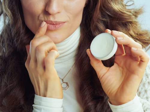
The vermilion border lacks sebaceous glands entirely; a specific structural gap that distinguishes it from every other area of facial skin. Whilst lips do receive some moisture contribution from the oral mucosa and benefit from a rich vascular supply that supports healing, they have no local lipid-producing infrastructure of their own. The surface protection, emolliency, and antimicrobial defence that sebum provides elsewhere on the face must come from external application on the vermilion border. In UV-exposed, mechanically stressed tissue with a thinner stratum corneum than surrounding facial skin, that specific absence makes the case for a stable, oxidatively resistant topical lipid – rather than a high-PUFA seed oil-based product – genuinely structural rather than cosmetic preference.
For clients who treat lip products as optional or cosmetically trivial, the framing that tends to reposition the conversation is straightforward: “Your lips don’t have the oil-producing glands the rest of your face has. They get some help from inside, but the surface lipid layer has to come from what you put on them.”
Acne and Sebum Dysregulation
Acne vulgaris is fundamentally a sebaceous gland condition – driven by androgen-stimulated sebum overproduction, follicular hyperkeratinisation, altered surface microbiome ecology, and the downstream inflammatory cascade. Understanding the DHT/type 1 5-alpha reductase mechanism clarifies why acne does not always correlate with measurably elevated systemic androgens: the dysregulation is occurring locally within the gland, at the level of intracellular DHT production, not necessarily at the level of circulating hormone. For clients with persistent adult acne who have been told their hormone levels are “normal,” this distinction is genuinely useful, it explains why systemic hormone normalisation does not always resolve the presentation.
The squalene oxidation point is worth holding as background knowledge: under UV stress, squalene in sebum generates peroxidated products that contribute to comedogenesis by promoting follicular hyperkeratinisation. Clients with acne-prone skin who also have significant UV exposure are dealing with a compounding mechanism – another reason photoprotection is not only an anti-ageing conversation.
Post-Procedure Surface Lipid Environment
In the days following barrier-disrupting procedures – microneedling, RF microneedling, chemical peels – the skin’s surface lipid film is compromised both by the procedure itself and by the temporary reduction in sebaceous output that accompanies acute epidermal stress. The recommendation of a stable, simple, low- PUFA topical in this window is not merely about occlusion – it is about providing the surface lipid environment the sebaceous glands are temporarily less able to supply. For clients with already-reduced sebaceous output (perimenopausal women, clients on oral retinoids, older male clients with late-decade testosterone decline), this temporary gap is more significant and the homecare guidance correspondingly more important.
References
Dawson TL Jr (2007). Malassezia globosa and restricta: breakthrough understanding of the etiology and treatment of dandruff and seborrheic dermatitis through whole-genome analysis. J Investig Dermatol Symp Proc, 12(2), 15-9 . doi.org/10.1038/sj.jidsymp.5650049
Downing DT, Stewart ME, Wertz PW, et al. (1986). Essential fatty acids and acne. J Am Acad Dermatol, 14(2 Pt 1), 221-5 . doi.org/10.1016/s0190-9622(86)70025-x
Draelos ZD, Matsubara A, Smiles K (2006). The effect of 2% niacinamide on facial sebum production. J Cosmet Laser Ther, 8(2), 96-101 . doi.org/10.1080/14764170600717704
Forton FMN, De Maertelaer V (2021). Which factors influence Demodex proliferation? A retrospective pilot study highlighting a possible role of subtle immune variations and sebaceous gland status. J Dermatol, 48(8), 1210-1220 . doi.org/10.1111/1346-8138.15910
Geissler SE, Michelsen S, Plewig G (2003). Very low dose isotretinoin is effective in controlling seborrhea. J Dtsch Dermatol Ges, 1(12), 952-8 . doi.org/10.1046/j.1365-2303.2003.00108.x-i1
Gollnick H (2003). Current concepts of the pathogenesis of acne: implications for drug treatment. Drugs, 63(15), 1579-96 . doi.org/10.2165/00003495-200363150-00005
Grice EA, Segre JA (2011). The skin microbiome. Nat Rev Microbiol, 9(4), 244-53 . doi.org/10.1038/nrmicro2537
Hou X, Wei Z, Zouboulis CC, et al. (2022). Aging in the sebaceous gland. Front Cell Dev Biol, 10, 909694 . doi.org/10.3389/fcell.2022.909694
Makrantonaki E, Ganceviciene R, Zouboulis C (2011). An update on the role of the sebaceous gland in the pathogenesis of acne. Dermatoendocrinol, 3(1), 41-9 . doi.org/10.4161/derm.3.1.13900
Makrantonaki E, Zouboulis CC (2007). Testosterone metabolism to 5alpha-dihydrotestosterone and synthesis of sebaceous lipids is regulated by the peroxisome proliferator-activated receptor ligand linoleic acid in human sebocytes. Br J Dermatol, 156(3), 428-32 . doi.org/10.1111/j.1365-2133.2006.07671.x
Rychlik K, Sternicka J, Nowicki RJ, et al. (2025). Significance of Demodex folliculorum and Demodex brevis in Pathogenesis of Dermatological Diseases-Current State of Knowledge. Medicina (Kaunas), 61(4) . doi.org/10.3390/medicina61040660
Also Known As
- sebaceous glands
Anatomical Relationships
Referenced in Conditions & Treatments
- this Stimulated by Oestrogen
- this Inhibited by Retinoid Evidence: Academic: Isotretinoin induces sebocyte apoptosis reducing sebaceous gland size; pmc.ncbi.nlm.nih.gov/articles/PMC2262030/
- this Affected by Cortisol Evidence: Academic: HPA axis/ cortisol modulates sebaceous gland activity; doi.org/10.25259/csdm_118_2025
- this Affected by Hallmarks of ageing Evidence: Deregulated nutrient sensing (IGF-1/mTOR), stem cell exhaustion, and altered intercellular communication affect sebocyte function and sebaceous gland renewal with ageing (PMC10874500; Aging Dis doi:10.14336/AD.2023.0321).
- this Affected by Oestrogen Evidence: Academic: Oestrogen modulates sebaceous gland activity; pmc.ncbi.nlm.nih.gov/articles/PMC2685269/
- this Affected by Oestrogen decline Evidence: As oestrogen declines with relative androgen persistence, sebaceous activity initially fluctuates; sebum excretion rate then falls progressively (~40% by 6th decade). PMC12374573; PMID 11834844.
- this Affected by Skin ageing Evidence: Sebum production declines with age; sebocyte function modulated by androgen decline and HPA axis changes with ageing. PMC3174533
- this Affected by Topical steroid withdrawal Evidence: High-potency TCS cause epidermal and adnexal atrophy including sebaceous gland atrophy; these structural effects persist into the withdrawal phase (PMC4228634 side-effects review).
- this Part of Hair follicle PMID: 29261946 Evidence: Sebaceous gland opens into follicle infundibulum; sebum lubricates the hair shaft and canal.
- this Part of Pilosebaceous Unit Evidence: Entity text explicitly states pilosebaceous unit includes the follicle, sebaceous glands, and arrector pili muscle. Sebaceous gland is a named constituent.
- this Part of Skin Evidence: Sebaceous glands are skin appendages within the dermis; entity text references sebum production in skin context.
- this Part of system Hypothalamic–pituitary–adrenal axis Evidence: Sebaceous glands are part of peripheral cutaneous HPA axis – sebocytes express CRH-R1 and melanocortin receptors. DOI:10.25259/csdm_118_2025
- this Associated condition Dermatitis Evidence: Seborrhoeic dermatitis is distributed at sebaceous gland-rich sites (scalp, nasolabial folds, glabella); Malassezia metabolises sebum triglycerides at these sites (PMC12562114).
- this Associated condition Perimenopausal skin changes Evidence: Sebaceous glands express ERα/ERβ; oestrogen/androgen ratio shift during perimenopause drives sebaceous dysregulation. Entity text; PMC12374573.
Learn More
This topic is discussed in 1 article:
-
Oil-producing glands present throughout facial skin but completely absent from the vermilion border of lips