
Hair follicle
The hair follicle is not simply a tube that produces hair, but a self-contained cycling organ with multiple distinct stem cell compartments, a dedicated perifollicular vascular system that expands and contracts with the growth cycle, an intimate two-way signalling relationship between its dermal and epithelial compartments, and the capacity to regenerate the epidermis during wound healing. Every entity in the hair biology knowledge base describes one component of this structure: the dermal papilla, the HFSC niche, the GAS6/ cortisol axis, the DHT/Wnt interaction in androgenetic alopecia, the telogen effluvium mechanism. All operate within and between the anatomical compartments described here.
The hair follicle is a skin appendage of remarkable biological complexity; a miniature cycling organ that the surrounding skin biology depends on for more than hair production. Scalp follicles extend from the epidermal surface through the full depth of the dermis and, in the case of terminal hair follicles, into the subcutis. [5] Vellus follicles – which replace terminal follicles in androgenetic alopecia – extend only to the upper reticular dermis, a structural difference that reflects the miniaturisation of the entire follicle unit, not just the hair shaft. The follicle does not exist in isolation: it is embedded within the pilosebaceous unit, a compound structure that includes the follicle itself, one or more sebaceous glands, and the arrector pili muscle, together forming an integrated functional unit rather than adjacent independent structures.
Longitudinal Anatomy: Three Segments, Two Portions
The hair follicle is divided longitudinally into three named segments with distinct functional identities. The infundibulum is the uppermost segment, running from the epidermal surface down to the opening of the sebaceous duct. [5] Below it, the isthmus runs from the sebaceous duct opening down to the bulge – the site of arrector pili muscle insertion. The inferior segment (also called the lower follicle or inferior segment) extends from the bulge to the base of the follicle, and is the only segment that cycles through anagen, catagen, and telogen. [5]
This last distinction is clinically significant: the infundibulum and isthmus together form the permanent portion of the follicle. They remain structurally intact throughout the entire hair cycle, across every anagen and telogen phase throughout a lifetime. The inferior segment, containing the bulb and its relationship with the dermal papilla, is the transient portion. It grows downward during anagen, retracts and involutes during catagen, and is regenerated from stem cell populations at the start of each new anagen. [9] The permanent portion’s integrity explains why even follicles that have shed their hair during telogen effluvium or undergone catagen-driven regression retain the structural capacity to regenerate the inferior segment fully; the architectural scaffold is preserved regardless of cycle phase.
The Anatomy of Regeneration
| Segment | Portion | Cycle Status | Clinical Significance |
|---|---|---|---|
| Infundibulum | Permanent | Static | The “exit” canal; accessible to topical treatments. |
| Isthmus | Permanent | Static | Houses Lrig1+ progenitors; vital for epidermal repair. |
| Bulge | Permanent | Static | The Stem Cell Niche. The target for regenerative signals. |
| Inferior Segment | Transient | Cycling | The part that regresses in AGA; requires growth factors to rebuild. |
The Bulb: Matrix, Dermal Papilla, and Melanocytes
At the base of the inferior segment sits the bulb, a flask-shaped expansion of the follicle epithelium that encases the dermal papilla. The follicular matrix, which lines the upper dome and sides of the bulb around the dermal papilla, contains the highest mitotic rate of any tissue in the human body. [5] Matrix keratinocytes proliferate, differentiate, and migrate upward to form the hair shaft and inner root sheath, driven by the proliferative and differentiation signals the dermal papilla provides.
The dermal papilla itself is a condensate of specialised fibroblastic cells at the very base of the follicle, connected to the rest of the dermis by the dermal sheath, a connective tissue sleeve that lines the epithelium from the bulge level downward and is contiguous with the DP base through a stalk. [9] The dermal compartment (DP and dermal sheath together) forms the mesenchymal half of the epithelial–mesenchymal partnership that controls the entire hair cycle. Neither compartment governs the cycle independently; the cycle is the product of their ongoing reciprocal signalling.
Melanocytes interspersed among matrix cells at the base of the bulb provide the hair shaft with pigment but their presence in the bulb is not independent of the follicle’s stem cell biology. Melanocyte stem cells also reside in the bulge alongside the HFSCs, migrating to the matrix to populate each new anagen cycle. [3] This co-location explains why follicle damage – whether from sustained oxidative stress, radiation, or aggressive inflammatory conditions – can produce concurrent hair loss and greying: the same stem cell niche supports both.
Root Sheaths: Structure and Function
Surrounding the hair shaft and matrix are two concentric epithelial cylinders that serve distinct structural functions. The inner root sheath (IRS) sits immediately around the growing hair shaft and guides it mechanically. Its role is to maintain hair shaft geometry during upward growth. The outer root sheath (ORS) envelops the IRS and is continuous with the interfollicular epidermis at the infundibulum. [5] The ORS is not structurally passive. This is the compartment from which VEGF is upregulated during anagen to drive perifollicular angiogenesis, and it contains the bulge, the primary stem cell niche of the follicle.
Stem Cell Compartments: Three Distinct Niches
The hair follicle houses at least three spatially and molecularly distinct stem cell populations, each with different functional contributions and different marker profiles:
- The bulge: located at the arrector pili insertion point within the ORS, the bulge is the primary HFSC niche. Bulge cells express CD34, keratin 15 (K15), and K19, are slow-cycling under normal conditions, and are activated at the start of each anagen cycle through DP-derived signals. [3] Bulge HFSCs also contribute to epidermal repair during wound healing, migrating toward wound edges and participating in re-epithelialisation when the interfollicular epidermis is damaged.
- The isthmus: between the bulge and sebaceous duct, the isthmus houses a molecularly distinct progenitor population expressing Lrig1, Lgr6, and MTS24, markers not expressed in the bulge. jci.org Isthmus cells contribute to sebaceous gland and epidermal lineages and respond differently to injury signals than bulge HFSCs. They are functionally distinct, not redundant.
- The secondary hair germ: a transitory structure in the lower follicle during telogen, directly adjacent to the DP. Recent evidence places the secondary hair germ as the primary target of anagen-inducing signals from the DP, not the bulge, which is protected from the DP signalling milieu by the hair germ acting as a barrier. [6] The hair germ’s lower portion forms all ascending follicle layers in the initial anagen subphase; its upper portion (bulge-derived cells) forms the ORS.
This multi-niche architecture has practical implications for hair treatment: therapies that reach the bulge and hair germ zones, including iPRF scalp injections placed at the appropriate depth, are delivering growth factor signals to the cell populations that actually initiate and sustain anagen, not simply to the superficial tissue above them.
The Hair Cycle: Signalling Architecture
The hair cycle is not simply a growth-rest oscillation, but a precisely timed signalling programme executed through reciprocal communication between the DP and epithelial compartments, governed by the balance of Wnt, BMP, and FGF pathway activity at each transition point.
Telogen to anagen is initiated when Wnt signalling activation in the DP and matrix suppresses BMP inhibitory signalling that keeps the bulge and hair germ quiescent. [3] The DP drives this transition through GAS6-mediated activation of HFSCs – the mechanism established in detail in the GAS6 entity – alongside Wnt ligand secretion and BMP inhibitor production. The inferior segment grows downward, re-encasing the DP in a new bulb, and matrix mitosis begins.
Anagen duration is governed by a molecular clock operating within the DP itself. Wnt agonists (Rspondins) and antagonists (Dkk2, Notum) are progressively and cooperatively altered in the DP across the anagen phase; as Wnt signalling in the matrix progressively falls in late anagen, catagen induction becomes inevitable. nature.com FGF5 expression in the lower ORS during anagen promotes matrix cell apoptosis at the anagen-catagen transition; IGF-1 promotes anagen maintenance through MAPK signalling in DPCs; EGF and its receptor promote catagen entry. [7] The balance between these signals determines how long anagen lasts in an individual follicle.
Catagen involves apoptosis initiating in the matrix and spreading upward, the inferior segment shortening and retracting, and the DP withdrawing along the epithelial strand to its telogen position beneath the permanent portion. nature.com In androgenetic alopecia, DHT acting through DP androgen receptors progressively shortens anagen by driving early Wnt withdrawal and TGF-β2 upregulation; a sustained shift in the DP molecular clock rather than an acute event.
The Molecular “Clock” of Hair Growth
| Phase | Dominant Signal | Action | Treatment Goal |
|---|---|---|---|
| Anagen (Growth) | Wnt, IGF−1, GAS6 | Matrix mitosis & shaft production. | Extend this phase as long as possible. |
| Catagen (Regression) | TGF−β2, FGF5 | Programmed cell death (apoptosis). | Delay this “off-switch.” |
| Telogen (Resting) | BMP | Quiescence (the “sleep” state). | Break this state using Wnt activation (CAP). |
Perifollicular Vasculature: Cycling With the Follicle
The hair follicle’s metabolic demands during anagen are enormous. The matrix, with the highest mitotic rate in the body, requires a correspondingly intensified vascular supply that telogen-phase tissue does not need. The perifollicular vasculature cycles in lockstep with the follicle: during anagen, perifollicular vessel area increases more than fourfold compared to telogen levels; during catagen and telogen it regresses rapidly back to baseline. [10]
This angiogenic expansion is driven by VEGF upregulated in ORS keratinocytes, not the dermal papilla, as the follicle enters anagen. [10] Blocking VEGF-mediated angiogenesis directly impairs hair growth; transgenic VEGF overexpression produces accelerated regrowth and larger follicles. The vascular support is not background infrastructure but rather an active, cycle-driven component without which anagen cannot be sustained at normal duration or intensity.
An additional vascular layer: lymphatic vessels run in close proximity to the bulge region, directly connecting individual follicles. Conditioned media from lymphatic endothelial cells stimulates DPC growth and increases DPC expression of IGF-1 and alkaline phosphatase. plos.org The perifollicular vasculature is therefore a composite system: arterial supply feeding the matrix, venous drainage removing metabolic waste, and lymphatic vessels modulating DPC signalling in the permanent portion, all coordinated within a few millimetres of tissue.
The Pilosebaceous Unit: Associated Structures
The follicle operates within the pilosebaceous unit alongside two associated structures whose positions are anatomically meaningful rather than coincidental. The sebaceous gland opens into the infundibulum – its sebum output lubricates the hair shaft and canal, and its Blimp1+ stem cell population in the sebaceous gland itself is molecularly distinct from the isthmus and bulge populations. [3] The arrector pili muscle inserts at the bulge – its contraction produces piloerection, but its structural attachment also physically demarcates the bulge from the isthmus above it, making the arrector pili insertion a reliable anatomical marker for the upper boundary of the stem cell niche. [1] Free nerve endings associated with each follicle provide mechanoreceptor input; the follicle is a sensory organ as well as a producing one, with deflection of the hair shaft registering as tactile information through these endings. kenhub.com
Clinical Application
Treatment depth and the stem cell niche
The bulge and secondary hair germ – the compartments that actually initiate each new anagen cycle – sit in the permanent portion of the follicle, roughly 1–1.5 mm below the scalp surface. [5] That depth matters for scalp iPRF injection: placing the growth factor payload at the correct dermal level means the PDGF, IGF-1, and VEGF components diffuse into the same zone as the stem cell niche, the dermal papilla, and the perifollicular vasculature – all three at once, rather than sitting above them in the superficial dermis where their reach is more limited.
The multi-niche architecture also explains why a treatment series produces progressively better results than a single session. One treatment delivers growth factors to the follicle environment at one moment in the cycle; a series spaced across the anagen induction window maintains elevated signalling availability through the weeks when DPC-to-HFSC communication is most active – nudging the DP’s molecular clock toward longer anagen rather than providing a single stimulus and stepping back.
Perifollicular vasculature and the VEGF rationale
The fourfold expansion of perifollicular vessels during anagen is not background biology but an active requirement for a tissue with the highest mitotic rate in the body. [10] In miniaturising follicles under androgenetic alopecia, anagen shortens progressively, which means the follicle spends less time in the phase that drives this vascular expansion, and the perifollicular network quietly degrades over successive cycles. By the time thinning becomes visible, the vascular support has already been compromised for some time.
iPRF-delivered VEGF compensates for this attrition. The follicle normally generates its VEGF signal from ORS keratinocytes during anagen; as anagen shortens, that signal weakens. Supplementing it through the iPRF payload supports vessel maintenance even as the intrinsic anagen-VEGF cycle is curtailed, and it is one reason early treatment, before miniaturisation has significantly progressed, works considerably better than later intervention when the vascular infrastructure has already regressed.
Cold atmospheric plasma: three converging mechanisms
The most interesting thing about CAP as a hair treatment is that it reaches the follicle through three independent mechanisms simultaneously, not variations on the same pathway, but genuinely distinct biological routes that converge on the same outcome.
The first, and most directly relevant, is Wnt/β-catenin activation in dermal papilla cells. CAP treatment upregulates Wnt pathway components directly in human DPCs – β-catenin, p-GSK3β, cyclin D1 – and increases the DPCs’ own secretion of Wnt ligands. [2] When researchers blocked the Wnt pathway in CAP-treated cells, the hair growth effects disappeared, confirming that Wnt activation is the actual mechanism, not a downstream consequence of something else. nature.com In the follicle cycle, Wnt/β-catenin in the DP is precisely the molecular switch that initiates anagen – the same pathway that DHT progressively suppresses in androgenetic alopecia. CAP is activating it directly, in the cells responsible for driving it.
The second mechanism is perifollicular angiogenesis through nitric oxide. Air-based plasma generates nitric oxide that penetrates to dermal depth – measurable directly by microelectrode sensor in the dermis – where it drives capillary tube formation and upregulates angiogenesis-related proteins. pubs.rsc.org This is the same perifollicular vascular expansion that VEGF drives in the iPRF pathway, but through a completely independent upstream signal. The outcome is the same; the route is different.
The third is stem cell differentiation through calcium influx. CAP induces cellular calcium influx in keratinocyte and stem cell populations – the same mechanism through which minoxidil is thought to promote hair follicle stem cell differentiation towards the hair-producing lineage. This activates the bulge stem cell population toward the matrix keratinocyte fate that anagen requires, at the same time as Wnt signalling is driving the DP side of the same partnership.
Used alongside scalp iPRF, CAP’s hair growth contribution is genuinely additive rather than repetitive. iPRF activates DPCs through growth factor receptor signalling – PDGF, IGF-1, the GAS6 pathway. CAP activates them through intracellular Wnt. iPRF delivers VEGF to drive perifollicular angiogenesis through growth factor receptors; CAP generates eNO to drive it through nitric oxide. Both address the same anagen initiation architecture from different biological angles, which is what makes the combination more productive than either treatment alone.
In vivo studies show significantly increased anagen induction, follicle length, and bulb diameter in CAP-treated models. [2] Clinical pilot data in androgenetic alopecia patients over six months of indirect CAP treatment found it well tolerated, with improvement confirmed by investigator assessment in the majority of participants. [4] A 2025 paper in the Journal of the American Academy of Dermatology reports hair growth induction from a CAP device in humans. jaad.org The evidence is solid enough to discuss with confidence, with the honest framing that it does not yet carry the weight of evidence that minoxidil or finasteride have accumulated over decades.
Terminal vs vellus: why timing matters
Miniaturisation in androgenetic alopecia is not simply the hair shaft becoming finer. The entire inferior segment of the follicle regresses to a shallower depth, with a smaller matrix, a smaller dermal papilla, and a less developed perifollicular vascular network to match. [5] By the time a vellus follicle has replaced a terminal one, the DP has lost cell count, signalling capacity, and its vascular support simultaneously. Treatments can slow and partially reverse this, but they are working against an established structural regression rather than preserving infrastructure that is still largely intact.
This is the strongest argument for early intervention. The further a follicle has miniaturised, the more of the biological machinery that treatments rely on has degraded. Catching clients at the stage where follicles are miniaturising – crown thinning becoming visible, density reducing at the hairline – means the DP, the stem cell niche, and the perifollicular vasculature are still functional enough that growth factor support and Wnt pathway activation have viable targets to work with.
Wound healing: the follicle as epidermal reservoir
Scalp skin heals more rapidly from superficial wounding than facial skin in follicle-sparse areas and the biology behind that observation is directly relevant to microneedling, RF microneedling, and thulium laser resurfacing performed across the face or scalp. During re-epithelialisation, bulge HFSCs migrate from their niche toward wound edges, contributing to epidermal closure through a physiologically established pathway that the interfollicular epidermis alone cannot replicate at the same speed. [3] Follicle density is therefore not just an aesthetic variable, it is a wound healing variable. The relatively rapid recovery of scalp skin post-treatment compared to lower-face or décolletage skin reflects this structural difference, and it is worth factoring into post-treatment advice and realistic recovery timelines for clients across different treatment zones.
Laser hair removal: targeting the follicle’s permanent structures
Everything discussed in this entity so far has been about supporting, extending, and restoring the follicle’s regenerative capacity. Laser hair removal works in the opposite direction and understanding the follicle anatomy above makes clear exactly why it works as it does, and why it requires multiple sessions to achieve lasting reduction.
Laser hair removal operates through selective photothermolysis: the laser emits a wavelength absorbed by melanin, which converts the light energy to heat and diffuses it outward to the surrounding follicular structures. [8] The two structures that need to be reached and damaged for lasting reduction are the hair matrix (which produces the current hair shaft) and the bulge stem cells, which would otherwise regenerate the inferior segment and produce new hair in subsequent cycles. Damaging the matrix alone produces temporary inhibition; the follicle can regenerate. Reaching the bulge eliminates that regenerative capacity. Both targets need to be hit for permanent reduction rather than a temporary growth delay. [8]
The bulge sits in the permanent portion of the follicle at the arrector pili insertion point, which is also the depth that iPRF targets for growth factor delivery. The same anatomical zone that makes iPRF effective at reaching the stem cell niche makes it the critical depth for laser energy to reach during hair removal. The difference is whether the follicle leaves that session with its regenerative capacity intact or compromised.
Anagen is the preferred treatment phase because melanin concentration in the matrix is at its highest, the follicle is at its full depth in the dermis, and the dermal papilla is maximally close to the melanin source, all of which maximise heat delivery to the targets. jcadonline.com The classical picture of laser working only in anagen was based partly on animal models where follicles lose their melanin almost entirely during telogen. In humans, follicles retain pigmentation throughout the cycle. The anagen advantage is one of degree rather than an absolute on/off effect, with higher melanin concentration in anagen simply producing more efficient energy absorption. thepmfajournal.com
The multiple-session requirement follows directly from the 15–20% anagen ratio at any given moment. Each session targets the proportion of follicles currently in their most treatable phase, and successive sessions progressively reduce the remaining active pool. Session spacing matched to the site’s typical cycle length – shorter intervals for facial hair with its faster cycling, longer for body areas with extended anagen – ensures each session reaches the maximum proportion of follicles that have transitioned into the optimal phase since the last treatment.
References
Goel P, Gerriets V (2026). Chloroquine. StatPearls Publishing. ncbi.nlm.nih.gov/books/NBK551512
Hwang JH, Lee HY, Chung KB, et al. (2021). Non-thermal atmospheric pressure plasma activates Wnt/β-catenin signaling in dermal papilla cells. Sci Rep, 11(1), 16125 . doi.org/10.1038/s41598-021-95650-y
Joulai Veijouye S, Yari A, Heidari F, et al. (2017). Bulge Region as a Putative Hair Follicle Stem Cells Niche: A Brief Review. Iran J Public Health, 46(9), 1167-1175 . PMC5632317
Khan A, Malik S, Walia J, et al. (2020). Tolerability of Six Months Indirect Cold (Physical) Plasma Treatment of the Scalp for Hair Loss. J Drugs Dermatol, 19(12), 1177-1180 . doi.org/10.36849/jdd.2020.5186
Martel JL, Miao JH, Badri T, et al. (2026). Anatomy, Hair Follicle. StatPearls Publishing. ncbi.nlm.nih.gov/books/NBK470321
Panteleyev AA (2018). Functional anatomy of the hair follicle: The Secondary Hair Germ. Exp Dermatol, 27(7), 701-720 . doi.org/10.1111/exd.13666
Rishikaysh P, Dev K, Diaz D, et al. (2014). Signaling involved in hair follicle morphogenesis and development. Int J Mol Sci, 15(1), 1647-70 . doi.org/10.3390/ijms15011647
Vaidya T, Hohman MH, Kumar D D (2026). Laser Hair Removal. StatPearls Publishing. ncbi.nlm.nih.gov/books/NBK507861
Yang CC, Cotsarelis G (2010). Review of hair follicle dermal cells. J Dermatol Sci, 57(1), 2-11 . doi.org/10.1016/j.jdermsci.2009.11.005
Yano K, Brown LF, Detmar M (2001). Control of hair growth and follicle size by VEGF-mediated angiogenesis. J Clin Invest, 107(4), 409-17 . doi.org/10.1172/jci11317
Also Known As
- follicle
- follicles
- hair follicles
Anatomical Relationships
Structural Connections
- Produces Keratin Evidence: Matrix keratinocytes differentiate upward to form the hair shaft composed of keratin; keratinisation is the primary anagen-phase output. Natarelli et al. (2023) J Clin Med 12(3):893. doi:10.3390/jcm12030893
- Produces Vascular endothelial growth factor Evidence: Entity text: VEGF upregulated in ORS keratinocytes as follicle enters anagen; this drives perifollicular angiogenesis. Yano et al. (2001) J Clin Invest 107(4):409. PMC199257
- Affects Androgenetic alopecia Evidence: Entity text: DHT-driven follicular miniaturisation (progressive shortening of anagen) is the proximate mechanism of AGA. Cardenas-Lopez et al. (2024) Int J Mol Sci 25(5):2542. doi:10.3390/ijms25052542
- Affects Catagen Evidence: The follicle undergoes apoptosis-driven regression during catagen; anagen duration in follicle directly determines catagen timing via DP molecular clock. Entity text describes this mechanism.
- Affects Telogen Evidence: Follicle enters relative quiescence during telogen; bulge HFSCs held quiescent by BMP signalling until DP signals re-initiate anagen. Plikus et al. (2012) J Invest Dermatol 132(8):1969. doi:10.1038/jid.2012.38
- Requires Anagen Evidence: Entity text: anagen is the phase during which follicle actively produces hair shaft; the follicle functionally requires anagen cycling to fulfil its biological role.
- Requires Vascular endothelial growth factor Evidence: Entity text: blocking VEGF-mediated angiogenesis directly impairs hair growth; perifollicular VEGF is an active requirement for anagen sustenance. PMC199257
- Connected to Dermis Evidence: Terminal follicles extend through the full dermis depth into the subcutis.
- Connected to Epidermis PMID: 29261946 Evidence: ORS is continuous with the interfollicular epidermis at the infundibulum
- Has sub-structure Dermal Papilla PMID: 29261946 Evidence: Bulb encases the dermal papilla at the base of the inferior segment
- Has sub-structure Melanocyte PMID: 29026781 Evidence: Melanocytes reside in the follicle bulb matrix and in the bulge HFSC niche
- Has sub-structure Sebaceous gland PMID: 29261946 Evidence: Sebaceous gland opens into follicle infundibulum; sebum lubricates the hair shaft and canal.
- Part of system Integumentary system Evidence: Hair follicle is a skin appendage; canonical component of the integumentary system.
- Androgenetic alopecia Evidence: DHT shortens follicle anagen via DP androgen receptors; structural miniaturisation defines AGA.
- Hair loss Evidence: Follicle miniaturisation and inferior segment regression are the structural substrate of all hair loss types.
- Psoriasis Evidence: Scalp psoriasis occurs most frequently at follicle-bearing scalp; bidirectional HF-psoriasis axis exists. Suzuki et al. (2021) Exp Dermatol 31(3):314. doi:10.1111/exd.14462
- Telogen effluvium Evidence: Telogen effluvium shifts follicles into telogen; permanent portion retains regenerative capacity.
- Thyroid dysfunction Evidence: Thyroid hormones regulate hair follicle cycling; hypothyroidism prolongs telogen and causes diffuse alopecia. Natarelli et al. (2023) J Clin Med 12(3):893. doi:10.3390/jcm12030893
- Cold Atmospheric Plasma therapy Evidence: Entity text: CAP reaches follicle via Wnt/beta-catenin in DPCs, NO-driven perifollicular angiogenesis, and stem cell calcium influx – three independent mechanisms. PMC8352944
- Microneedling Evidence: Microneedling stimulates follicular Wnt/beta-catenin and VEGF pathways; promotes hair growth. Kim et al. (2016) Ann Dermatol 28(5):586 demonstrate Wnt3a upregulation. doi:10.5021/ad.2016.28.5.586
- Platelet-rich plasma therapy Evidence: PRP scalp injections deliver growth factors to bulge/hair germ zones to initiate anagen.
- Polynucleotides Evidence: Polynucleotides stimulate FGF and tissue regeneration pathways in follicular environment. Gessese et al. (2024) Scientific World J 2024:5259055 confirm FGF as essential for follicular morphogenesis. doi:10.1155/tswj/5259055
Referenced in Conditions & Treatments
- this Stimulated by Dermal Papilla Evidence: The dermal papilla… is the primary organising centre for the hair follicle, directing its development, cyclical activity, and response to systemic signals
- this Stimulated by Microneedling Evidence: Scalp microneedling upregulates growth factors in follicular environment and enhances transdermal minoxidil delivery via microchannels; 12 RCTs SMD 1.32 hair count. PMC11890238.
- this Stimulated by Platelet-derived growth factor Evidence: PDGF activates DPCs via PDGFR-beta explaining iPRF effectiveness in hair follicle stimulation; PDGF signalling required for hair follicle dermal stem cell maintenance. PMC9917549; PMC5665619.
- this Stimulated by Polynucleotides Evidence: PN stimulates dermal papilla fibroblast paracrine signals for follicular regeneration, extends anagen via VEGF vascularisation, reduces follicular inflammatory burden. PMID 39951159.
- this Stimulated by Vascular endothelial growth factor Evidence: VEGF drives expansion of perifollicular capillary plexus supplying growing hair follicle; transgenic VEGF overexpression increases follicle size. PMC12153676; Yano 2001 PMC199257.
- this Inhibited by Cortisol Evidence: Academic: Glucocorticoids/cortisol promote early catagen in hair follicles; doi.org/10.25259/csdm_118_2025
- this Contains Claudin-1 Evidence: Claudin-1 expressed in the outer root sheath of hair follicle forming a TJ barrier at the follicular canal; governs transdermal absorption depth via follicular route (Nature Sci Rep 2018 doi:10.1038/s41598-018-30341-9).
- this Affected by Adipocyte Evidence: Adipocyte layer thickens/thins with anagen/telogen; PDGF from preadipocytes activates follicle stem cells. Entity text; PMC4221925.
- this Affected by Hallmarks of ageing Evidence: Stem cell exhaustion (COL17A1 proteolysis in hair follicle stem cells) is an explicit hallmark driving hair thinning, greying, and reduced anagen duration in aged skin (PMC10676801 stem cell exhaustion section).
- this Affected by Oestrogen Evidence: Academic: Oestrogen modulates hair follicle cycling; pmc.ncbi.nlm.nih.gov/articles/PMC2685269/
- this Affected by Oestrogen decline Evidence: Declining oestrogen shortens anagen phase and reduces follicular growth factor stimulation; 50% of women experience hair thinning during menopausal transition. PMC10669803, Biomedicines 2023.
- this Affected by Tissue regeneration Evidence: Hair follicle regeneration is one of the primary models of tissue regeneration; anagen phase entry recapitulates developmental signalling; wound-induced hair follicle neogenesis demonstrates regenerative healing (PMC3663196; PMC3772651).
- this Connected to Dermis Evidence: Terminal follicles extend through the full dermis depth into the subcutis.
- this Connected to Epidermis PMID: 29261946 Evidence: ORS is continuous with the interfollicular epidermis at the infundibulum
- this Part of Pilosebaceous Unit Evidence: Entity text states the hair follicle is embedded within the pilosebaceous unit; the pilosebaceous unit is the parent composite structure containing the follicle.
- this Part of Skin Evidence: Hair follicles are skin appendages embedded within dermal layers; entity text discusses follicles as integral skin components.
- this Part of system Hypothalamic–pituitary–adrenal axis Evidence: Hair follicles have fully autonomous HPA-equivalent system (CRH, ACTH, cortisol with feedback). PMC3381079
- this Associated condition Perimenopausal skin changes Evidence: ERβ is highly expressed in all hair follicle compartments including bulge region; oestrogen withdrawal affects anagen/telogen cycling, contributing to hair thinning. Entity text, citing PMID 15024720.
Learn More
This topic is discussed in 10 articles:
-

Skin structure that produces hair, cycles through growth phases
-

Struggling with thinning hair? Discover natural, effective treatments at Creative Touch. We offer iPRF therapy, PRP, and scalp micropigmentation in Rotherham, Sheffield and South Yorkshire.
-

Miniature organs cycling through anagen (2-7 years growth), catagen (2-3 weeks transition), and telogen (2-4 months rest) phases
-

Complex miniature organs embedded in the dermis and subcutis that cycle through growth (anagen), transition (catagen), and rest (telogen) phases throughout life.
-
Complex miniature organs embedded in the dermis and subcutis that cycle through growth (anagen), transition (catagen), and rest (telogen) phases throughout life.
-
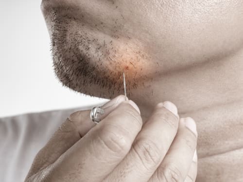
Ingrown hairs got you down? Laser hair removal can be your BFF! Discover how laser treatments tackle the root cause of ingrown hairs for smoother, irritation-free skin.
-

Is waxing a drag? Ditch the rashes and frequent appointments! Laser hair removal offers a long-lasting solution for smooth, hair-free skin. Learn how laser compares to waxing and if it’s right for you.
-
Searching for the best lip balm for dry lips? Discover which ingredients have real evidence behind them, why your lips get dry, and how to choose products that actually work.
-

While many patients experience positive weight loss results from Mounjaro injections, here’s what you should know about tiredness as a potential side effect of Mounjaro.
-
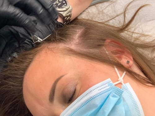
Learn how scalp micropigmentation can help you restore your hair density and confidence. Find out what causes hair loss, how the treatment works, and what results you can expect.