
Scalp
The scalp is not simply the skin on top of the head but a structurally distinct, five-layered tissue with a richer blood supply than almost any skin on the body, a follicle density that drives wound healing capacity well above what facial skin can achieve, and an architecture that makes it simultaneously the most forgiving and the most complex region to treat. Understanding its layers clarifies why injection depth matters, why scalp skin recovers faster than décolletage skin, and why treatments targeting follicle biology need to reach a specific anatomical zone rather than simply penetrating the surface.
The scalp extends from the supraorbital ridges anteriorly to the superior nuchal lines posteriorly, and laterally to the temporal lines. Its five layers are often taught using the mnemonic SCALP: Skin, Connective tissue, Aponeurosis (galea aponeurotica), Loose areolar connective tissue, and Pericranium. [4] The first three layers are tightly bound to each other and move as a single unit, sliding freely over the pericranium via the loose areolar fourth layer – a plane of clinical significance in both trauma and surgery.
Layer 1: The Skin
Scalp skin is among the thickest on the body at 3–4 mm, and it carries a follicle density – approximately 120,000 hair follicles across the scalp – that makes it structurally distinct from skin elsewhere. Each follicle is part of a pilosebaceous unit: the follicle itself, a sebaceous gland that secretes sebum into the follicle channel, and an arrector pili muscle that connects the follicle to the dermis. [3] The sebaceous glands are androgen-sensitive – testosterone and DHT stimulate sebum production, which is why sebum output increases at puberty and why scalp conditions such as seborrhoeic dermatitis have a hormonal component. The dermis of the scalp skin contains Pacinian corpuscles – deep pressure receptors – providing the scalp with tactile sensitivity to pressure and vibration. kenhub.com
Layer 2: Dense Connective Tissue
Immediately beneath the skin lies a dense fibrous connective tissue layer that binds the skin firmly to the galea. This is not merely structural – it is the layer that houses the primary blood vessels, lymphatics, and sensory nerves supplying the scalp. Hair follicles extend downward from the skin into this layer, placing the follicle bulb, dermal papilla, and bulge stem cell niche within or at the base of this vascular and neural-rich environment. The dense connective tissue layer is where the blood supply travels. Because it is tightly septate (divided by fibrous partitions), vessels in this layer cannot retract when cut, which is why scalp lacerations bleed disproportionately even when superficial.
Layer 3: The Galea Aponeurotica
The galea aponeurotica (epicranial aponeurosis) is a tough, flat fibrous sheet approximately 1–2 mm thick that connects the frontalis muscle anteriorly to the occipitalis muscle posteriorly, forming the structural foundation of the scalp’s mobile unit. It is relatively avascular compared to the layers above and below it, and it is the structural layer responsible for distributing the mechanical tension of facial expression across the scalp. When the frontalis contracts, raising the eyebrows, the galea transmits that movement across the entire scalp surface.
Layer 4: Loose Areolar Connective Tissue
The loose areolar layer is the functional sliding plane of the scalp. It contains no tight fibrous attachments and allows the upper three layers to move freely over the pericranium during normal scalp motion. In surgical contexts this is the “danger space” – infections or haematomas that reach this layer can spread rapidly across the entire scalp without the fibrous partitions that contain spread in the dense connective tissue above. It also contains blood vessels that contribute to scalp circulation.
Layer 5: Pericranium
The pericranium is the periosteum of the skull – the dense fibrous membrane covering the outer surface of the cranial bones. It is firmly adherent to the bone except at suture lines, and it contains osteogenic cells capable of contributing to bone repair. For scalp treatment purposes, the pericranium is simply the floor, the anatomical boundary below which no topical or injectable treatment is directed.
Blood Supply: The Vascular Density Advantage
The scalp has one of the richest blood supplies of any region of the body. Five pairs of arteries supply it from both the external carotid (superficial temporal, posterior auricular, occipital) and internal carotid (supratrochlear, supraorbital) systems, anastomosing freely across the vertex. [4] This vascular density is why scalp skin heals more rapidly from superficial wounding than facial skin in follicle-sparse areas – a distinction relevant to recovery timelines after any energy-based or injectable treatment across the scalp versus the lower face or décolletage.
Clinical Application
The scalp’s anatomy is not background knowledge for treatment conversations – it is the direct explanation for why treatments work as they do, why depth matters, and why the scalp responds differently to interventions than other skin regions.
Why injection depth is not arbitrary
The dense connective tissue layer – the second of the five layers – is where the blood supply, lymphatics, and lower follicle structures all converge. Scalp iPRF injections targeting the 1–1.5 mm dermal depth are delivering growth factors into this environment: the same zone where the perifollicular vasculature travels, where the dermal papilla sits at the base of anagen follicles, and where the bulge stem cell niche is positioned. [4] Placing the payload here is not a rough approximation – it is the specific anatomical location where PDGF, IGF-1, and VEGF have the shortest diffusion distance to all three targets simultaneously. Injections sitting in the most superficial dermis or epidermis place growth factors above this zone, not within it.
Why scalp skin heals faster
Clients often notice that scalp treatments recover faster than equivalent procedures on the lower face, neck, or décolletage. The explanation is structural: follicle density drives epidermal wound healing capacity through bulge HFSC migration to wound edges, and the scalp’s exceptional vascular density accelerates nutrient and growth factor delivery to the repair site. [2] Décolletage skin – sparse in follicles, with a less dense vascular network – cannot draw on either of these advantages. This structural difference is worth communicating clearly in post-treatment advice: a scalp that looks red for 24 hours post-treatment is not experiencing anything unusual; it is simply responding with the speed its architecture allows.
CAP and the scalp surface
Cold atmospheric plasma applied to the scalp acts through three mechanisms – Wnt/β-catenin activation in dermal papilla cells, nitric oxide generation driving perifollicular angiogenesis, and calcium influx activating bulge stem cells – all of which require the energy to reach the connective tissue layer where these structures reside. The scalp’s relatively thin epidermis (approximately 1.5 mm) allows plasma-generated reactive species to penetrate to dermal depth within the treatment zone, making the scalp more accessible to CAP effects than thicker-skinned body regions. [1]
Scalp micropigmentation: working with the scalp’s architecture
Scalp micropigmentation (SMP) takes a fundamentally different approach to hair loss than regenerative treatments – rather than attempting to restore follicle function, it works with the scalp’s appearance as it currently exists, using the visual language of follicle density to create the impression of fuller, thicker hair.
The technique deposits pigment into the upper dermis of the scalp using a fine needle, placing stippled dots that replicate the cross-sectional appearance of hair follicle openings at the scalp surface. Pigment is placed shallower than conventional tattooing – deeper placement would produce colour migration and blurring as the pigment disperses through the loose connective tissue – and the dot pattern is calibrated to match the client’s natural hair colour and existing follicle distribution. Results are typically visible after the first session and reach their intended density over 2–3 sessions.
At Creative Touch, SMP is offered as a hair density treatment – filling in areas of thinning and scalp show-through, strengthening the hairline, and covering scarring – in addition to the full shaved-head replication seen in male pattern baldness SMP. SMP at Creative Touch Results last three to five years, with occasional touch-up sessions to maintain colour saturation as the pigment gradually fades with UV exposure and natural skin turnover.
The clinical positioning of SMP alongside regenerative treatments is straightforward and honest: iPRF, CAP, and minoxidil work to restore and support biological follicle function over months; SMP delivers an immediate visual improvement that is independent of follicle biology entirely. For clients whose density has reduced to the point where iPRF results will take time to become visible, or for those with scarring that regenerative treatment cannot address, SMP provides an aesthetic outcome that does not require waiting for the follicle environment to respond. The two approaches are not in competition, they are addressing different aspects of the same concern.
References
Hwang JH, Lee HY, Chung KB, et al. (2021). Non-thermal atmospheric pressure plasma activates Wnt/β-catenin signaling in dermal papilla cells. Sci Rep, 11(1), 16125 . doi.org/10.1038/s41598-021-95650-y
Joulai Veijouye S, Yari A, Heidari F, et al. (2017). Bulge Region as a Putative Hair Follicle Stem Cells Niche: A Brief Review. Iran J Public Health, 46(9), 1167-1175 . PMC5632317
Martel JL, Miao JH, Badri T, et al. (2026). Anatomy, Hair Follicle. StatPearls Publishing. ncbi.nlm.nih.gov/books/NBK470321
Tajran J, Gosman AA (2026). Anatomy, Head and Neck, Scalp. StatPearls Publishing. ncbi.nlm.nih.gov/books/NBK551565
Anatomical Relationships
Referenced in Conditions & Treatments
- this Affected by Microneedling Evidence: Scalp microneedling creates microchannels enabling drug delivery (minoxidil, PDRN) and stimulates perifollicular growth factor environment for hair restoration. PMC11890238.
- this Associated condition Psoriasis Evidence: Scalp is explicitly one of the four primary anatomical sites of psoriatic plaque formation alongside elbows, knees, and lower back (entity text; PMC5796008).
Learn More
This topic is discussed in 4 articles:
-

Struggling with thinning hair? Discover natural, effective treatments at Creative Touch. We offer iPRF therapy, PRP, and scalp micropigmentation in Rotherham, Sheffield and South Yorkshire.
-

Experiencing hair loss on Mounjaro? Discover why it happens, plus evidence-based prevention strategies, and treatment options. Most cases are temporary and reversible.
-
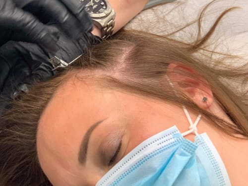
Learn how scalp micropigmentation can help you restore your hair density and confidence. Find out what causes hair loss, how the treatment works, and what results you can expect.
-
