
Pilosebaceous Unit
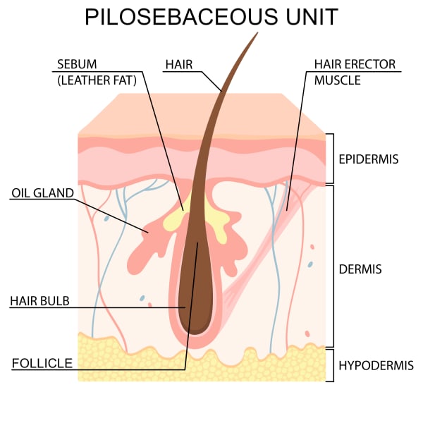
The pilosebaceous unit (PSU) is not simply a hair follicle with associated structures – it is an integrated multi-tissue system performing functions that span structural, secretory, immunological, and regenerative biology simultaneously. Its three components are the hair follicle (producing the hair shaft and housing the follicular stem cell niche), the sebaceous gland (synthesising and secreting sebum into the follicular canal), and the arrector pili muscle (providing the mechanical attachment that defines the bulge region’s anatomical boundaries). The PSU is present across virtually all skin surfaces with the exception of the palms, soles, and glabrous genital skin – but its size, proportions, and relative component dominance vary considerably by site, producing the spectrum from the large terminal follicles of the scalp to the large sebaceous follicles of the face with barely visible hair shafts. Understanding the PSU as a single integrated unit rather than three separate structures is what makes several clinical mechanisms – acne pathogenesis, androgenic hair miniaturisation, laser hair removal targeting, and scalp treatment combinations – legible at the mechanistic level rather than descriptive.
The pilosebaceous unit is an epidermal invagination – a downward projection of the epidermis into the dermis – that develops embryonically from ectodermal epithelium (hair follicle cells and sebaceous gland) and mesodermal contributions ( dermal papilla, inner and outer root sheaths, dermal sheath). [6] All three components of the PSU – follicle, gland, and muscle – are present from the second trimester of foetal development, with the sebaceous gland completing its structural development when the common excretory duct forms the first pilosebaceous canal. [4]
The Three Components
The hair follicle is the structural core of the PSU – a tubular invagination extending from the epidermal surface through the dermis and, for terminal follicles, into the subcutaneous fat. It is divided into four anatomical zones stacked vertically from the skin surface downward:
- Infundibulum: The uppermost segment, from the follicular opening at the skin surface down to the entry point of the sebaceous duct. This zone communicates directly with the skin surface environment and is where sebum, shed corneocytes, and environmental microbiota meet. It is the site of comedone formation in acne.
- Isthmus: From the sebaceous duct entry down to the attachment point of the arrector pili muscle. The lower portion of the isthmus – the bulge – is the most biologically significant zone of the entire PSU, housing the follicular stem cell niche.
- Suprabulbar zone: The mid-follicular region above the hair bulb, where the inner root sheath is maturing and the hair shaft is taking form.
- Bulb: The deepest zone, containing the hair matrix cells – the most mitotically active cells in the body during active growth – and the dermal papilla, the mesenchymal condensate that orchestrates the hair growth cycle through signalling to the surrounding matrix. [7]
The sebaceous gland is a holocrine secretory gland – meaning it releases its product (sebum) through complete cell disintegration rather than active secretion. Sebocytes mature from peripheral undifferentiated progenitor cells through progressive lipid accumulation, eventually rupturing and releasing their entire lipid contents into the sebaceous duct, which empties into the follicular canal at the infundibulum-isthmus junction. Sebum composition is unique to the sebaceous gland and distinct from the intercellular epidermal lipids: triglycerides and fatty acids constitute 57.5% of total sebum lipids, wax esters 26%, squalene 12%, and cholesterol and cholesterol esters the remaining 4.5%. [9] The squalene and wax ester fractions are specific to sebaceous secretion – they are not present in epidermal surface lipids – and their unique properties make them central to both sebum’s protective functions and its role in acne pathogenesis.
The arrector pili muscle is a smooth muscle bundle innervated by the autonomic nervous system that runs obliquely from the dermal-epidermal junction to the bulge region of the outer root sheath. Its visible function – causing piloerection in response to cold or autonomic stimulation – is biologically minor in humans. Its structural significance is anatomically defining: the arrector pili insertion point marks the lower boundary of the isthmus and the upper boundary of the bulge, creating the physical niche that protects the stem cell population. [10] The muscle also encircles the sebaceous gland, providing mechanical compression that assists sebum expression into the ductal system during contraction.
The Bulge: The Stem Cell Niche
The bulge region – anatomically defined as the outer root sheath protrusion at the arrector pili insertion – is the most clinically significant zone of the PSU from a regenerative medicine perspective. It houses two distinct stem cell populations: hair follicle stem cells (HFSCs) and melanocytic stem cells (McSCs). [3]
HFSCs are label-retaining cells – slow-cycling under normal conditions but capable of rapid activation at the onset of each new hair cycle. During telogen (the follicular resting phase), the dermal papilla migrates upward toward the bulge, enabling direct DPC-to-HFSC contact. This physical proximity is the signalling event that activates the stem cells – DPCs release WNT ligands and other activation signals that initiate HFSC proliferation and the onset of anagen. [5] If the dermal papilla cannot reach the bulge during the catagen regression phase – due to follicle miniaturisation, fibrosis, or structural disruption – cycling terminates and the follicle is permanently lost. The bulge also serves as a reservoir for epidermal keratinocyte progenitors that contribute to wound re-epithelialisation, making the PSU an active participant in surface skin repair as well as hair production.
McSCs in the bulge provide the melanocytes that populate the hair matrix and are responsible for hair shaft pigmentation during each anagen phase. Their progressive depletion through successive cycles – accelerated by UV damage and oxidative stress – is the cellular mechanism of hair greying.
PSU Subtypes
PSUs are not anatomically uniform. Four functional subtypes exist, distinguishable by the relative proportions of their components:
| Subtype | Hair Shaft | Follicle Depth | Sebaceous Gland | Primary Sites |
|---|---|---|---|---|
| Terminal follicle | Thick, pigmented, long | Deep dermis to subcutis | Moderate | Scalp, beard, axillae, pubis |
| Vellus follicle | Fine, short, minimally pigmented | Superficial dermis | Small | Face, trunk, limbs |
| Sebaceous follicle | Thin vellus-type shaft | Moderate | Very large (dominant component) | Nose, forehead, cheeks |
| Lanugo follicle | Fine, non-pigmented | Superficial | Absent/minimal | Foetal skin; replaced post-birth |
The sebaceous follicle subtype is the primary PSU type involved in acne – its disproportionately large sebaceous gland relative to the hair shaft means the follicular canal is lipid-rich and narrow relative to the volume of sebum it must transport, creating the retention conditions that precede comedone formation.
The Hair Growth Cycle
The PSU does not continuously produce hair – it cycles through phases of active growth, programmed regression, and rest, each phase involving different cell populations and different spatial relationships between the bulge, dermal papilla, and hair matrix. The cycle has four phases:
- Anagen (growth): 2–7 years on the scalp; weeks to months on body sites. The most mitotically active phase; matrix cells divide every 24–72 hours, producing the hair shaft. Melanocytes in the matrix are active and pigmenting the cortex. The follicle is at its maximum depth.
- Catagen (regression): Approximately 2 weeks. Programmed apoptosis of the lower follicle; the dermal papilla condenses and begins migrating upward; the hair shaft moves toward a club hair structure; the follicle shortens significantly.
- Telogen (rest): 2–3 months. The dermal papilla rests adjacent to the bulge; HFSCs are in quiescence; no new hair shaft is produced; the existing club hair is retained.
- Exogen (shedding): The club hair detaches and sheds – not a passive process but an active enzyme-mediated event distinct from telogen. A new anagen then begins. [7]
The hair cycle phase has direct clinical consequences in treatment: the laser hair removal target (melanin in the matrix adjacent to the dermal papilla) is only present and vulnerable during anagen; hair count measurements for efficacy assessment are only valid when the same proportion of follicles are in anagen; and scalp microneedling growth factor delivery to perifollicular tissue is most productive when stimulating a follicle population in active cycling.
Androgenic Regulation
The PSU is one of the most androgen-responsive tissue systems in the body. Androgen receptors are expressed in sebocytes, follicular keratinocytes, and dermal papilla cells. The critical enzyme is 5α-reductase, present at high concentrations in sebaceous gland tissue and follicular keratinocytes of androgen-sensitive sites, which converts testosterone (T) to dihydrotestosterone (DHT) – the significantly more potent androgen. [11]
DHT drives two divergent responses in two different PSU subtypes that are central to the clinical presentations of androgenic alopecia and acne:
- Sebaceous follicles (face, chest, back): DHT stimulates sebocyte proliferation and sebum production, enlarging the gland and increasing sebum output – the fundamental androgenic driver of acne-prone oily skin.
- Terminal scalp follicles (in genetically predisposed individuals): DHT binds androgen receptors in dermal papilla cells, triggering a signalling cascade that shortens the anagen phase progressively, producing shorter, finer hair with each successive cycle until the terminal follicle miniaturises to a vellus or non-cycling state – androgenic alopecia.
The same hormone produces opposite visible outcomes in different PSU subtypes because the androgen receptor signalling pathways activated in sebocytes (lipogenic differentiation) differ from those activated in scalp DPCs (cycle shortening, miniaturisation).
The Follicular Microenvironment and Cutibacterium acnes
The infundibulum and upper follicular canal of the sebaceous follicle create a microenvironment with specific physical and biochemical properties: anaerobic (oxygen-limited), lipid-rich, and immunologically distinct from the skin surface. These conditions are the natural niche for Cutibacterium acnes – an obligate anaerobe that metabolises sebaceous triglycerides through lipase activity, releasing free fatty acids that lower follicular pH and provide its own energy substrate. In healthy skin, C. acnes is a commensal member of the follicular microbiome, coexisting with other species and performing barrier-supportive functions through antimicrobial peptide production.
In acne, the microenvironment shifts: increased sebum production increases the available lipid substrate; follicular hyperkeratinisation narrows the infundibular opening, reducing oxygen tension and creating conditions that favour C. acnes outgrowth over competitors. This dysbiotic shift is not simply an increase in C. acnes numbers but a change in strain distribution – specifically, a dominance shift toward phylotype IA1 strains, which carry greater biofilm-forming capacity and more potent virulence factors. [1] IA1-dominant biofilms chronically colonise the follicular wall, triggering TLR2-mediated innate immune activation in keratinocytes, sebocytes, and immune cells, producing the IL-1β, TNF-α, and IL-6 cascade that drives inflammatory acne lesion formation.
Clinical Application
The pilosebaceous unit is the anatomical target or mechanistic site for several distinct treatment categories, each interacting with a different component or phase:
Acne: The Four-Pillar PSU Pathogenesis
Acne vulgaris is not caused by any single PSU dysfunction – it is the result of four converging disruptions that are individually insufficient but cumulatively sufficient to produce disease: [8]
- Follicular hyperkeratinisation: Abnormal desquamation in the infundibulum produces a retention of corneocytes that narrows the follicular opening – the microcomedone, the invisible precursor to all visible acne lesions.
- Sebum overproduction: Androgenic stimulation increases sebaceous gland output, filling the narrowed follicular canal with lipid substrate and providing the metabolic environment that favours anaerobic bacterial growth.
- Microbial dysbiosis: Shift from diverse commensal microbiota to C. acnes IA1 phylotype dominance and biofilm formation, triggering the innate immune activation cascade.
- Immune-inflammatory response: TLR2 and TLR4 activation by C. acnes virulence factors triggers IL-1β, TNF-α, and IL-6 production in keratinocytes and sebocytes; neutrophil and macrophage recruitment to the follicular wall produces the rupture of the follicle and the spread of keratin debris and bacteria into the surrounding dermis – the transition from comedone to inflammatory papule, pustule, or nodule.
Squalene peroxidation deserves specific mention: in acne-prone sebum, squalene – the 12% component of sebum unique to sebaceous secretion – is oxidised to squalene peroxide, which is directly comedogenic and pro-inflammatory independent of the C. acnes pathway. [9] Reduced antioxidant capacity in acne-prone sebaceous secretion (lower vitamin E) accelerates squalene peroxidation, adding an oxidative lipid chemistry pathway to the inflammatory cascade. This is one reason that antioxidant support is clinically rational in acne management beyond its general skin health benefits.
Laser Hair Removal: Targeting the PSU
Laser hair removal exploits the PSU’s structural architecture through selective photothermolysis: a laser wavelength absorbed preferentially by melanin (the chromophore in the hair shaft and matrix cells) converts to thermal energy that diffuses from the melanin-containing structures to the adjacent non-pigmented follicular targets. [12]
The two biological targets for permanent hair reduction within the PSU are:
The dermal papilla: A 2022 mechanism study confirmed that the primary route to permanent hair reduction is thermal diffusion from melanin-containing precortex cells in the hair bulb to the adjacent non-pigmented DPCs – producing thermal necrosis of approximately 24% of dermal papilla cells per anagen-phase laser session. [2] Critically, this study found that bulge stem cell numbers remained unchanged after laser delivery – the bulge is not the primary permanent reduction target. When the laser was delivered during telogen (no melanin in the precortex adjacent to the DPCs), no DPC necrosis and no hair miniaturisation occurred. This confirms that anagen-phase treatment is not merely preferred but mechanistically required for dermal papilla damage and progressive permanent reduction.
The bulge: The bulge region at 1–1.5mm depth is a secondary target – thermal energy from the hair shaft melanin diffuses proximally to the bulge, with the extended theory of selective photothermolysis accounting for damage to non-pigmented bulge stem cells through heat diffusion from the more superficial shaft. Bulge damage alone produces temporary alopecia (the bulge regrows without DPC input) – permanent reduction requires DPC damage.
The clinical implications: multiple sessions are required because only anagen-phase follicles are vulnerable, and on any given body site only 15–30% of follicles are in anagen simultaneously; session spacing must match the site-specific cycle length; and follicles with reduced melanin (grey, white, fine vellus) are resistant because the thermal diffusion from melanin to DPCs cannot occur without the melanin chromophore present.
Hair Loss Treatments: Supporting the PSU
Scalp-targeted treatments in the Creative Touch portfolio interact with the PSU at the follicular and perifollicular level:
iPRF and scalp microneedling: Growth factors delivered to the perifollicular environment – PDGF, IGF-1, VEGF, and FGF from iPRF – act on DPCs and perifollicular vasculature. VEGF and FGF drive angiogenesis in the perifollicular vascular plexus, improving nutrient and oxygen supply to the bulb during anagen; IGF-1 directly activates DPC signalling that sustains anagen duration. Scalp microneedling delivers these factors through microchannels whilst simultaneously stimulating a WNT-pathway wound response in DPCs that independently promotes anagen initiation.
Cold atmospheric plasma (CAP): The antimicrobial RONS generated by CAP at the scalp surface selectively disrupt the follicular microbiota balance, reducing pathogenic load in the perifollicular environment without antibiotic resistance risk – whilst separately stimulating VEGF expression and perifollicular angiogenesis through nitric oxide-mediated pathways.
1927nm thulium FTL (scalp): Fractional laser to the scalp delivers the follicular growth factor stimulus and endogenous IGF-1 restoration that supports DPC activity – confirmed by prospective study showing significant increase in hair count following FTL monotherapy. jkslms.or
Summary: PSU as Clinical Anchor
| Treatment Context | PSU Target | Mechanism |
|---|---|---|
| Laser hair removal | Hair matrix melanin → DPC thermal diffusion | Selective photothermolysis; anagen-phase DPC necrosis |
| Acne treatments | Infundibulum, sebaceous gland, C. acnes biofilm | Four-pillar disruption; sebum normalisation; antimicrobial |
| iPRF (scalp) | Dermal papilla, perifollicular vasculature | DPC growth factor activation; VEGF angiogenesis |
| Scalp microneedling | Perifollicular environment, DPC zone | WNT activation; growth factor delivery via microchannels |
| CAP (scalp) | Follicular microbiota, perifollicular vasculature | RONS antimicrobial; nitric oxide VEGF stimulation |
| 1927nm FTL (scalp) | Perifollicular tissue | IGF-1 restoration; follicular collagenesis |
| Androgenic hair loss | DPC androgen receptor, 5α-reductase | DHT-driven cycle shortening; follicle miniaturisation |
References
Cavallo I, Sivori F, Truglio M, et al. (2022). Skin dysbiosis and Cutibacterium acnes biofilm in inflammatory acne lesions of adolescents. Sci Rep, 12(1), 21104 . doi.org/10.1038/s41598-022-25436-3
Chiu HY, Wang WH, Kuan CH, et al. (2022). Depilatory laser miniaturizes hair by inducing bystander dermal papilla cell necrosis through thermal diffusion. Lasers Surg Med, 54(6), 916-927 . doi.org/10.1002/lsm.23533
Han J, Lin K, Choo H, et al. (2023). Distinct bulge stem cell populations maintain the pilosebaceous unit in a β-catenin-dependent manner. iScience, 26(1), 105805 . doi.org/10.1016/j.isci.2022.105805
Hoover E, Aslam S, Krishnamurthy K (2026). Physiology, Sebaceous Glands. StatPearls Publishing. ncbi.nlm.nih.gov/books/NBK499819
Lin X, Zhu L, He J (2022). Morphogenesis, Growth Cycle and Molecular Regulation of Hair Follicles. Front Cell Dev Biol, 10, 899095 . doi.org/10.3389/fcell.2022.899095
Martel JL, Miao JH, Badri T, et al. (2026). Anatomy, Hair Follicle. StatPearls Publishing. ncbi.nlm.nih.gov/books/NBK470321
Natarelli N, Gahoonia N, Sivamani RK (2023). Integrative and Mechanistic Approach to the Hair Growth Cycle and Hair Loss. J Clin Med, 12(3) . doi.org/10.3390/jcm12030893
Oliveira MB, Maurício AC, Barros AN, et al. (2025). Rebalancing the Skin: The Microbiome, Acne Pathogenesis, and the Future of Natural and Synthetic Therapies. Molecules, 30(24) . doi.org/10.3390/molecules30244684
Ottaviani M, Camera E, Picardo M (2010). Lipid mediators in acne. Mediators Inflamm, 2010 . doi.org/10.1155/2010/858176
Poblet E, Jiménez F, Ortega F (2004). The contribution of the arrector pili muscle and sebaceous glands to the follicular unit structure. J Am Acad Dermatol, 51(2), 217-22 . doi.org/10.1016/j.jaad.2004.01.054
Sánchez-Pellicer P, Navarro-Moratalla L, Núñez-Delegido E, et al. (2022). Acne, Microbiome, and Probiotics: The Gut-Skin Axis. Microorganisms, 10(7) . doi.org/10.3390/microorganisms10071303
Vaidya T, Hohman MH, Kumar D D (2026). Laser Hair Removal. StatPearls Publishing. ncbi.nlm.nih.gov/books/NBK507861
Anatomical Relationships
Structural Connections
- Has sub-structure Hair follicle Evidence: Entity text states the hair follicle is embedded within the pilosebaceous unit; the pilosebaceous unit is the parent composite structure containing the follicle.
- Has sub-structure Sebaceous gland Evidence: Entity text explicitly states pilosebaceous unit includes the follicle, sebaceous glands, and arrector pili muscle. Sebaceous gland is a named constituent.
Referenced in Conditions & Treatments
- this Connected to Dermis Evidence: Pilosebaceous units are embedded in and structurally supported by the dermis. Niemann & Horsley Semin Cell Dev Biol 2012 doi:10.1016/j.semcdb.2012.08.010