

You’ve probably seen the treatment photos. Perhaps you’ve read testimonials from clients who’ve experienced hair regrowth or skin rejuvenation. You may have gained an understanding of the basics.
Now it’s time to go deeper into the science of PRP and iPRF treatments:
- What exactly are these growth factors everyone mentions?
- Why does the processing method matter so much?
- What’s actually happening at a cellular level when platelets trigger regeneration?
We aren’t trying to overwhelm you with biochemistry, but we do want to satisfy genuine curiosity about the mechanisms driving these treatments. When you understand the science, the treatment timeline makes sense. The protocol decisions become logical. The reasons for individual variation become clear.
Let’s explore what happens when we harness your body’s own regenerative capacity, from the moment blood is drawn to the cellular conversations that ultimately rebuild tissue.
Your blood: A sophisticated system
Before we can understand platelet-rich plasma therapies, we need to appreciate what blood actually contains. And why platelets, specifically, hold such regenerative potential.
The components working together
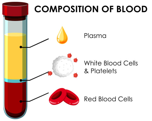
Your blood is a complex tissue containing multiple cell types, each with distinct functions.
Red blood cells (erythrocytes) comprise about 40% to 45% of blood volume [1]. They’re the oxygen carriers, filled with haemoglobin and lacking nuclei to maximise space for their primary job. Simple, efficient, focused on one task.
White blood cells make up less than 1% of blood volume [1] but play crucial roles in immune function. There are several types (neutrophils, lymphocytes, monocytes) each with specific defensive capabilities. They’re your internal security system, constantly patrolling.
Platelets (thrombocytes) are tiny cell fragments. About 150,000 to 400,000 per microlitre of blood in healthy individuals [2]. They’re best known for blood clotting, but that barely scratches the surface of their capabilities.
Plasma is the liquid component, roughly 55% of blood volume [1]. It contains proteins, nutrients, hormones, and countless dissolved substances that maintain bodily functions.
Why platelets are remarkable
Here’s something interesting: platelets aren’t even complete cells. They’re cellular fragments.
They originate from megakaryocytes in your bone marrow, which essentially break apart to release thousands of platelets into circulation. A bit like a cellular piñata, if you will.
What makes them extraordinary for regenerative medicine? It’s what they carry inside.
Each platelet contains specialised storage compartments called alpha granules. These granules are packed with over 300 different proteins [3], many of which function as signalling molecules that coordinate healing.

When tissue injury occurs, platelets don’t just form clots. They release these stored proteins in a carefully orchestrated sequence, essentially broadcasting instructions to surrounding cells about what needs to happen next. These instructions initiate the entire wound healing cascade.
The insight driving PRP and iPRF therapies is elegant: if we concentrate these platelets and trigger their release mechanisms, we can amplify the body’s natural healing signals where they’re needed the most.
Think about that for a moment. We’re not introducing anything foreign. We’re just increasing the volume on signals your body already uses.
The molecular pharmacy inside platelets
Let’s open those alpha granules and examine what’s actually inside.
Understanding these specific molecules helps explain why platelet therapies work for such diverse applications. Once you see what’s in here, the whole thing starts making sense.
Growth factors: The primary signalling molecules
Platelet-Derived Growth Factor (PDGF)
PDGF exists in several isoforms, but all share a primary function: stimulating fibroblast proliferation and migration.
Fibroblasts are the cells responsible for producing collagen, elastin, and the structural proteins that give skin its framework and strength. Your skin’s construction workers.

When PDGF binds to receptors on fibroblast surfaces, it triggers intracellular signalling pathways that tell the cell to divide and get to work. This is why PDGF is crucial during the proliferative phase of wound healing, ensuring you have enough working cells to rebuild damaged tissue.
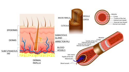
For hair restoration, PDGF influences dermal papilla cells. These are the specialised fibroblasts at the base of hair follicles (think of them as the command centre) that regulate the hair growth cycle.
Transforming Growth Factor-Beta (TGF-β)
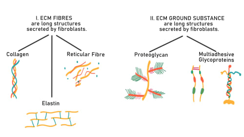
TGF-β might be the most important growth factor for collagen synthesis. It stimulates fibroblasts to produce extracellular matrix components—the structural scaffolding between cells that gives tissue its mechanical properties.
But here’s the thing. TGF-β is a double-edged sword.
Whilst it drives collagen production essential for healing, excessive TGF-β signalling can lead to fibrosis (excessive scarring with disorganised collagen). The body normally tightly regulates TGF-β activity, and the controlled delivery in PRP/ iPRF maintains this balance.
In skin rejuvenation, TGF-β is responsible for the gradual improvement in firmness and thickness that emerges months after treatment as new collagen accumulates and matures. This is why patience is required.
Vascular Endothelial Growth Factor (VEGF)
VEGF specialises in angiogenesis—the formation of new blood vessels.
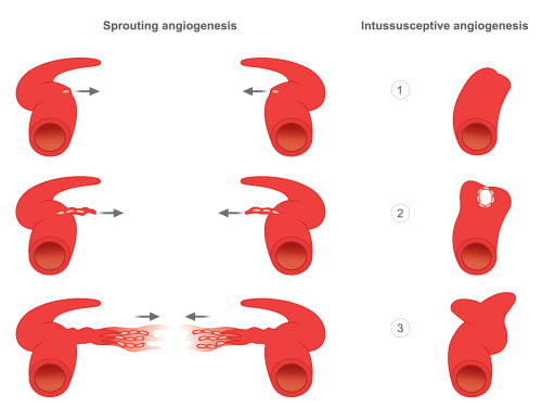
Adequate blood supply is essential for tissue health. Hair follicles, in particular, are highly vascular structures requiring robust circulation to support the metabolically demanding process of hair production. Growing hair requires a lot of energy.
VEGF signals endothelial cells (which line blood vessels) to proliferate and migrate, forming new capillary networks. This improved vascularisation enhances nutrient delivery and waste removal, supporting the long-term health of treated tissue.
Epidermal Growth Factor (EGF)
EGF primarily targets keratinocytes—the predominant cell type in your epidermis. It promotes their proliferation and migration, accelerating wound closure and supporting skin barrier restoration.
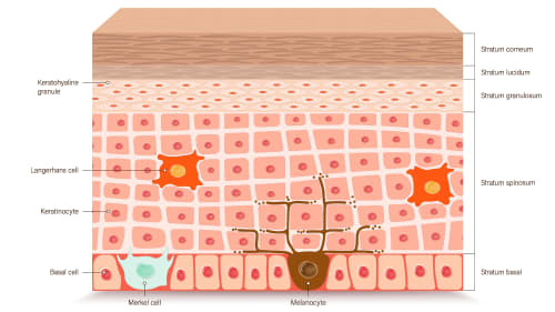
For facial rejuvenation, EGF contributes to improved skin texture and the renewal of the skin surface, complementing the deeper structural effects of other growth factors.
Insulin-like Growth Factor (IGF)
IGF-1, the main form in platelets, promotes cell survival and supports tissue regeneration. It works synergistically with other growth factors, often amplifying their effects.
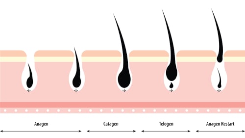
In hair follicles, IGF-1 appears to prolong the anagen (active growth) phase of the hair cycle whilst delaying entry into catagen (regression phase), resulting in longer, thicker hair shafts.
Cytokines and other signalling proteins
Beyond classical growth factors, platelets contain numerous cytokines that modulate inflammation and immune responses.
Some are pro-inflammatory (necessary for initiating healing), whilst others are anti-inflammatory (important for resolution once repair is underway). It’s all about balance and timing.
Platelets also release chemokines that attract stem cells and other reparative cells to the treatment site, essentially calling in reinforcements for the regenerative process.
Structural proteins
The fibrin and fibronectin released from platelets don’t just signal—they physically structure the healing environment, providing scaffolding for cells to attach, migrate across, and organise themselves during tissue reconstruction.
Architecture matters.
PRP processing: Concentrating regenerative components
Standard Platelet-Rich Plasma (PRP) involves centrifuging whole blood to separate components by density.
Understanding this process helps clarify why technique matters so much (and it does matter).

The separation physics
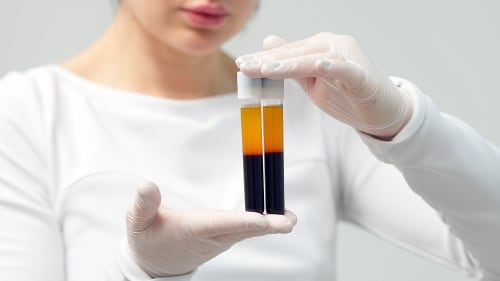
When blood spins in a centrifuge, centrifugal force causes denser components to move outward (or downward, depending on centrifuge orientation), creating distinct layers:
The red blood cell layer (heaviest) settles at the bottom. These cells are denser due to their haemoglobin content.
The buffy coat forms a thin layer above the red cells. This is where white blood cells and platelets concentrate, sitting at the density interface. This is what we’re after.
Platelet-poor plasma remains at the top—the lightest component.
The goal is to harvest the buffy coat and some surrounding plasma to concentrate platelets whilst removing most red blood cells. Simple in concept, precise in execution.
Processing variables that matter
Centrifugation speed and duration: Higher speeds and longer spins create cleaner separation but may damage platelets through excessive force. Too aggressive, and you break what you’re trying to harvest. PRP preparation protocols vary, with centrifugation speeds typically ranging from approximately 1,000-3,500 rpm and times from 5-20 minutes, depending on the specific protocol and equipment used.
Anticoagulant choice: Most protocols use anticoagulants like citrate to prevent clotting during processing. However, anticoagulants chelate calcium, which platelets need for optimal activation. This is why activation steps often add calcium chloride back.
Single vs double spin: Some protocols perform a second, gentler spin to further concentrate platelets from the initial harvest. This increases platelet concentration, but this adds processing time and the potential for platelet activation during handling. There are trade-offs.
Activation: The crucial final step
Concentrated platelets must be activated to release their growth factors. Several activation methods exist:
Calcium chloride: Restores calcium ions that trigger degranulation (release of alpha granule contents).
Thrombin: Mimics the body’s natural clotting cascade, providing a strong activation signal.
Collagen or physical trauma: The injection process itself can partially activate platelets through contact with tissue.
Non-activated PRP releases growth factors more gradually as platelets encounter activation triggers in tissue. Activated PRP delivers an immediate burst of factors. Each approach has theoretical advantages depending on the application.
What you end up with
PRP can achieve 3-5 times baseline platelet concentration [4]. The preparation consists primarily of platelets suspended in plasma, with minimal white blood cells and virtually no red blood cells when processed well.
Once injected, these concentrated platelets release their growth factors within hours. Proliferation of these factors peak in the first 24-72 hours before degrading or being cleared [5].
This is why PRP protocols often involve multiple treatments. Each provides a finite pulse of signalling. One treatment, one pulse. Three treatments, sustained momentum.
iPRF processing: Preserving the regenerative package
Injectable Platelet-Rich Fibrin (iPRF) represents a fundamental shift in processing philosophy.
Rather than maximum separation and concentration, iPRF prioritises preservation of components that work together synergistically.

Low-speed centrifugation preserves complexity
Here’s where iPRF gets interesting. It uses significantly lower centrifugation speeds—typically around 700 rpm for just 3-4 minutes [6].
This gentler processing creates less distinct separation between layers. That’s not a bug, it’s a feature.
The result is a preparation that retains not only platelets but also:
White blood cells at significant concentrations. Whilst some consider these inflammatory, research suggests they may contribute beneficially to the regenerative process through immune modulation and their own growth factor production. Turns out they’re not just along for the ride.
Circulating stem cells including mesenchymal stem cells and haematopoietic progenitor cells. These cells potentially contribute to tissue regeneration directly, not just through signalling.
Fibrin network: The lower speed and absence of anticoagulants allows natural fibrin formation. This creates a three-dimensional scaffold with remarkable properties. We’ll get to that.
The fibrin matrix: More than just structure
Fibrin isn’t simply holding everything together. The matrix creates a sustained-release system for growth factors.
Think about it this way: when growth factors are released from platelets, many become physically trapped within the fibrin mesh. The fibrin network slowly degrades, gradually releasing trapped growth factors over 7 to 14 days [7] as it dissolves.
This creates fundamentally different kinetics compared to PRP. Instead of hours of growth factor presence, iPRF maintains bioactive concentrations for well over a week. Your cells receive continuous signalling throughout the critical early regenerative period.
Research using immunoassays to measure growth factor release confirms this difference. PRP shows peak concentrations within hours, declining rapidly. iPRF shows sustained release curves extending beyond 10 days.
No anticoagulants: Purer biology
Because iPRF doesn’t require anticoagulants, the preparation contains only what came from your blood. There’s no citrate chelating calcium, no additives that might theoretically interfere with cellular responses.
Just your blood’s regenerative components, concentrated and preserved.
The absence of anticoagulants also means calcium remains available, supporting optimal platelet function without requiring re-addition during activation.
Concentration differences
Here’s where it gets paradoxical. Whilst PRP can achieve higher platelet concentration (perhaps 5-fold vs 2-3 fold for iPRF), iPRF often produces superior clinical results.
Concentration isn’t everything.
The sustained release kinetics, preserved cellular diversity, and fibrin scaffold make the difference. Quality over pure quantity.
Cellular conversations: How growth factors change behaviour
Understanding growth factor release is one thing. Understanding what happens when they reach target cells reveals why regeneration takes time and why results build progressively.
This is where it gets really fascinating.
Receptor binding and signal transduction
Growth factors don’t enter cells. They bind to receptors on cell surfaces, triggering intracellular signalling cascades that ultimately reach the nucleus and alter gene expression.
Most growth factors bind receptor tyrosine kinases—proteins that span the cell membrane with an external binding domain and internal enzymatic activity. When growth factors bind externally, the receptor’s internal portion activates, phosphorylating (adding phosphate groups to) downstream signalling proteins.

These phosphorylated proteins activate further proteins in branching pathways—like MAPK, PI3K, and JAK/STAT pathways. Each pathway influences different cellular processes: proliferation, migration, survival, differentiation, or protein synthesis.
It’s a molecular relay: the growth factor triggers the receptor, which activates pathway A, which activates proteins B and C, which activate effector proteins D, E, and F, which finally alter gene transcription in the nucleus.
A cascade of falling dominoes, but in three dimensions and with branching pathways.
This multi-step amplification means a relatively small number of growth factor molecules can trigger substantial cellular responses. However, it also means the response takes time—hours for initial effects, days for meaningful changes in cell behaviour.
Biology doesn’t rush.
Gene expression changes
When signalling pathways reach the nucleus, they activate transcription factors—proteins that bind DNA and increase or decrease the transcription of specific genes.
For collagen synthesis, TGF-β signalling activates SMAD proteins that enter the nucleus and increase transcription of collagen genes (COL1A1, COL1A2, COL3A1). The cell begins producing collagen mRNA, which ribosomes translate into collagen protein.
But producing collagen protein is just the start. Collagen molecules must be secreted from cells, assembled into fibrils in the extracellular space, and cross-linked to form a functional structural protein.
This entire process takes weeks to months, explaining why you don’t see firmness improvements immediately after treatment. You’ve initiated a construction project that unfolds on cellular timescales.
PDGF, meanwhile, activates genes controlling cell cycle progression—CDK4, Cyclin D—that allow fibroblasts to exit their resting state and begin dividing. A single stimulated fibroblast becomes two, then four, then eight, exponentially increasing the number of collagen-producing cells over several cell cycles spanning days.
The proliferation and migration dance
Growth factors don’t just tell cells to make proteins. They coordinate complex behaviours.
PDGF triggers fibroblast migration toward the growth factor source (chemotaxis). Cells extend protrusions, attach to the surrounding matrix, and pull themselves forward. This is essential for wound healing, where fibroblasts must physically move into damaged areas.
EGF promotes keratinocyte migration across wounded surfaces during re-epithelialisation. Without this coordinated cell movement, wounds wouldn’t close properly.
VEGF orchestrates the multi-step process of angiogenesis: endothelial cells must degrade their existing basement membrane, migrate through surrounding tissue, proliferate, organise into tubular structures, and establish a new basement membrane and blood flow.
This sequence requires days to complete. New blood vessels don’t appear overnight.
Duration of response
Here’s a important point: cellular responses to growth factors outlast the growth factors themselves.
Once a fibroblast receives PDGF signalling that pushes it into active cell cycling, it continues dividing for several cycles even after PDGF disappears. The decision to proliferate, once made, persists for days.
Similarly, once collagen synthesis genes are activated, increased collagen production continues for some time even after the initial TGF-β signal fades. Momentum builds.
However, sustained or repeated signalling produces more robust responses than brief pulses. This is why iPRF’s prolonged growth factor release (maintaining signals for over a week rather than hours) tends to produce more pronounced regenerative effects than standard PRP.
Duration matters as much as intensity.
Application-specific mechanisms: Why the same treatment works differently for hair and skin
The same growth factors produce distinct outcomes depending on the target tissue.
Understanding these differences helps explain treatment protocols and timelines. Context is everything.
Hair follicle biology and regeneration
Hair follicles are miniature organs cycling through distinct phases [8]:
Anagen is the active growth phase, lasting 2-7 years for scalp hair. Follicle cells at the bulb divide rapidly, producing the hair shaft. This phase requires tremendous metabolic energy and robust blood supply. Growing hair is hard work.
Catagen is a brief transitional phase (2-3 weeks) where growth stops and the follicle begins regressing. Intermission.
Telogen is the resting phase (2-4 months) where the follicle essentially shuts down before either re-entering anagen or dying. Hibernation.
In androgenetic alopecia (pattern baldness), androgen hormones gradually miniaturise follicles. The anagen phase shortens, producing finer, shorter hairs. Eventually, follicles may enter prolonged telogen or die altogether.
Not a pleasant process.
How PRP/iPRF intervenes in hair loss
Growth factors influence multiple aspects of follicle function.
Dermal papilla stimulation: The dermal papilla is a specialised group of cells at the follicle base that orchestrates the hair growth cycle. These cells express receptors for PDGF, VEGF, and IGF-1. Growth factor signalling stimulates dermal papilla cells to produce factors that maintain follicles in anagen.
Angiogenesis around follicles: VEGF improves blood supply to the metabolically demanding follicle. Enhanced circulation delivers nutrients and removes waste more efficiently. Better infrastructure, better function.
Anti-apoptotic effects: IGF-1 and other factors promote cell survival, potentially rescuing follicles that might otherwise undergo apoptosis (programmed cell death). Life support for struggling follicles.
Stem cell activation: Hair follicles contain stem cells in the bulge region that must activate to regenerate the follicle for each anagen phase. Growth factors appear to support this activation.
Possible anti-inflammatory effects: Cytokines in PRP/iPRF may modulate inflammation that can damage follicles in some types of hair loss. Calming the environment.
Why hair results take months
Even if treatment successfully stimulates a follicle to enter anagen, you won’t see hair immediately. The follicle must reconstruct its growth structures, begin producing a new shaft, and grow that shaft long enough to emerge from the scalp.
This entire process takes 2-3 months minimum.
Additionally, follicles are at different cycle phases at any given time. Treatment may immediately influence actively growing follicles, whilst follicles in telogen won’t respond until they’re ready to re-enter anagen.
This is why hair restoration protocols typically involve 3-4 treatments over several months. You’re giving repeated signals as different follicles become receptive, and reinforcing effects in follicles that responded to earlier treatments.
Patience isn’t optional here. It’s built into the biology.
Skin rejuvenation mechanisms
For facial skin, the goals differ: improving firmness through collagen renewal, enhancing hydration, refining texture, and reducing visible signs of ageing.
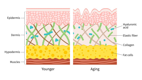
Fibroblast activation in aged dermis: Dermal fibroblasts become less active with age, producing less collagen and more collagen-degrading enzymes ( matrix metalloproteinases). Growth factors can reactivate these senescent fibroblasts, ramping up their synthetic activity.
Collagen remodelling: TGF-β doesn’t just increase collagen quantity. It influences collagen organisation. Properly organised collagen fibres provide better mechanical support than disorganised scar-like collagen.
Elastin synthesis: Elastin fibres provide skin’s elastic recoil—that quality where skin bounces back when pinched. Aged skin loses elastin, becoming lax. Growth factors support new elastin production, though this is a slower process than collagen synthesis.
Hyaluronic acid production: Fibroblasts also produce hyaluronic acid, the glycosaminoglycan that holds moisture in skin. Increased hyaluronic acid synthesis improves hydration and plumpness.
Dermal thickness: One measurable outcome from PRP/iPRF facial treatments is increased dermal thickness seen on ultrasound imaging. This reflects accumulated collagen and ground substance filling the dermis, physically thickening skin and smoothing surface irregularities.
You can actually see the difference in imaging.
Why skin results also require patience
Collagen synthesis is slow. Even after fibroblasts activate, they must produce collagen molecules, secrete them, allow assembly into fibrils, and wait for enzymatic cross-linking that stabilises the structures.
This process unfolds over 2-3 months.
The collagen present when you receive treatment continues degrading at its normal rate (about 1% daily turnover), so early on, you’re essentially breaking even. Only as synthesis overtakes degradation do you see net improvement.
By 3-6 months post-treatment, the new collagen has matured, properly cross-linked, and integrated into the dermal architecture. This is when results typically peak.
Timing, dosing, and sustained release
The temporal aspects of growth factor therapy are just as important as the factors themselves.
Timing is everything, really.
Optimal concentration thresholds
Growth factors follow dose-response curves. Too little produces no effect because you don’t reach the threshold for receptor activation. Too much can paradoxically reduce effectiveness or trigger different cellular responses.
There’s a sweet spot.
Research suggests growth factors follow dose-response curves, with optimal PDGF concentrations for fibroblast proliferation in the nanogram per millilitre range. Concentrations that are too low produce minimal effects, whilst excessively high concentrations may paradoxically reduce effectiveness.
PRP typically contains growth factors in the optimal range, which is why attempting to create “super concentrated” preparations doesn’t necessarily improve outcomes and might worsen them.
More isn’t always better. Sometimes it’s just more.
The sustained signal advantage
Here’s where iPRF’s prolonged release becomes particularly significant.
Cellular responses depend not just on peak concentration but on duration of exposure. A cell exposed to growth factors for 6 hours activates certain immediate-response genes but may not fully commit to long-term behavioural changes.
Brief encounter versus sustained relationship.
A cell receiving continuous signalling for 7-10 days undergoes more stable phenotypic changes—the effects of gene expression changes accumulate, leading to more dramatic shifts in cell function.
Studies comparing PRP to iPRF consistently show more robust and lasting outcomes with iPRF despite lower peak growth factor concentrations. The sustained signal duration appears more important than maximum concentration.
Quality of signal matters more than peak intensity.
Why repeated treatments work
A single treatment provides finite signalling. Cells respond, but as growth factor concentrations decline, the stimulus fades.
A second treatment, 4-6 weeks later, catches cells while they’re still in an activated state. Rather than starting from scratch, you’re amplifying and extending the response already underway. Building momentum.
By the third treatment, you’ve established sustained regenerative momentum. This is why treatment courses typically involve 3-4 sessions. You’re building effects cumulatively.
For maintenance, once yearly treatments keep cells receiving periodic regenerative signals before regression occurs, preserving improvements long-term.
Think of it like compound interest for your skin.
The evidence: What research tells us
Understanding mechanisms is valuable, but clinical evidence determines whether treatments actually work.
Let’s look at what the studies show.
PRP efficacy studies
For androgenetic alopecia, several randomised controlled trials demonstrate that PRP increases hair density and shaft thickness. Multiple meta-analyses have demonstrated that PRP significantly increases hair density in androgenetic alopecia, with studies showing mean increases of 14-25 hairs per square centimetre over 3-6 months [9].
Real results, measured objectively.
The studies show heterogeneous results, partly due to variations in PRP preparation protocols. Studies using higher platelet concentrations and activation methods tend to show stronger effects.
For facial rejuvenation, evidence includes histological studies showing increased dermal collagen density following PRP treatments, and clinical trials demonstrating improvements in wrinkle scores and skin elasticity measurements [11]. A 2021 systematic review of 36 studies involving over 3,000 patients concluded that whilst PRP shows beneficial effects for facial rejuvenation, the lack of uniformity in PRP preparation protocols and standardised assessment methods limits the ability to draw definitive conclusions, noting that large-scale randomised controlled trials with standardised protocols are warranted [10].
The evidence is there, though there’s room for more rigorous research.
iPRF clinical evidence
iPRF research is newer but growing rapidly. Comparative studies suggest iPRF produces superior outcomes to standard PRP for hair restoration, with higher percentages of patients achieving clinically significant improvement.
Makes sense given the sustained-release mechanism.
For facial applications, studies using ultrasound to measure dermal thickness show greater increases with iPRF compared to PRP, supporting the sustained-release hypothesis.
The evidence base, whilst encouraging, remains moderate in quality. Most studies are relatively small, and long-term outcome data beyond 6-12 months are limited. Larger randomised trials with standardised protocols would strengthen conclusions.
But the trend is clear: iPRF shows promise.
Safety profile
Both PRP and iPRF show excellent safety profiles in published studies. Adverse events are primarily minor: transient pain, bruising, and swelling.
Serious complications such as infection or allergic reactions are exceedingly rare, likely because the treatments use autologous (your own) material. Hard to be allergic to yourself.
The main “risk” is inadequate response, i.e., treatments simply not working for some individuals, rather than causing harm.
Why individual responses vary
If you’ve researched these treatments, you’ve probably noticed that some clients achieve remarkable results whilst others see modest improvement.
Several biological factors explain this variation. And honestly, it’s one of the most frustrating aspects for practitioners and clients alike.
Baseline platelet characteristics
Not all platelets are created equal.
Platelet counts vary between individuals (normal range: 150,000-400,000 per microlitre). Someone with naturally high platelet counts may achieve better concentration in their PRP.
More importantly, platelet function varies. Some individuals’ platelets contain higher concentrations of growth factors or release them more efficiently upon activation. These “high responder” phenotypes show better outcomes.
Unfortunately, there’s no simple way to predict this beforehand. Platelet function testing exists but isn’t routinely performed for aesthetic applications. We’re basically flying blind on this variable.
Age-related factors
Younger individuals tend to respond more robustly. This likely reflects several factors:
Fibroblasts from younger skin show greater proliferative capacity when stimulated. They’re not yet senescent. More eager to respond.
Growth factor production may be higher in platelets from younger individuals, but research on this is limited.
Tissue healing capacity generally declines with age via multiple mechanisms: reduced stem cell numbers, accumulated cellular damage, and reduced inflammatory responses.
This doesn’t mean older clients won’t benefit - they absolutely can. However, the magnitude of responses may be somewhat reduced compared to younger individuals. That’s just reality, not pessimism.
Medications and health status
Certain medications impair platelet function.
Aspirin irreversibly inhibits COX-1 enzymes in platelets, reducing their ability to aggregate and potentially affecting growth factor release [12].
Other NSAIDs, certain antidepressants, and blood thinners can affect platelet function to varying degrees.
Chronic health conditions affecting circulation, immunity, or healing capacity may reduce treatment effectiveness. Poorly controlled diabetes, for instance, impairs multiple aspects of wound healing and tissue regeneration.
Your baseline health matters.
Tissue health and receptor sensitivity
Severely damaged or heavily scarred tissue may respond less effectively. Growth factor signals have fewer viable cells to act upon if dermal fibroblasts are depleted or the extracellular matrix is too degraded.
Growth factors can’t signal to cells that aren’t there.
Conversely, tissue with mild to moderate age-related changes typically shows robust responses because functional cells are still present and capable of responding. They just need the right stimulus.
Treatment protocol variables
Seemingly minor variations in technique can affect outcomes:
Injection depth and distribution matter. Growth factors must reach the appropriate tissue layers and distribute adequately. Too superficial or too deep, and you’re not hitting the target.
Platelet activation status—whether iPRF is mechanically activated during injection or activates gradually in tissue—may influence results.
Post-treatment care, though less critical than for other procedures, still matters. Avoiding activities that cause excessive sweating or potentially introduce bacteria to injection sites supports optimal healing.
The devil’s in the details.
Alternative pathways to regeneration
PRP and iPRF work by delivering concentrated growth factors directly to tissue. It’s worth noting that other regenerative treatments achieve similar outcomes through different mechanisms.
Growth Factor Concentrate: a different approach to the same goal
A newer autologous treatment has been attracting attention: Growth Factor Concentrate (GFC). Like PRP and iPRF, it begins with your own blood. But where both those approaches concentrate the biological machinery (platelets, cells, fibrin) that delivers growth factors, GFC takes the process a step further. It isolates the growth factor molecules themselves.
The preparation uses a specialist activation kit containing a platelet-activating solution. Your blood is mixed with this solution and left to stand for roughly 30 minutes, during which the platelets degranulate and release their contents into the surrounding plasma. A subsequent centrifuge spin at higher speed then separates out a clear, cell-free layer. No red blood cells, no white blood cells, no platelets, just the concentrated signalling proteins that platelet degranulation produced.
If you’ve followed the processing sections above, you’ll recognise what this means mechanistically. PRP concentrates platelets and injects them with their growth factors still packaged inside, releasing them in a burst over hours once in tissue. iPRF preserves the fibrin matrix and cellular diversity, releasing growth factors slowly over 7 to 14 days. GFC pre-releases the growth factors before injection and delivers them in concentrated, immediately bioavailable form with no waiting for in-tissue activation.
The potential advantage is reduced post-injection inflammation. Since white blood cells are removed entirely, the pro-inflammatory cytokines they carry aren’t present in the injectate. Some practitioners report less post-treatment swelling and scalp sensitivity compared to leukocyte-rich PRP preparations. The potential trade-off is the loss of the broader biological environment: no fibrin scaffold for sustained release, and none of the leukocyte-mediated immune modulation or stem cell contribution that makes iPRF particularly compelling.
Early clinical data looks promising. A 2024 prospective study published in Cureus confirmed improved hair density trichoscopically across four treatment sessions, with negative hair pull tests at eight weeks and no adverse events reported. [8]
It’s worth noting that GFC represents a reductionist philosophy: strip everything back and deliver only the end product. That’s a coherent approach. But whether the removed components (the fibrin, the leukocytes, the stem cell signalling) were passengers or active contributors to outcomes is still being worked out by the research community. The answer probably varies by application and patient.
We don’t currently offer GFC at Creative Touch. If it’s something you’ve been reading about, the differences above should help you understand where it sits relative to the treatments we do provide.
Polynucleotides
Polynucleotides stimulate your cells to produce their own growth factors by activating specific cellular receptors (particularly adenosine receptors), ultimately triggering many of the same regenerative pathways—collagen synthesis, angiogenesis, and tissue remodelling—but through endogenous (internally produced) signalling rather than exogenous (externally delivered) growth factors.
Different route, similar destination.
Understanding that multiple pathways can achieve regenerative outcomes helps explain why treatment combinations sometimes work synergistically and why certain approaches may suit specific situations better than others.
Discover how polynucleotides support skin regeneration through cellular activation.
Working with your body’s regenerative intelligence
PRP and iPRF represent a sophisticated approach to aesthetic medicine. Oone that doesn’t override your biology but rather amplifies what your body already knows how to do.
The science reveals why these treatments work: growth factors trigger proven regenerative pathways, cellular responses accumulate over appropriate timescales, and sustained signalling produces more robust outcomes than brief pulses.
It also reveals why patience is essential. You’re not applying a topical that sits on the surface or injecting a filler that provides immediate structure. You’re initiating a biological process that unfolds according to cellular timelines measured in weeks and months.
Biology moves at its own pace, not yours.
The evidence supports both PRP and iPRF as effective regenerative treatments, with iPRF’s sustained-release kinetics offering theoretical and practical advantages for many applications.
Understanding these mechanisms doesn’t just satisfy curiosity, it sets realistic expectations and helps you appreciate the complexity of what’s occurring beneath your skin after treatment. You’re witnessing cellular biology in action, orchestrated by signals your body already uses for healing.
That’s rather remarkable when you think about it.
Ready to explore whether PRP or iPRF therapy suits your specific concerns? Discover our regenerative treatment options or book a consultation to discuss your personalised approach.
References
American Society of Hematology (2025). Blood Basics. American Society of Hematology. hematology.org/education/patients/blood-basics (Accessed: 2025-10-24)
Cleveland Clinic (2021). What Is a Platelet Count?. Cleveland Clinic. my.clevelandclinic.org/…21782-platelet-count (Accessed: 2025-10-24)
Whiteheart, S. (2011). Platelet granules: surprise packages. Blood, 118(5), 1190-1191. doi.org/10.1182/blood-2011-06-359836
doi: 10.1182/blood-2011-06-359836Machado, E., Leite, R., dos Santos, C., Artuso, G., Gluszczak, F., de Jesus, L., Caldas, J., Bredemeier, M. (2019). Turn down - turn up: a simple and low-cost protocol for preparing platelet-rich plasma. Clinics, 74(), e1132. doi.org/10.6061/clinics/2019/e1132
doi: 10.6061/clinics/2019/e1132Gorodilova, A., Kharisova, C., Osinnikova, M., Kitaeva, K., Filin, I., Mayasin, Y., Solovyeva, V., Rizvanov, A. (2024). The Well-Forgotten Old: Platelet-Rich Plasma in Modern Anti-Aging Therapy. Cells, 13(21), 1755. doi.org/10.3390/cells13211755
doi: 10.3390/cells13211755Hassan, H., Quinlan, D., Ghanem, A. (2020). Injectable platelet‐rich fibrin for facial rejuvenation: A prospective, single‐center study. Journal of Cosmetic Dermatology, 19(12), 3213-3221. doi.org/10.1111/jocd.13692
doi: 10.1111/jocd.13692Pavlovic, V., Ciric, M., Jovanovic, V., Trandafilovic, M., Stojanovic, P. (2021). Platelet-rich fibrin: Basics of biological actions and protocol modifications. Open Medicine, 16(1), 446-454.
Bhargava, A., Singh, V., Tiwari, R., Arya, A., Chokshi, K. (2024). Revitalizing Hair Growth: A New Regimen Utilizing Growth Factor Concentrate for Hair Loss Treatment. Cureus, . doi.org/10.7759/cureus.63354
doi: 10.7759/cureus.63354