
Vitamin E
Vitamin E’s skin mechanism is more specific than general antioxidant protection implies. α-Tocopherol is actively secreted onto the skin surface via sebum. Facial stratum corneum contains significantly higher α-tocopherol concentrations than body skin, directly because sebaceous output is higher on the face. At the surface, it intercepts UV-driven squalene photooxidation: squalene is the most UV-sensitive lipid in skin surface lipids, and its oxidation products (SqPx) directly activate AhR and EGFR-linked inflammatory pathways in keratinocytes. α-Tocopherol’s chain-breaking antioxidant activity halts this cascade before inflammatory signalling begins but is consumed in the process, and requires vitamin C for regeneration. This C-E recycling cycle is the functional unit of lipid-phase skin antioxidant defence.
Vitamin E encompasses eight structurally related fat-soluble compounds – four tocopherols (α, β, γ, δ) and four tocotrienols (α, β, γ, δ) – that share a chromanol ring head group but differ in their isoprenoid tail structure and, consequently, their biological activity. In human physiology, α-tocopherol dominates almost completely: the α-tocopherol transfer protein (α-TTP) in the liver selectively retains α-tocopherol for redistribution into plasma lipoproteins, with α-, β-, γ-, and δ-tocopherols having relative affinities of approximately 100%, 38%, 9%, and 2% respectively. [4] The biological predominance of α-tocopherol is therefore not dietary coincidence but the result of a selective retention mechanism that filters out the other forms.
In skin, this preferential α-tocopherol distribution is further amplified by a delivery route unique to the face and sebaceous-rich body sites.
The Sebaceous Delivery Route
The skin surface lipid (SSL) film that covers the stratum corneum is a mixture of epidermal lipids and sebum. Sebum contains α-tocopherol at concentrations that significantly exceed the levels delivered by passive diffusion from plasma, and its secretion onto the skin surface is the primary physiological route by which vitamin E reaches the outermost skin layers. [7] A study confirming this mechanism found that upper facial stratum corneum contained significantly higher α-tocopherol levels than corresponding arm stratum corneum layers, a distribution pattern that maps precisely to facial sebaceous gland density. Dietary α-tocopherol intake directly influences sebum vitamin E content, making systemic vitamin E status the upstream driver of surface antioxidant protection on sebaceous skin. [1]
This delivery mechanism has a direct clinical implication: topical vitamin E supplementation and systemic vitamin E status are complementary, not redundant. Topical application deposits α-tocopherol directly into the stratum corneum lipid bilayers, providing immediate local antioxidant capacity. Adequate systemic status ensures ongoing replenishment through sebum secretion; a sustained release mechanism that topical application alone cannot replicate.
The Squalene Photooxidation Cascade
Squalene, a polyunsaturated hydrocarbon, is the most UV-sensitive lipid in skin surface lipids and the quantitatively dominant lipid in human sebum. Under UVA and UVB exposure, squalene undergoes photooxidation to generate squalene peroxides (SqPx) – reactive lipid oxidation products that diffuse into the viable epidermis and directly activate the aryl hydrocarbon receptor (AhR) and EGFR-linked inflammatory pathways in keratinocytes, producing TNF-α, IL-6, and MMP upregulation. [5] SqPx exposure in primary human keratinocyte cultures reproduces the majority of UVA+UVB inflammatory effects, confirming that squalene photooxidation is not a side reaction but a primary UV signal transduction mechanism in the skin surface.
α-Tocopherol interrupts this cascade at the photooxidation step, functioning as a chain-breaking antioxidant in the lipid phase of the SSL. By donating a hydrogen atom to squalene-derived lipid peroxyl radicals, it converts them to stable lipid hydroperoxides before they can generate the SqPx that would otherwise activate AhR and EGFR. academic.oup.com The protection is pre-inflammatory: it prevents the inflammatory signal from forming rather than suppressing it after activation. A 2004 in vivo study confirmed that topical α-tocopherol treatment significantly inhibited squalene photooxidation in human skin surface lipids following UV exposure, with increased α-tocopherol deposition maintained for at least 24 hours. [2]
This mechanism also explains why vitamin E is disproportionately effective on sebaceous skin: the face, chest, and back, where squalene-rich sebum is in greatest supply as the photooxidation substrate. On low-sebum skin (forearms, legs), the squalene pool is smaller and the mechanism proportionally less relevant, though the membrane lipid peroxidation protection in cell membranes beneath the surface remains.
The Vitamin C Regeneration Cycle
α-Tocopherol is consumed when it donates a hydrogen atom to a lipid peroxyl radical – it becomes a tocopheroxyl radical in the process, which is relatively stable but no longer antioxidant-active. The restoration of tocopheroxyl radical back to active α-tocopherol requires vitamin C (ascorbic acid), which donates an electron to the tocopheroxyl radical, regenerating active vitamin E and becoming dehydroascorbic acid in the process. [6]
This recycling reaction is the mechanistic basis for the synergy between vitamins C and E in skin antioxidant protection. They are not performing the same function in parallel; they are operating in series, with vitamin E as the lipid-phase radical interceptor and vitamin C as the aqueous-phase regenerator that keeps it active. Without adequate vitamin C, spent tocopheroxyl radicals accumulate and net antioxidant capacity declines faster under UV load than either vitamin alone would suggest. The practical implication is that topical vitamin E applied without adequate vitamin C availability (either topical or systemic) delivers less sustained antioxidant protection than the C+E combination.
Glutathione completes the cycle: it regenerates oxidised vitamin C (dehydroascorbic acid) back to ascorbic acid. The full antioxidant network is therefore α-tocopherol → vitamin C → glutathione, with each component dependent on the availability of the next for regeneration.
Membrane Protection: The Lipid Peroxidation Chain Reaction
Beyond the SSL surface, α-tocopherol is incorporated into cell membrane phospholipid bilayers, particularly in mitochondrial membranes and nuclear membranes of keratinocytes and fibroblasts. Here it performs the same chain-breaking function, intercepting lipid peroxyl radicals before they propagate through the membrane in an autocatalytic oxidation cascade. [4] A single tocopherol molecule can break the peroxidation chain before it propagates – its physical location in the membrane, with the chromanol head positioned at the water-lipid interface and the phytyl tail anchoring it in the hydrophobic core, gives it direct access to the lipid peroxyl radicals it intercepts.
This membrane antioxidant function is the basis for α-tocopherol’s role in maintaining keratinocyte and fibroblast viability under oxidative stress, which is distinct from the surface SSL squalene mechanism. Both are relevant but at different anatomical levels of the skin.
Tocotrienols: Emerging Skin Relevance
The tocotrienol subfamily – less studied than tocopherols and not retained preferentially by α-TTP – has emerging skin evidence worth noting without overstating. Tocotrienols have been shown to modulate melanogenesis in some models, and a systematic review found suggestive evidence for limiting lipid and protein oxidation, DNA damage, and telomere shortening in keratinocytes. casi.org The evidence tier is lower than for α-tocopherol – predominantly in vitro and animal models – and tocotrienols are not retained in human skin through the same sebaceous route. Their primary clinical interest currently is in oral supplementation contexts rather than topical application. They are noted here for completeness rather than as a current clinical priority.
Wound Healing: A Nuanced Picture
Vitamin E’s role in wound healing is genuinely complex and worth addressing honestly rather than defaulting to the general “antioxidant supports healing” framing. Evidence for vitamin E’s anti-inflammatory and CTGF-modulating effects is real: α-tocopherol modulates connective tissue growth factor (CTGF) expression, influences fibroblast activation, and has demonstrated activity against MRSA-infected wounds. [3]
However, there is also evidence that vitamin E in high concentrations can inhibit wound healing, specifically by reducing collagen accumulation and wound tensile strength in animal models, an effect attributed to its anti-inflammatory activity suppressing the proliferative phase of healing. [8] The clinical implication is concentration-dependent: physiological vitamin E levels in a well-nourished client support healing through antioxidant and anti-inflammatory mechanisms; supraphysiological topical application to healing wounds may impair rather than accelerate collagen deposition. This is a reason for caution with high-dose vitamin E topicals on active wound sites, including immediately post-procedure, rather than a reason to avoid vitamin E in homecare formulations at cosmeceutical concentrations.
Clinical Application
The clinical question vitamin E creates is primarily a pairing and positioning question, specifically, whether it is present at adequate concentration alongside vitamin C to function as a complete antioxidant system, and whether its topical application timing and concentration is appropriate for the client’s current skin state.
Morning Antioxidant Protocol: C+E as the Functional Unit
As the antioxidant network regeneration mechanism establishes, vitamins C and E should be considered as a system rather than interchangeable alternatives. A morning topical protocol containing both – vitamin C in an aqueous phase at appropriate pH and concentration (as detailed in the Vitamin C entity), alongside vitamin E in the lipid phase – provides antioxidant protection that operates across both the aqueous and lipid compartments of the stratum corneum and upper epidermis simultaneously.
The sequencing rationale is the same as established for vitamin C: both are providing pre-inflammatory protection by intercepting the ROS and lipid peroxidation cascades before they activate downstream inflammatory signalling (AP-1/ MMP-1 for vitamin C; AhR/EGFR for vitamin E). Both are more effective applied before UV exposure than after. A morning C+E application before SPF is the most complete photoprotection homecare preparation currently supported by the evidence.
For clients using retinoids in their evening routine, this morning C+E protocol provides the complementary antioxidant half of the protocol – retinoids suppress MMP transcription via AP-1/c-Jun downregulation at the nuclear level; morning C+E interrupts the ROS that would otherwise trigger that same AP-1 activation at the cellular signalling level.
Post-Procedure: Timing Matters
The wound healing nuance – high-concentration vitamin E potentially impairing collagen deposition – is directly relevant to the post-procedure context. In the acute recovery phase following microneedling, RF microneedling, or laser, the wound-healing inflammatory cascade is the mechanism through which the treatment produces its collagen remodelling benefit. Applying high-dose vitamin E topicals in the immediate post-procedure window risks attenuating exactly the proliferative phase response that the treatment has initiated.
The practical recommendation: maintain the standard C+E morning antioxidant protocol up to the day before treatment; during the acute recovery phase (typically five to seven days), use formulations that prioritise barrier support ( ceramides, dexpanthenol, hyaluronic acid) over antioxidant loading; resume the full C+E protocol once the acute healing phase is complete and remodelling is underway. The antioxidant protection benefit of vitamin E is most valuable at the remodelling stage – protecting the newly synthesised collagen from ongoing UV-driven oxidative degradation – not during the acute wound response.
Vitamin E in Tallow-Based Topicals
Unlike vitamin B12, vitamin E is fat-soluble and heat-stable; it survives the tallow rendering process at meaningful concentrations. Grass-fed tallow contains α-tocopherol and mixed tocopherols from the animal’s own fat-soluble vitamin profile, and these remain bioavailable in the rendered product. Applied topically, tallow-derived tocopherols provide genuine membrane lipid peroxidation protection and contribute to the SSL antioxidant pool. The concentration is modest and variable compared to a standardised tocopherol-enriched cosmeceutical, but the presence is chemically real – consistent with the statement made in the Vitamin B12 entity that tallow’s genuine topical benefit lies in its fat-soluble vitamin content including vitamin E.
References
Ekanayake-Mudiyanselage S, Kraemer K, Thiele JJ (2004). Oral supplementation with all-Rac- and RRR-alpha-tocopherol increases vitamin E levels in human sebum after a latency period of 14-21 days. Ann N Y Acad Sci, 1031, 184-94 . doi.org/10.1196/annals.1331.017
Ekanayake-Mudiyanselage S, Tavakkol A, Polefka TG, et al. (2005). Vitamin E delivery to human skin by a rinse-off product: penetration of alpha-tocopherol versus wash-out effects of skin surface lipids. Skin Pharmacol Physiol, 18(1), 20-6 . doi.org/10.1159/000081682
Hobson R (2016). Vitamin E and wound healing: an evidence-based review. Int Wound J, 13(3), 331-5 . doi.org/10.1111/iwj.12295
Keen MA, Hassan I (2016). Vitamin E in dermatology. Indian Dermatol Online J, 7(4), 311-5 . doi.org/10.4103/2229-5178.185494
Kostyuk V, Potapovich A, Stancato A, et al. (2012). Photo-oxidation products of skin surface squalene mediate metabolic and inflammatory responses to solar UV in human keratinocytes. PLoS One, 7(8), e44472 . doi.org/10.1371/journal.pone.0044472
Pullar JM, Carr AC, Vissers MCM (2017). The Roles of Vitamin C in Skin Health. Nutrients, 9(8) . doi.org/10.3390/nu9080866
Thiele JJ, Weber SU, Packer L (1999). Sebaceous gland secretion is a major physiologic route of vitamin E delivery to skin. J Invest Dermatol, 113(6), 1006-10 . doi.org/10.1046/j.1523-1747.1999.00794.x
Unknown Author. PMC: PMC1355132. PMC1355132
Learn More
This topic is discussed in 2 articles:
-
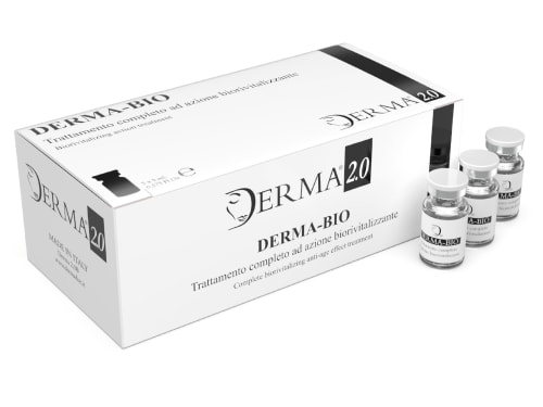
Discover the science behind Derma Bio, a mesotherapy product designed to combat signs of aging and achieve a brighter, smoother, and more youthful appearance.
-
Antioxidant providing protection against environmental damage; present in shea butter and tallow