
Stratum corneum
The stratum corneum is the skin’s definitive interface, functioning as a “bricks and mortar” system where anucleate corneocytes (the bricks) are embedded in a lamellar lipid matrix (the mortar). Clinical integrity depends on more than just lipid quantity; it requires a precise acidic pH (4.5–5.5) to activate the enzymes responsible for ceramide processing and controlled desquamation. Effective barrier restoration is a dual-track process: we must support the upstream keratinocyte differentiation programme while maintaining the acidic surface environment that allows the barrier to self-assemble and shed correctly.
The stratum corneum is the terminal product of keratinocyte differentiation – the layer at which the elaborate upward journey from basal cell to corneocyte concludes. It is thin (10–40 μm depending on body site), structurally dense, and functionally non-negotiable: without an intact stratum corneum, the epidermis cannot regulate water loss, exclude environmental threats, or maintain the acid pH environment on which its own lipid-processing enzymes depend. It is the layer that all the upstream biology of the Keratinocyte, Filaggrin, Ceramide, and Free Fatty Acid entities is ultimately building toward.

Two Components, Two Functions
The stratum corneum operates through two structurally distinct components whose functions are complementary but independent. [5]
Corneocytes are the anucleate, keratin-filled cells that provide mechanical reinforcement – the physical resistance to abrasion, UV damage, and pathogen penetration. Each corneocyte is surrounded by a cornified envelope, a highly cross-linked protein shell replacing the original plasma membrane during terminal differentiation. Inside, keratin filaments are compacted within a filaggrin protein matrix, giving the cell its characteristic flat, dense architecture. Corneocytes remain connected to adjacent cells by corneodesmosomes – modified desmosomes that are progressively degraded by serine proteases toward the surface, orchestrating the controlled shedding process of desquamation.
The extracellular lipid matrix fills the intercellular spaces between corneocytes in a highly organised lamellar arrangement – ceramides, cholesterol, and free fatty acids stacked in repeating bilayer phases that create the primary waterproof seal of the barrier. This is the component responsible for regulating transepidermal water loss (TEWL) and for controlling the selective permeability that determines what the barrier lets through and what it excludes. The lipid matrix is not simply a passive sealant. It is the source of the acidic pH environment – generated through free fatty acid release by secretory phospholipase A2 – that activates the ceramide-processing enzymes essential for its own maintenance. It also hosts antimicrobial peptides that contribute to surface-level pathogen defence.

These two components fail through different mechanisms and require different approaches to restoration, which is the primary reason that treating stratum corneum dysfunction as a single problem produces incomplete outcomes. Corneocyte quality depends on the completeness of the keratinocyte differentiation programme, the filaggrin content of the cell, and the integrity of the cornified envelope. Lipid matrix quality depends on the synthesis capacity of differentiating keratinocytes, the availability of ceramide, cholesterol, and free fatty acid precursors, and the acid pH environment in which lamellar body secretion and lipid processing occur. [5]
The “Bricks and Mortar” Anatomy
| Component | Biological Role | Deficit Presentation | Restorative Focus |
|---|---|---|---|
| Corneocytes (The Bricks) | Mechanical shield, UV protection, keratin compaction. | Thinning, “fragile” feel, poor resilience. | Differentiation boosters (Thulium, Retinoids). |
| Lipid Matrix (The Mortar) | TEWL regulation, waterproof seal, antimicrobial. | Tightness, dehydration, “invisible” water loss. | Lipid substrate & pH management. |
| Corneodesmosomes (The Glue) | Cell-to-cell adhesion and desquamation control. | Dullness, “flaky” patches, rough texture. | PHA/AHA (to support enzyme-driven shedding). |
The Stratum Disjunctum: The Desquamation Zone
The stratum disjunctum is the upper portion of the stratum corneum – the zone in which corneocytes are progressively losing their intercellular adhesion and moving toward active shedding from the skin surface. It is not a structurally distinct layer visible in histology but a functional zone defined by the progressive degradation of the corneodesmosomes that, in the stratum compactum below, are still intact and load-bearing.
The transition from compactum to disjunctum is driven by a serine protease cascade centred on two kallikrein-related peptidases, KLK5 and KLK7, secreted as inactive precursors from lamellar granules in the upper stratum granulosum and activated in the intercellular space of the stratum corneum. KLK5 is the cascade initiator – it self-activates and then cleaves and activates KLK7 and KLK14; KLK14 feeds back positively to amplify KLK5 activity, creating a self-reinforcing proteolytic cascade. [6] Activated KLK5 and KLK7 then degrade the three structural proteins of the corneodesmosome – desmoglein-1 (DSG1), desmocollin-1 (DSC1), and corneodesmosin (CDSN) – progressively dissolving the intercellular adhesion that held those cells in place. Once corneodesmosomal integrity is lost, the corneocyte can no longer be retained, and desquamation follows.
The enzyme cascade is regulated at the stratum disjunctum level by the serine protease inhibitor LEKTI (lympho-epithelial Kazal-type-related inhibitor, encoded by SPINK5), which binds KLK5 to hold the cascade in check. LEKTI’s inhibitory grip on KLK5 is directly pH-dependent: as the epidermal pH gradient acidifies toward the surface, LEKTI dissociates from KLK5, releasing active protease and permitting corneodesmosomal cleavage to occur precisely in the most superficial stratum corneum layers. [2] Desquamation is therefore pH-gated: the acid mantle’s maintenance of the correct surface pH is not just an antimicrobial function but an active regulator of the timing and location of corneodesmosomal cleavage.
When this system is dysregulated – through SPINK5 mutation (Netherton syndrome), KLK5/7 upregulation ( atopic dermatitis and psoriasis), or alkaline pH disruption of the gradient that normally confines protease activity to the disjunctum – desquamation accelerates uncontrollably, barrier integrity collapses, and the inflammatory cascade that follows produces the clinical presentations of those conditions. [1] The same mechanism explains why sustained alkaline pH exposure – harsh surfactants, hard water, repeated alkaline cleanser use – produces clinical barrier dysfunction even in skin without underlying genetic risk: the acid gradient that confines KLK activity to the disjunctum is disrupted, and proteolytic activity spreads downward into the compactum.
Clinical significance for topical exfoliation: Glycolic acid’s exfoliating action is specifically targeted to the stratum disjunctum. Biopsy evidence confirms that enhanced corneodesmosomal breakdown under glycolic acid treatment is restricted to the disjunctum – accelerating the KLK-driven dissolution that is already occurring there – whilst desmosomes in the stratum compactum remain entirely unaffected. [3] This selectivity is why well-formulated chemical exfoliants do not strip the barrier: they amplify the disjunctum’s own controlled desquamation process without penetrating to the compactum’s intact adhesion structures.
The Stratum Compactum: The Barrier Zone
The stratum compactum is the lower portion of the stratum corneum – the mechanically robust, biochemically intact zone that performs the primary barrier functions of TEWL regulation, selective permeability, and physical resistance. Where the disjunctum above it is characterised by progressive corneodesmosomal dissolution, the compactum is characterised by intact corneodesmosomes, fully organised lamellar lipid bilayers, and the highest corneocyte density in the stratum corneum.
It is the compactum that most directly corresponds to the “bricks and mortar” model described in the section above. The lamellar lipid bilayers – ceramides, cholesterol, and free fatty acids in repeating organised phases – reach their most complete and tightly ordered arrangement in the compactum, having been fully processed from lamellar body precursors by the acidic sphingomyelinase and beta-glucocerebrosidase enzymes whose activity is maintained by the acid pH generated in this zone. The corneodesmosomes here are unprocessed by the KLK cascade: LEKTI inhibition of KLK5 at the lower stratum corneum’s relatively less acidic pH holds protease activity in check, ensuring that corneodesmosomal integrity is maintained at exactly the layer where barrier structural function requires it. [4]
Understanding the compactum as the zone of intact barrier function changes how barrier disruption and recovery should be interpreted clinically. Mild exfoliation, well-calibrated chemical peels, and even low-energy fractional laser treatments disrupt the disjunctum – removing accumulated surface cells and temporarily thinning the overall stratum corneum – whilst the compactum remains structurally competent, lipid matrix intact, and capable of rapid barrier recovery. Recovery is fast because the structural foundation is untouched. By contrast, over-exfoliation that penetrates into the compactum – through excessive concentration, very low pH, or frequency beyond the barrier’s regeneration capacity – disrupts the lipid lamellar organisation, permits the KLK cascade to spread beyond the disjunctum, and produces the barrier failure characterised by elevated TEWL, increased irritant sensitivity, and the prolonged recovery that clients describe as their skin “breaking down.” [3]
Disjunctum vs. Compactum: Functional Summary
| Property | Stratum Disjunctum | Stratum Compactum |
|---|---|---|
| Location | Upper SC (surface-facing) | Lower SC (epidermis-facing) |
| Corneodesmosome status | Progressively degraded by KLK5/7 | Intact; LEKTI-inhibited KLK cascade |
| Lipid organisation | Loosening; desquamation-ready | Fully organised lamellar bilayers |
| Primary function | Controlled shedding of spent corneocytes | TEWL regulation; barrier integrity |
| Response to exfoliants | Targeted – accelerates natural KLK cascade | Unaffected at cosmetic concentrations |
| Response to over-exfoliation | First zone disrupted; threshold exceeded | Lipid lamellae disrupted; recovery prolonged |
| pH regulation role | Surface acid mantle confines KLK activity here | Slightly less acidic; LEKTI active; protease inhibited |
The Acid Mantle and pH Dependency
Healthy stratum corneum surface pH sits between approximately 4.5 and 5.5. This is not incidental, it is the pH range at which the two enzymes responsible for the final processing of ceramide precursors into active ceramides, acidic sphingomyelinase and beta-glucocerebrosidase, operate at optimal activity. At neutral or alkaline pH, both enzymes become significantly less active, and ceramide production from the lamellar body precursor pool slows measurably even when upstream synthesis is intact. The acid mantle is therefore not a standalone antimicrobial feature, it is an active prerequisite for the barrier’s own lipid maintenance machinery.
Sources of pH disruption that impair this system include alkaline surfactants in cleansers, hard water mineral exposure, and filaggrin deficiency – which reduces the urocanic acid and pyrrolidone carboxylic acid that filaggrin degradation contributes to the acid mantle. The Filaggrin page describes this relationship in detail, and the cross-dependency between filaggrin status and stratum corneum lipid processing is one of the clearest examples in barrier biology of how two apparently separate components are mechanistically coupled.
The pH “Switch” (Enzyme Activity)
| Surface pH | Enzyme Status | Barrier Result |
|---|---|---|
| 4.5 – 5.5 (Acidic) | Optimal: Ceramides are processed; desquamation is smooth. | Healthy, resilient, glowing barrier. |
| 6.0 – 7.0 (Neutral) | Suppressed: Ceramide synthesis slows; “glue” remains sticky. | Flaky, congested skin; chronic dehydration. |
| 7.5+ (Alkaline) | Failed: Barrier lipids cannot form; microbiome shifts. | Irritation, inflammation, and barrier collapse. |
How the Stratum Corneum Changes with Age
The stratum corneum undergoes measurable changes through both chronological ageing and hormonal transition that impair its barrier performance without necessarily being visible at the surface.
Corneocyte transit time through the stratum corneum increases with age – from approximately 20 days in younger adults to 30 days or more in older adults – meaning that the surface is occupied by older, more structurally compromised cells for longer before desquamation removes them. Lipid composition shifts with age: ceramide content declines, and the chain length distribution of free fatty acids changes in ways that reduce lamellar phase organisation. Post-menopause, oestrogen withdrawal reduces the activity of enzymes involved in ceramide synthesis, contributing an additional hormone-mediated route to the lipid matrix deterioration described in the Ceramides page.
The result is a stratum corneum that retains its basic architecture but performs it less efficiently: higher TEWL, greater vulnerability to irritant penetration, slower recovery following disruption, and a compromised acid mantle that further impairs the lipid processing the barrier needs to self-maintain.
Aging and the Stratum Corneum
| Feature | Youthful SC | Mature/Post-Menopausal SC |
|---|---|---|
| Renewal Rate | ~20 days (Rapid turnover) | 30+ days (Slower renewal) |
| Lipid Volume | High ceramide/FFA content | 30%–50% reduction in total lipids |
| pH Buffer | Strong (Resists alkaline shock) | Weak (Slow to recover acid pH) |
| Appearance | Compact, translucent, smooth | Thickened (hyperkeratosis), dull, opaque |
Clinical Application
The stratum corneum is the endpoint of every treatment aimed at barrier function, and the lens through which barrier outcomes should be assessed. TEWL measurement and surface hydration are the clinical proxies for stratum corneum integrity, and they reflect the state of both the corneocyte and lipid matrix components. The treatment framework for the stratum corneum therefore organises around which of the two components is the primary deficit, whether the underlying cause is synthesis suppression, renewal programme impairment, or the inflammatory environment driving both.
Restoring the Lipid Matrix
Where the primary stratum corneum deficit is lipid matrix disorganisation – the common presentation in dry, dehydrated, post-menopausal, or atopic-tendency skin – the treatment logic follows the three-route ceramide restoration framework established in the Ceramides entity: resolve inflammatory suppression of synthesis (CAP, polynucleotides, omega-3), stimulate synthesis directly ( niacinamide, microneedling-driven SPTLC3 upregulation), and supply topical and oral substrate (ceramide-cholesterol-FFA formulations in physiological ratios, phytoceramides orally).
The acid mantle dimension adds a fourth consideration: any treatment or product that raises stratum corneum pH impairs ceramide processing at the enzymatic step, undermining lipid matrix restoration regardless of how much substrate is being supplied. Surfactant selection in cleansers and water quality are practical clinical factors that belong in client homecare guidance alongside the lipid supplementation strategy.
Restoring Corneocyte Quality
Where the primary deficit is corneocyte quality – incomplete differentiation, reduced filaggrin content, impaired cornified envelope formation – the treatment logic centres on the differentiation programme that the Keratinocyte entity describes. Treatments that activate the keratinocyte differentiation cascade (thulium laser, microneedling, exosome-assisted topical delivery) address corneocyte quality from the production end. Homecare actives that support differentiation programme completion – niacinamide for EDC gene upregulation, retinoids for improved turnover and differentiation quality – maintain corneocyte quality between professional treatments.
The filaggrin pathway is particularly relevant here: where filaggrin expression is suppressed by IL-4 and IL-13, corneocytes arrive at the stratum corneum with reduced NMF precursor content and reduced keratin compaction quality. Addressing filaggrin suppression – through CAP, polynucleotides, or inflammatory environment management – improves corneocyte quality at the point of production rather than at the surface.
Desquamation Rate and Stratum Corneum Turnover
For clients where the surface presentation is dull, uneven, or congested – reflecting an accumulation of retained corneocytes beyond their optimal retention time – supporting appropriate desquamation rate is a distinct clinical priority from lipid restoration or corneocyte quality. AHAs support the serine protease activity that drives corneodesmosome degradation and controlled shedding, accelerating the removal of surface corneocytes whose retention is impairing surface appearance and treatment penetration. This is not barrier disruption when applied appropriately but restoration of the desquamation rate that maintains stratum corneum surface homeostasis.
The clinical caution is that in a compromised lipid matrix, accelerating surface shedding without simultaneously restoring the lipid matrix increases TEWL and can worsen barrier function. Exfoliation and lipid restoration should be sequenced or combined deliberately, not treated as independent interventions.
References
Borgoño CA, Michael IP, Komatsu N, et al. (2007). A potential role for multiple tissue kallikrein serine proteases in epidermal desquamation. J Biol Chem, 282(6), 3640-52 . doi.org/10.1074/jbc.m607567200
Deraison C, Bonnart C, Lopez F, et al. (2007). LEKTI fragments specifically inhibit KLK5, KLK7, and KLK14 and control desquamation through a pH-dependent interaction. Mol Biol Cell, 18(9), 3607-19 . doi.org/10.1091/mbc.e07-02-0124
Fartasch M, Teal J, Menon GK (1997). Mode of action of glycolic acid on human stratum corneum: ultrastructural and functional evaluation of the epidermal barrier. Arch Dermatol Res, 289(7), 404-9 . doi.org/10.1007/s004030050212
Kim MY, Lee SE, Chang JY, et al. (2011). Retinoid Induces the Degradation of Corneodesmosomes and Downregulation of Corneodesmosomal Cadherins: Implications on the Mechanism of Retinoid-induced Desquamation. Ann Dermatol, 23(4), 439-47 . doi.org/10.5021/ad.2011.23.4.439
Murphrey MB, Miao JH, Zito PM (2026). Histology, Stratum Corneum. StatPearls Publishing. ncbi.nlm.nih.gov/books/NBK513299
Zani MB, Sant’Ana AM, Tognato RC, et al. (2021). Human Tissue Kallikreins-Related Peptidases Are Targets for the Treatment of Skin Desquamation Diseases. Front Med (Lausanne), 8, 777619 . doi.org/10.3389/fmed.2021.777619
Also Known As
- horny cell layer
- horny layer
- SC
- Skin barrier
Anatomical Relationships
Structural Connections
- Inhibits Transepidermal water loss PMID: 30020671 Evidence: Intact SC lipid matrix reduces transepidermal water loss
- Produces Natural moisturising factor Evidence: NMF components (pyrrolidone carboxylic acid, urocanic acid) are produced within corneocytes via filaggrin degradation in the stratum corneum; entity text describes this explicitly.
- Affects Skin microbiome Evidence: SC hosts antimicrobial peptides and maintains acidic pH that shapes microbial colonisation; alkaline SC pH shifts microbiome as described in full_description.
- Requires Ceramides Evidence: Ceramides are one of three obligate lipid components of the SC extracellular lipid matrix creating the waterproof seal; ~50% of SC lipid by weight (PMC11450438).
- Requires Cholesterol Evidence: Cholesterol is one of three obligate SC lipid matrix components in repeating bilayer phases; ~25% of SC lipid by weight; enzyme activity for ceramide processing depends on SC pH (PMC11450438).
- Requires Free fatty acids Evidence: Free fatty acids are the third obligate SC lipid matrix component; ~15% of SC lipid by weight; also generate acidic pH via phospholipase A2 activity (PMC11450438).
- Connected to Stratum granulosum Evidence: Stratum corneum is directly contiguous with the stratum granulosum; lamellar bodies secreted from upper SG into SC; anatomically adjacent layers of the epidermis (NBK513299).
- Has sub-structure Corneocyte PMID: 29262154
- Part of system Integumentary system PMID: 29262154
- Dermatitis Evidence: KLK5/7 upregulation in atopic dermatitis and SPINK5 mutation (Netherton syndrome) cause desquamation dysregulation and barrier collapse; explicitly named in full_description.
- Psoriasis Evidence: KLK5/7 upregulation in psoriasis causes dysregulated desquamation and barrier collapse described explicitly in full_description; [PMC3229936].
- Skin barrier dysfunction Evidence: The SC is definitionally the skin barrier; its dysfunction is skin barrier dysfunction: without intact SC the epidermis cannot regulate water loss (full_description; NBK513299).
- Microneedling Evidence: Microneedling named as a treatment activating the keratinocyte differentiation cascade addressing corneocyte quality in the stratum corneum (clinical_context_summary).
- Thulium Fractional Laser Evidence: Thulium laser listed as a differentiation booster addressing corneocyte quality in the bricks-and-mortar table: “Differentiation boosters (Thulium, Retinoids)” (full_description).
Referenced in Conditions & Treatments
- this Stimulated by Niacinamide Evidence: Niacinamide upregulates EDC genes improving corneocyte quality and stimulates ceramide synthesis supporting SC lipid matrix (clinical_context_summary).
- this Affected by Ceramides Evidence: Text: Ceramide composition and chain length determine SC barrier performance; pmc.ncbi.nlm.nih.gov/articles/PMC10841493/
- this Affected by Filaggrin
- this Affected by Glycolic acid Evidence: Biopsy evidence cited: glycolic acid enhances corneodesmosomal breakdown restricted to the stratum disjunctum, accelerating desquamation without affecting the stratum compactum (PMID:9248619).
- this Affected by Hard water Evidence: Hard water mineral exposure explicitly named as a source of alkaline pH disruption that impairs SC ceramide-processing enzymes and produces clinical barrier dysfunction (full_description).
- this Affected by Oestrogen decline Evidence: Post-menopause, oestrogen withdrawal reduces ceramide synthesis enzyme activity, contributing a hormone-mediated route to SC lipid matrix deterioration (full_description).
- this Affected by Retinoid Evidence: Retinoids improve SC turnover and differentiation quality; listed as differentiation boosters for corneocyte quality in the stratum corneum (clinical_context_summary).
- this Affected by Transepidermal water loss Evidence: Elevated TEWL causes stratum corneum hydration decrease; dry corneocytes shrink, lamellar structure loosens, increasing permeability further. Outgoing direction. Entity text explicit.
- this Part of Epidermis PMID: 29262154
- this Part of Skin Evidence: Epidermis culminates in stratum corneum – terminal barrier layer within skin.
- this Associated condition Dermatitis Evidence: The stratum corneum is the primary barrier locus; its compromise by genetic protein deficiency, allergens, or irritants is the shared mechanism across all three dermatitis types (entity full_description).
- this Associated condition Skin barrier dysfunction Evidence: Stratum corneum is the primary anatomical site of skin barrier dysfunction – lamellar lipid bilayers and corneocyte envelope form the regulated permeability barrier. Entity text explicit.
- this Associated condition Topical steroid withdrawal Evidence: High-potency TCS suppress ceramide synthesis and filaggrin expression, thinning the epidermis; TSW leaves a compromised stratum corneum that is highly susceptible to TEWL (PMC8481181).
Learn More
This topic is discussed in 12 articles:
-

The outermost layer of the epidermis consisting of corneocytes embedded in a lipid matrix. Acts as the primary protective barrier against environmental stressors, pathogens, and water loss. Maintains approximately 3:1:1 ratio of ceramides, cholesterol, and fatty acids for optimal function.
-

The outermost layer of the epidermis, composed of 15–20 layers of anucleate corneocytes embedded in a structured lipid matrix. Functions as the primary permeability barrier.
-

The outermost layer of the epidermis, composed of 15–20 layers of anucleate corneocytes embedded in a structured lipid matrix. Functions as the primary permeability barrier.
-

The outermost protective layer of skin; only 3-5 cell layers thick on lips compared to 15-20 layers on facial skin, contributing to higher moisture loss
-

The science behind polynucleotides – a revolutionary treatment that offers a comprehensive approach to skin rejuvenation. How does it compare?
-

Getting adequate rest is the best investment you can make into your beauty. We take a look at some of your body’s natural skin rejuvenation processes that occur during sleep.
-

The outermost layer of the epidermis, composed of 15–20 layers of anucleate corneocytes embedded in a structured lipid matrix. Functions as the primary permeability barrier.
-
The outermost layer of the epidermis, composed of 15–20 layers of anucleate corneocytes embedded in a structured lipid matrix. Functions as the primary permeability barrier.
-

Discover the surprising ways air pollution can damage your skin. Learn how to protect your complexion from environmental pollutants and achieve a healthier, more radiant glow.
-
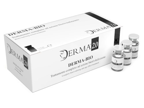
Discover the science behind Derma Bio, a mesotherapy product designed to combat signs of aging and achieve a brighter, smoother, and more youthful appearance.