
Microneedling
Microneedling – also known as percutaneous collagen induction therapy – is a minimally invasive treatment in which fine needles create thousands of controlled micro-injuries in the epidermis and dermis, initiating the body’s intrinsic wound repair cascade. The resulting release of TGF-β, PDGF, and VEGF activates fibroblast migration and stimulates new Type I and III collagen, elastin, and glycosaminoglycan synthesis. The collagen formed integrates as an organised basket-weave lattice that persists for five to seven years. A distinct and clinically significant secondary function is transient dermal microporation – the open microchannels substantially increase skin permeability, enabling direct dermal delivery of topical actives such as polynucleotides, HA preparations, and vitamin C. Microneedling carries an excellent safety profile across all Fitzpatrick skin types and is the most versatile platform treatment in the portfolio for skin quality improvement, scar management, and combination delivery protocols.
Microneedling – formally termed percutaneous collagen induction therapy – is a minimally invasive procedure in which multiple fine needles create controlled micro-injuries to the epidermis and dermis. The treatment does not ablate tissue; it preserves the epidermis whilst creating micro-wounds that initiate the skin’s natural healing cascade, producing new structural proteins and improving skin quality through entirely endogenous biological processes. [2] Device formats range from manual dermarollers (192+ needles in cylindrical arrays) to motorised automated pen devices such as the Dermapen, which allow precise depth control from 0.5mm at the superficial end to 3mm for scar and body applications. The pen format produces a stamping motion that minimises epidermal trauma between needle points compared to rolling – a clinically meaningful difference for safety across darker skin types.
The Wound Healing Cascade
The mechanism begins the moment a needle punctures tissue. The three-phase wound healing response is initiated:
Phase 1 – Inflammation (0–3 days): Micro-injury activates platelets and neutrophils, which release TGF-β (transforming growth factor beta), PDGF (platelet-derived growth factor), and CTAP (connective tissue activation protein) into the wound site. These signals recruit fibroblasts and keratinocytes and establish the initial growth factor environment that drives the subsequent phases. [2]
Phase 2 – Proliferation (3 days – 2 weeks): Fibroblasts migrate to the wounded area and begin producing Type III collagen (early repair collagen), elastin, glycosaminoglycans, and proteoglycans. VEGF drives angiogenesis simultaneously – new capillary formation improves nutrient and oxygen supply to the treated tissue, supporting the sustained synthesis activity that follows. At approximately five days, a fibronectin matrix forms from β-fibroblast arrangement. This fibronectin scaffold determines the spatial pattern of new collagen deposition, and it is this organised matrix that produces the basket-weave collagen architecture characteristic of successfully treated skin rather than the parallel-bundle arrangement of scar tissue. [2]
Phase 3 – Remodelling (2 weeks – 6 months): Type III collagen is progressively converted to structurally stronger Type I collagen via collagenases and matrix metalloproteinases in a controlled remodelling process. The TGF-β3 isoform predominance during this phase specifically suppresses the parallel-fibre arrangement associated with fibrosis, instead directing the organised basket-weave matrix that produces skin quality improvement. The resulting collagen network remains functionally integrated for five to seven years before natural degradation. [2]
The Collagen Timeline
| Phase | Timing | Key Event |
|---|---|---|
| Inflammation | Days 0–3 | TGF-β, PDGF, CTAP release; fibroblast recruitment |
| Proliferation | Days 3–14 | Fibronectin matrix formation; Type III collagen; VEGF-driven angiogenesis |
| Remodelling | Weeks 2–24 | Type III → Type I conversion; basket-weave architecture established |
| Sustained integration | Months 3–6 | Peak collagen and elastin; improvement measurable by profilometry |
| Longevity | 5–7 years | New collagen persists before natural degradation |
Elastin: The Timeline That Requires Explanation
Elastin follows a distinctly different timeline to collagen, and one that is worth understanding for client consultation. A statistically significant decrease in total elastin is measurable at one month post-treatment – an initial degradation of existing disorganised elastic fibres that appears to be a precursor step to new synthesis. By three months, elastin formation has increased significantly above baseline. [2] A client reviewed at four weeks who asks whether their skin feels less elastic is not reporting an adverse event; they are describing the midpoint of a two-phase elastin remodelling process. This is worth communicating at consultation so it does not become an unnecessary concern.
Drug Delivery: The Microchannel Advantage
Beyond its structural collagen effects, microneedling has a second, clinically independent function that is as significant as the wound healing cascade in a treatment portfolio context: transient dermal microporation.
Each needle pass creates microchannels through the stratum corneum – the primary barrier to topical active penetration. These channels remain open for anywhere from a few minutes to several hours depending on needle depth and individual skin characteristics, during which time the skin’s permeability for hydrophilic water-soluble molecules is substantially increased. [2] Actives that would otherwise be blocked by the intact stratum corneum can reach the vascularised dermis directly, bypassing the absorption limitation that constrains topical-only application.
This mechanism is the basis for the microneedling delivery system used with Lumi-PN (topical PDRN applied through microchannels) and Kiara Reju (the device-compatible HA skin booster formulation), both of which are designed specifically for microneedling-assisted dermal delivery rather than standalone topical use. It also makes microneedling the natural platform treatment for vitamin C serum delivery in clients where stratum corneum penetration of ascorbic acid is otherwise limited by concentration and vehicle. The drug delivery function operates in addition to – not instead of – the wound healing cascade; both occur simultaneously within the same treatment session.
Barrier and Filaggrin Effects
A less widely discussed but clinically relevant outcome of microneedling is its positive effect on epidermal barrier function – not simply a downstream consequence of improved dermal structure but a directly measurable upstream change. Controlled micro-injury upregulates transglutaminase-1 (TGM-1), the key enzyme that cross-links filaggrin proteins to form the cornified envelope surrounding corneocytes. [2] Ki-67 (a proliferation marker) and filaggrin expression both increase significantly six days post-treatment in ex vivo models, indicating genuine barrier reconstruction rather than temporary surface improvement. Additionally, upregulation of SPTLC3 – the primary enzyme in ceramide synthesis – increases ceramide concentrations measurably, contributing to improved stratum corneum hydration and reduced TEWL over a treatment course. The initial microporation transiently increases TEWL; by 90 minutes this normalises, and over a treatment course, TEWL improves beyond baseline as dermal and epidermal density increase. [2]
Evidence Base
The evidence for microneedling is established rather than emerging, though quality is variable across indication areas. A 2025 narrative review drawing on 70 peer-reviewed studies (PubMed, Web of Science, Embase) confirmed the core mechanistic pathways – collagen and elastin production, angiogenesis, transient permeability increase, and post-treatment epidermal barrier enhancement – as the principal physiological mechanisms. [2]
Acne scarring carries the strongest evidence base. Eight RCTs comparing microneedling to fractional laser resurfacing, PRP, and TCA-CROSS found comparable clinical outcomes with a meaningfully lower risk of side effects – specifically, less post-inflammatory hyperpigmentation than ablative alternatives and safe use across FST I–VI. Icepick and boxcar scars respond more reliably than rolling scars; a multi-patient series found 80% of participants showed 50–75% scar improvement after an average of 2.5 sessions.
Photoaging shows consistent benefit across profilometry, elasticity measurement, and epidermal density assessment. Monthly sessions produce significant reductions in wrinkle depth and improvement in skin tightness; improvements accumulate with successive sessions and are measurable at three months post-treatment completion. [2]
Hair loss: Microneedling combined with minoxidil consistently outperforms minoxidil alone in evidence-based comparison, through two routes – enhanced transdermal minoxidil delivery through microchannels, and growth factor stimulation in the perifollicular environment. This is one of the few genuinely synergistic device-drug pairings in aesthetics practice with consistent controlled evidence.
Stretch marks show 55% moderate-to-excellent improvement after three to four sessions at four-week intervals, comparable to laser therapy with fewer adverse effects.
Microneedling vs RF Microneedling
Standard microneedling and RF microneedling share the wound healing cascade but diverge in every other clinically meaningful dimension. A 2025 Seoul National University split-face RCT (n=29, female, age ≥60) – the first large prospective head-to-head comparison – found that MNRF produced statistically significant improvements in wrinkle parameters, gross elasticity, hydration, and TEWL at both three and five months; standard microneedling reached significance in most parameters only at five months, and improvements were consistently less pronounced. [1] Critically, MNRF significantly reduced the proportion of p16^INK4A^-positive senescent fibroblasts in the dermis – replacing them with newly proliferating non-senescent fibroblasts – whilst standard microneedling did not produce this effect. The correlation between senescent fibroblast reduction and collagen density improvement in MNRF responders suggests this is a mechanism difference with direct outcome consequences, not just an incremental improvement. Standard microneedling’s advantage remains its drug delivery function, its excellent FST I–VI safety profile, and its appropriate positioning for skin quality maintenance, scarring, and combination delivery – with RF microneedling as the escalation pathway when structural laxity, more significant photoageing, or senescent fibroblast burden becomes the primary clinical concern.
Clinical Application
Microneedling occupies a well-defined and genuinely useful position in the Creative Touch treatment portfolio, but that position is most productive when it’s understood accurately: microneedling is a skin quality, texture, and scar treatment, and a drug delivery platform. It is not primarily a structural laxity treatment, and recommending it for clients whose primary concern is significant laxity, volume loss, or deep structural collagen decline is setting them up for a result that won’t match what they came in hoping for. The honest positioning serves both the client and the clinic’s outcomes.
What microneedling does exceptionally well is restore skin quality – improving texture, reducing pore visibility, addressing early photodamage, managing scar appearance, and providing the microchannel delivery route that makes certain active combinations significantly more effective than they would be topically. For clients in the skin quality maintenance category, it is one of the most complete single-session tools in the portfolio: one treatment simultaneously stimulates collagen synthesis, delivers topical actives to the dermis, and progressively improves barrier integrity over the treatment course.
Who benefits most
Clients with acne scarring – particularly icepick and boxcar subtypes – where microneedling represents a genuinely evidence-based first-line intervention with an excellent safety profile across all skin tones. For FST IV–VI clients specifically, where laser options carry PIH risk, microneedling offers comparable scar improvement outcomes without the pigmentation risk that would follow ablative treatment.
Younger clients (late 20s–40s) with early photoageing and skin quality concerns. Before structural decline accumulates, microneedling addresses the texture, pore, and quality changes that clients notice first. It is a proportionate intervention for a proportionate concern – and one that builds the collagen foundation that delays the need for more intensive structural treatments.
Clients requiring hair restoration support. Scalp microneedling + minoxidil is the evidence-based combination for androgenic hair loss support, producing measurably superior outcomes to minoxidil alone. Adding Lumi-PN via microneedling during scalp treatment sessions layers PDRN cellular support onto the drug delivery and growth factor stimulation the treatment is already generating.
Clients in active combination protocols where microneedling’s drug delivery function is being used deliberately: Lumi-PN sessions for PN delivery without injection, Kiara Reju for HA dermal absorption, or vitamin C at clinical concentrations. In these applications, the microneedling device is as much a delivery platform as a standalone treatment, and that should be communicated as part of the treatment rationale.
Setting expectations honestly
The timeline is gradual and requires managing. Visible improvement in skin quality – texture, luminosity, overall freshness – begins around three to four weeks after the first session as collagen synthesis gains momentum. The full cumulative effect of a course builds across three to five sessions at four-to-six week intervals, with peak improvement measurable at three to six months after the final session. Acne scarring requires the same patience: initial softening of edges occurs within the first two sessions, but meaningful reduction in scar depth is a three-to-six month outcome.
There is one timeline nuance worth covering at consultation specifically: elastin may initially decrease before it increases. A client who notices their skin feels slightly less elastic at their four-week review is not responding badly to treatment – they are in the degradation phase that precedes new elastin formation, which will be measurably improved by month three. Setting this expectation avoids an unnecessary concern that might otherwise undermine confidence in the treatment course.
Where the combinations are most productive
Microneedling + Lumi-PN is the most complete non-injectable combination for skin quality and texture improvement. The microneedling wound healing cascade and the PDRN regenerative signal are additive: one drives synthesis through the growth factor cascade; the other drives it through the A2AR–macrophage axis. Both are active simultaneously in the same session. This is also the natural choice for clients who want the regenerative benefits of polynucleotides but prefer to avoid injections.
Microneedling + iPRF delivers growth factors directly into the dermal microchannels at maximum bioavailability – PRP and iPRF applied during or immediately after needling reach concentrations in the dermis that topical-only application cannot achieve. The timing matters: apply during needling or within the brief open-channel window, not after barrier recovery has begun.
Microneedling + polynucleotides (injectable) – the microneedling session provides the wound healing stimulus and the drug delivery function; the concurrent injectable PN course provides the sustained MMP-suppressive and tissue environment benefits that outlast the treatment session. These are additive rather than overlapping: one session can realistically combine both for a comprehensive protocol.
Microneedling as RF microneedling preparation. For clients who have not previously had energy-based treatments, a course of standard microneedling first – improving skin quality, reducing the inflammatory burden, and establishing a baseline collagen synthesis response – produces a more responsive dermal environment for RF microneedling when that escalation becomes appropriate.
The Protocol Reference
| Treatment Goal | Recommended Approach | Combination Logic |
|---|---|---|
| Skin quality / texture | 3–5 sessions, 4–6 week intervals | + Lumi-PN or Kiara Reju same session |
| Acne scarring | 4–6 sessions, 4–6 week intervals | + subcision for rolling scars if needed |
| Hair restoration | 4–6 scalp sessions, 2–4 week intervals | + minoxidil; + Lumi-PN for PDRN delivery |
| PN delivery (needle-phobic) | 3–4 sessions with Lumi-PN | Replaces injectable PN where appropriate |
| Pre-RF microneedling preparation | 2–3 sessions, 4-week intervals | Optimise skin quality before escalating |
| Recovery-phase support | Single session post-intensive treatment | Kiara Reju or HA for barrier + hydration support |
References
Hwang JM, Lee SH, Baek EJ, et al. (2025). Comparison of the effects of fractional microneedle radiofrequency and microneedling on modulating the senescent fibroblast milieu in aged skin. Sci Rep, 15(1), 18296 . doi.org/10.1038/s41598-025-02545-3
Tehrani L, Tashjian M, Mayrovitz HN (2025). Physiological Mechanisms and Therapeutic Applications of Microneedling: A Narrative Review. Cureus, 17(3), e80510 . doi.org/10.7759/cureus.80510
Also Known As
- collagen induction therapy
- dermarolling
- micro-needling
- percutaneous collagen induction therapy
- skin needling
Therapeutic Relationships
Therapeutic Context
- Stimulates Collagen Evidence: Microneedling wound cascade releases platelet/fibroblast TGF-beta1, driving procollagen I and III synthesis over subsequent weeks; collagen response documented in biopsy studies (PMC11497551; Collagen entity).
- Stimulates Corneocyte
- Stimulates Dermal Papilla Evidence: Scalp microneedling stimulates perifollicular growth factor release (VEGF, PDGF, TGF-β) in dermal papilla microenvironment, augmenting follicular anagen signalling. PMC11890238.
- Stimulates Elastin Evidence: Microneedling wound healing cascade supports tropoelastin transcription via TGF-beta signalling as part of general repair response. PMC11965193.
- Stimulates Filaggrin Evidence: Ex vivo: statistically significant increase in filaggrin expression at day 6 post-microneedling alongside Ki-67 and TGM-1, indicating barrier protein reconstruction. PMC11993440.
- Stimulates Hair follicle Evidence: Scalp microneedling upregulates growth factors in follicular environment and enhances transdermal minoxidil delivery via microchannels; 12 RCTs SMD 1.32 hair count. PMC11890238.
- Stimulates Tissue regeneration Evidence: Microneedling creates a controlled wound-healing cascade including platelet activation, TGF-beta1 release, and fibroblast recruitment; the tissue remodelling response produces new collagen and ECM, constituting a regenerative response (PMC11497551).
- Stimulates Vascular endothelial growth factor Evidence: VEGF expression increases during wound healing proliferative phase initiated by microneedling, driving angiogenesis and new capillary formation in treated dermis. PMC11993440.
- Inhibits Cellular senescence Evidence: 2025 Seoul National University split-face RCT (n=29): RF microneedling significantly reduced p16INK4A-positive senescent fibroblasts; standard MN noted comparatively. PMC12106790.
- Inhibits Transepidermal water loss Evidence: TEWL transiently increases post-procedure but decreases below baseline over course as SPTLC3-driven ceramide synthesis and epidermal density improve. PMC11993440.
- Treats Androgenetic alopecia Evidence: Meta-analysis 12 RCTs n=631: microneedling+minoxidil significantly improved hair count (SMD 1.32) and diameter vs minoxidil alone in AGA. PMC11890238, 2025.
- Treats Hyperpigmentation Evidence: Review of 64 studies confirmed microneedling reduces melanin density and enhances depigmenting active delivery in melasma/hyperpigmentation; significant MASI reductions. PMC11680975, 2024.
- Treats Perimenopausal skin changes Evidence: Microneedling explicitly positioned for perimenopausal skin quality/texture treatment in entity text; collagen induction and drug delivery rationale for post-menopausal protocol. Entity text PMC11993440.
- Treats Rosacea Evidence: Fractional microneedling RF reduced erythema index 13.6% and suppressed rosacea inflammatory/angiogenesis markers; standard MN used for rosacea management. PMID 27608206; PMID 39606600.
- Treats Skin ageing Evidence: 2025 review of 70 studies confirmed microneedling improves profilometry, elasticity, and reduces wrinkle depth; established mechanism for photoageing treatment. PMC11993440.
- Treats Telogen effluvium Evidence: Microneedling wound-healing cascade (TGF-β, PDGF, VEGF) supports hair follicle anagen re-entry in telogen effluvium; documented in hair loss protocols. PMID 40056230, 2025.
- Affects Melanocyte Evidence: Microneedling reduces melanin density and corrects prolapsed melanocytes in melasma, normalising melanocyte function and distribution. Review 64 clinical studies PMC11680975.
- Affects Scalp Evidence: Scalp microneedling creates microchannels enabling drug delivery (minoxidil, PDRN) and stimulates perifollicular growth factor environment for hair restoration. PMC11890238.
- Requires Skin Evidence: Microneedling requires intact skin as operational substrate; fine needles create micro-injuries specifically in epidermis and dermis to initiate wound healing cascade. Entity text.
Indications & References
- this Dermal Papilla Evidence: Microneedling channels facilitate growth factor delivery to DP level (1-1.5mm depth); entity text describes this as mechanistic rationale. Kim et al. (2016) Ann Dermatol 28(5):586. doi:10.5021/ad.2016.28.5.586
- this Dermis Evidence: Superficial microneedling… primarily acts at the papillary level, targeting the uppermost dermal remodelling zone
- this Fibroblast Evidence: Microneedling activates fibroblasts through the controlled injury cascade
- this Hair follicle Evidence: Microneedling stimulates follicular Wnt/beta-catenin and VEGF pathways; promotes hair growth. Kim et al. (2016) Ann Dermatol 28(5):586 demonstrate Wnt3a upregulation. doi:10.5021/ad.2016.28.5.586
- this Keratinocyte Evidence: Microneedling activates the keratinocyte differentiation gene programme through the wound-healing response, with gene expression analysis documenting upregulation across cornification
- this Skin Evidence: Microneedling activates keratinocyte differentiation cascade; addresses corneocyte quality from production end.
- this Stratum corneum Evidence: Microneedling named as a treatment activating the keratinocyte differentiation cascade addressing corneocyte quality in the stratum corneum (clinical_context_summary).
- this May treat Perimenopausal skin changes Evidence: Microneedling addresses skin quality, texture, drug delivery; explicitly positioned for perimenopausal skin maintenance; proportionate intervention for early-phase structural support. Entity text.
Learn More
This topic is discussed in 6 articles:
-

Fine lines and wrinkles? Say hello to your skin’s new BFFs: microneedling and RF microneedling! Discover the difference and find your perfect treatment at Creative Touch.
-

Creates controlled micro-injuries stimulating collagen and barrier repair responses. For use after barrier stabilisation, not during active damage.
-

A minimally invasive skin treatment using fine needles to create controlled micro-injuries that trigger the body’s wound healing cascade, stimulating collagen and elastin synthesis.
-

Elevate your autumn skincare routine with our expert advice and innovative treatments: achieve a radiant complexion as the seasons change and the leaves fall.
-
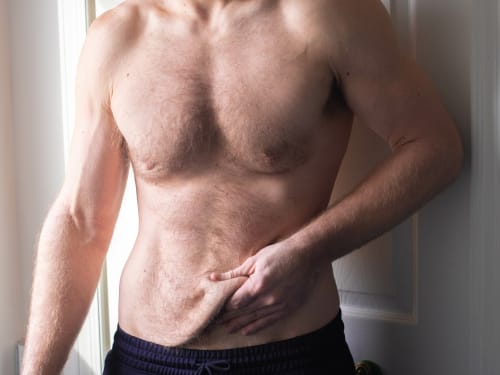
Don’t let loose skin overshadow your weight loss success. Learn why skin sags after significant weight loss and explore our advanced skin tightening and volume restoration treatments.
-
The science behind polynucleotides – a revolutionary treatment that offers a comprehensive approach to skin rejuvenation. How does it compare?