
Dermis
The dermis is the skin’s structural engine, organised into two functional zones: the papillary dermis (DEJ integrity) and the reticular dermis (volumetric strength). Dermal ageing is a triad of net collagen loss, solar elastotic accumulation, and ground substance depletion. Effective rejuvenation requires zone-specific targeting: restoring the “slack” mechanical environment of the deep reticular dermis while repairing the signalling interface of the superficial papillary zone.
The dermis is the structural core of the skin, lying beneath the dermo-epidermal junction and extending down to the subcutaneous tissue. Far thicker than the epidermis – ranging from approximately 1mm on the eyelid to 4mm on the back – it provides the mechanical support, vascular supply, and cellular machinery that determines how skin ages, how it responds to treatment, and how completely it recovers from either. For aesthetics purposes, the dermis is where most of the clinically significant changes of chronological and hormonal ageing occur, and it is the primary target of the majority of professional skin rejuvenation treatments.
Two Zones, Two Treatment Targets
The dermis is not uniform in structure, and the distinction between its two zones has direct practical relevance for treatment selection.
The papillary dermis sits immediately beneath the epidermis, comprising loosely arranged collagen and elastic fibres, a rich capillary network, and a higher concentration of the proteoglycan ground substance. It is the papillary dermis that forms the rete ridge interdigitations with the overlying epidermis, creating the structural interface through which nutrient and signalling exchange occurs. Age-related flattening of these rete ridges reduces surface area contact between the two layers, impairing nutrient delivery to basal keratinocytes and reducing the mechanical anchoring of the epidermis to its substrate. Superficial professional treatments – thulium fractional laser, superficial microneedling, and CAP – primarily act at the papillary level, targeting the uppermost dermal remodelling zone whilst preserving the reticular structure below. [7]
The reticular dermis is the deeper, thicker zone containing the densely packed collagen fibre bundles, the major elastic fibre network, and the larger vessels that supply the papillary capillary network. Its greater structural depth means it is less accessible to topical treatments but is the zone where the most mechanically significant collagen and elastin changes of ageing occur. RF microneedling, skin boosters delivered at mid-dermal depth, and deep polynucleotide injection protocols target this zone specifically, delivering their thermal, mechanical, or biochemical signals where the structural remodelling opportunity is greatest. [6]
Fibroblasts: The Dermal Engine
The dominant functional cell of the dermis is the fibroblast, responsible for synthesising and remodelling all major extracellular matrix components: collagen Types I, III, and V, elastin, fibronectin, and the glycosaminoglycans of the ground substance. Fibroblasts are not a uniform population. Research has demonstrated that papillary, reticular, and perifollicular fibroblast subpopulations have distinct gene expression profiles and respond differently to ageing and to treatment stimuli. Papillary fibroblasts are more proliferative, more responsive to TGF-β, and more active in their crosstalk with overlying keratinocytes; reticular fibroblasts produce the bulk of the structural collagen and elastin. [3]
Fibroblast activity declines with chronological ageing through several converging mechanisms: reduced TGF-β responsiveness, progressive cellular senescence, decreased cell density, and accumulation of mechanically fragmented collagen in the extracellular environment. This last mechanism is particularly relevant: fibroblasts are mechanosensitive cells that respond to physical tension in the surrounding matrix, and when collagen degradation creates a physically slack environment, fibroblast gene expression shifts away from synthesis, even in otherwise healthy cells. This is one of the mechanisms through which collagen loss becomes self-reinforcing: degraded matrix reduces the physical signal that would stimulate replacement production. [3]
The Fibroblast Phenotype (Cellular Logic)
| Feature | Papillary Fibroblasts | Reticular Fibroblasts |
|---|---|---|
| Growth Rate | High (Proliferative) | Low (Slower turnover) |
| Main Output | Type III Collagen, Decorin, Tenascin-C | Type I Collagen, Versican |
| Crosstalk | Strong signals to Keratinocytes (Epidermal Health) | Mostly ECM-focused (Structural Strength) |
| Wound Healing | Rapid “First Responders” | Slow, deliberate “Structural Remodellers” |
| Clinical Value | Governs glow, texture, and epidermal thickness. | Governs “bounceback,” lift, and facial contour. |
The Ground Substance
Alongside collagen and elastin, the dermis contains a gel-like ground substance composed of glycosaminoglycans (GAGs), proteoglycans, and glycoproteins. Hyaluronic acid is the most clinically relevant GAG: it holds water at up to 1,000 times its own weight, contributing to the skin’s hydrated volume, turgor, and the tissue environment in which fibroblasts function. Hyaluronic acid content in the dermis declines with age – post-menopausal skin demonstrates measurably reduced dermal HA compared with premenopausal skin – and this decline contributes both to visible volume loss and to the less hydrated, more mechanically vulnerable fibroblast microenvironment that further reduces synthesis activity. [17]
Versican, decorin, and fibromodulin are the principal dermal proteoglycans, with decorin specifically involved in regulating the lateral growth and organisation of collagen fibrils. Ultraviolet exposure degrades decorin, which disorganises the collagen fibril architecture even before the collagen itself is degraded by MMPs. This provides a mechanistic explanation for why photoaged collagen looks disorganised and coarse rather than simply reduced: the assembly-regulating proteoglycan is damaged before or alongside the structural protein. [9]
The Dermo-Epidermal Junction
The dermo-epidermal junction (DEJ) is not a passive boundary but a functionally active structure containing Type IV and Type VII collagen, laminin, nidogen, and the hemidesmosomal anchoring complexes that attach the basal keratinocytes of the epidermis to the dermal substrate. The DEJ mediates the bidirectional signalling between dermis and epidermis – including the fibroblast-to-keratinocyte TGF-β signals and the keratinocyte-to-fibroblast integrin signals described in the collagen and epidermis entities. Age-related changes at the DEJ include Type VII collagen reduction, reduced anchoring fibril density, and the flattening of the rete ridge architecture – all of which reduce epidermal stability and impair the cellular communication that sustains both keratinocyte differentiation quality and fibroblast activity. [5]
For treatments targeting dermal rejuvenation, the DEJ represents both a structural restoration target in its own right and the interface through which dermal treatment effects communicate upward to improve epidermal renewal quality.
How the Dermis Ages
Dermal ageing operates through three compounding mechanisms that feed back into each other:
Net collagen loss, driven by the interplay of reduced synthesis and increased MMP-driven degradation described in the Collagen entity. The Type I:III ratio shifts as Type III production declines disproportionately, progressively reducing the fibroblast activation feedback that hybrid fibrils provide. Collagen density decreases by approximately 1% per year from early adulthood, accelerating after menopause. [3] PLLA addresses net collagen loss through the longest horizon of any treatment in this group. Its M2 macrophage polarisation and direct PI3K/AKT fibroblast activation build new Type I and III collagen over months, with durability of 24–36 months reflecting genuine architectural restoration rather than the presence of an implanted material. Most appropriately positioned for clients with significant structural collagen decline where a gradual, durable outcome is the clinical goal. [1]
Elastic fibre fragmentation, through MMP-12-driven degradation of functionally intact fibres and accumulation of non-functional solar elastotic material in photodamaged skin. The fibrillin scaffold that would support new tropoelastin assembly degrades alongside the elastin, progressively impairing the skin’s recoil and mechanical resilience. [2]
Ground substance depletion, in which declining HA, decorin, and proteoglycan content reduces both the visible volume and the tissue environment that fibroblasts require for normal activity. The mechanical slack created by HA loss compounds the mechanosensitive signal reduction already occurring from collagen degradation. [17]
These three processes are not independent; they share upstream drivers in UV exposure, cortisol elevation, oestrogen decline, and the downstream consequences of barrier dysfunction. Perimenopause represents the clinical convergence point at which hormonal, chronological, and often cumulative environmental damage drivers arrive simultaneously, which explains the frequently reported experience of skin quality changing more rapidly in the 40–55 age range than the preceding decades. [15]
The Elastin Paradox (Aging Mechanics)
| Feature | Papillary (Oxytalan Fibres) | Reticular (Mature Elastic Fibres) |
|---|---|---|
| Orientation | Perpendicular (Vertical) | Parallel (Horizontal) |
| Aging Pattern | Depletion: Fibres break and disappear. | Accumulation: “Solar Elastosis” (toxic buildup). |
| Visual Result | Fine “paper-like” wrinkles, fragile surface. | Deep, coarse “leathery” wrinkles; loss of recoil. |
| Treatment Goal | Restore: Build new anchoring fibres. | Clear & Rebuild: Remove “clogged” elastin via heat. |
| Key Modality | Thulium, Polynucleotides, Tretinoin. | RF Microneedling (Thermal clearance). |
Dermal Layer Targeting
| Feature | Papillary Dermis (Superficial) | Reticular Dermis (Deep) |
|---|---|---|
| Primary Architecture | Fine collagen (Type III), loose fibers, high HA content. | Thick collagen (Type I) bundles, dense elastic network. |
| Key Cell Response | Rapid proliferation, high TGF-β sensitivity, DEJ crosstalk. | Massive ECM synthesis, structural remodeling, slower turnover. |
| Clinical Presentation of Decline | Fine lines, loss of “glow,” thinning at the surface, pigment shifts. | Sagging, deep folds, loss of “bounce” (recoil), structural thinning. |
| Aesthetic Goal | Texture refinement, pore reduction, DEJ “anchoring.” | Volumetric lift, structural tightening, dermal “thickness.” |
| Best-in-Class Treatments | Thulium Laser, Superficial Microneedling, CAP. | RF Microneedling, Deep Polynucleotides, Skin Boosters. |
The Dermis in the Structural Cascade
The dermis as visible endpoint of a multi-layer process
Intrinsic dermal biochemistry – collagen turnover, elastin network integrity, ground substance volume – determines how skin behaves in isolation. But the topography the dermis drapes over is not static. Beneath it, subcutaneous fat compartments atrophy, migrate, and descend; beneath them, the skeletal framework remodels. The dermis does not change in isolation: it is continuously reshaped by what happens in the layers supporting it.
Rohrich & Pessa (2007) demonstrated through cadaveric dissection that the subcutaneous fat of the face is partitioned into multiple independent anatomical compartments – the nasolabial fat, medial/middle/lateral malar fat, orbital fat, and jowl fat – each with discrete septal boundaries, each ageing at different rates and in different directions. Shearing between adjacent compartments as boundaries weaken is proposed as a mechanism of soft-tissue malposition: the dermis does not simply “fall” as a composite sheet, but is displaced heterogeneously as the architecture it overlies disorganises. [12]
Deeper still, Shaw & Kahn (2007) used three-dimensional CT volumetric reconstruction in a cohort of 60 subjects across three age decades to demonstrate significant age-related decreases in the glabellar and maxillary angles – objective evidence that the bony scaffold of the midface undergoes measurable dimensional change with age. An increasing pyriform aperture area in the transition from youth to middle age reflects progressive maxillary retrusion. [13] Mendelson & Wong (2012) synthesised the clinical implications from the structural anatomy literature: midface maxillary resorption, superomedial and inferolateral orbital rim changes, and prejowl mandibular resorption each occur in specific, predictable patterns that alter the skeletal foundation the overlying soft tissue depends upon – and that soft-tissue lifting and redraping alone cannot address. [11]
The practical implication for the dermis is this: the topographic contour the dermis occupies is the sum of its intrinsic biochemical status and the structural integrity of every layer beneath it. A dermis that has lost ground substance volume, collagen density, and elastic recoil will present very differently if it rests over a preserved skeletal and fat-compartment scaffold than if it rests over a depleted one. Surface skin quality – texture, reflectivity, fine line depth – is primarily a dermal property. Surface topography – the position of the nasolabial fold, the descent of the jowl, the depth of the tear trough – is a whole-architecture property in which the dermis is the visible endpoint, not the origin of change.
The SMAS relationship and the significance of injection depth
The dermis lies superficial to the subcutaneous fat, which in turn overlies the superficial musculoaponeurotic system (SMAS). The SMAS is a continuous fibromuscular layer that connects the facial mimetic muscles, separates the subcutaneous fat above from the deep facial fascia below, and provides the mechanical framework for facial expression. Its anatomical relationships directly govern where injectable agents act, and at what risk.
The facial soft-tissue architecture is conventionally described as a series of concentric layers: skin, subcutaneous fat, superficial facial fascia (SMAS), mimetic muscle, deep facial fascia (parotidomasseteric fascia), and the plane containing the facial nerve branches, parotid duct, and buccal fat. The key architectural rule with clinical significance is the relationship between the facial nerve and the deep facial fascia: in the cheek, facial nerve branches lie deep to the parotidomasseteric fascia, not within the SMAS or above it. The nerves, blood vessels, and lymphatics that transition between deep and superficial fascia do so via anatomical fusion zones between the two fascial layers – providing the basis for understanding which vascular structures are accessible at different injection depths.
For injectable treatments, injection depth is therefore not a stylistic choice but a safety and mechanism determinant. The superficial surface of the SMAS carries axial blood vessels and cutaneous sensory nerves supplying the overlying skin; the deep surface of the SMAS is where motor branches of the facial nerve approach the deep surface of the mimetic muscles they innervate. Injecting at the subdermal plane (below dermis, above SMAS) targets the subcutaneous fat compartments – appropriate for structural fillers requiring deep volumetric support. Injecting within the dermis itself (intradermal plane) targets the dermal matrix – appropriate for skin quality agents including low-viscosity hyaluronic acid skin boosters, polynucleotides, and superficial biostimulators. Injecting below the SMAS carries increased risk of encountering motor nerve branches and is generally reserved for specific surgical indications.
The dermis therefore occupies the shallow margin of the injectable target zone. Correctly distinguishing intradermal from subdermal injection – and subdermal from sub-SMAS – is the anatomical literacy that underpins both treatment efficacy and patient safety.
Why dermal-targeted treatment alone is insufficient in advanced ageing
Biostimulatory treatments – radiofrequency microneedling, polynucleotides, collagen-stimulating fillers such as poly-L-lactic acid, energy-based treatments – address intrinsic dermal mechanisms: collagen neosynthesis, elastin remodelling, fibroblast activation, ground substance restoration. These are legitimate, evidence-supported targets. However, they act within a tissue that sits atop an architecture.
When significant skeletal resorption has reduced the structural support beneath the midface, or when fat compartment descent has altered the surface topography in ways unrelated to dermal laxity, restoring the dermis’s intrinsic biochemistry will improve skin quality – texture, hydration, reflectivity – without necessarily addressing the topographic change. The nasolabial fold deepens not primarily because dermal collagen fails (though it does), but because the medial cheek fat descends as its supporting boundaries weaken, and because the maxillary skeleton that once provided forward projection retreats – a clinical synthesis of the structural anatomy literature. [11] The dermis in that region has acquired a new topographic position; restoring its biochemistry does not reposition it.
This is the rationale for combining intrinsic dermal treatments with structural support. Volumising fillers placed at the subdermal or supraperiosteal plane restore the skeletal and compartmental architecture the dermis drapes over. Biostimulators work on the dermal tissue itself. Energy-based treatments tighten the dermis and the SMAS. These are complementary, not redundant: each addresses a distinct layer of a multi-layer structural process. The most clinically effective approach to advanced facial ageing treats the architecture as a whole – and that requires understanding the dermis not as the problem, but as the endpoint of a cascade that begins far beneath it.
Clinical Application
The description of the dermis establishes three distinct compounding mechanisms of dermal ageing: net collagen loss, elastic fibre fragmentation and solar elastotic accumulation, and ground substance depletion. Most clinical conversations about professional skin rejuvenation treatments focus on the first mechanism only. The most complete dermal rejuvenation protocols address all three, and the treatments available at Creative Touch address different subsets of this triad through substantially different mechanisms. Knowing which combination a client most needs determines the protocol, not the treatment menu alone.
Restoring the Ground Substance: The Underaddressed Mechanism
Ground substance depletion – declining hyaluronic acid, decorin, and proteoglycan content – is the dermal ageing mechanism most consistently underweighted in aesthetics conversations yet most directly addressable with clinical treatment. It matters for two reasons beyond simple hydration or volume.
First, fibroblasts are mechanosensitive: they respond to the physical tension of their surrounding matrix environment, not merely to growth factor signals. When HA loss creates a mechanically slack extracellular environment, fibroblasts reduce their collagen and elastin synthesis even when their TGF-β responsiveness is otherwise intact. Restoring the physical tension of the dermal matrix through HA delivery is not simply cosmetic volume replacement; it reinstates part of the mechanical signalling environment that drives fibroblast synthesis activity. [3]
Second, decorin degradation by UV-induced MMPs disorganises collagen fibril architecture before the collagen itself is quantitatively depleted. Photoaged dermis loses its structural organisation through proteoglycan damage before the collagen loss that follows. Treatments that reduce MMP activity protect the organisational infrastructure of the dermal matrix, not just the structural proteins themselves. [9]
Skin boosters address the HA ground substance deficit directly. Injected cross-linked HA delivered at mid-dermal depth provides the volumetric and mechanical support that restores fibroblast tension, with the mechanoreceptive TGF-β activation detectable within one week and the resulting procollagen I synthesis measurable in biopsy tissue at four weeks. The fibroblast activation is sustained for six to nine months whilst the HA remains structurally integrated, giving skin boosters a fibroblast activation timeline considerably longer than any surface treatment. For clients where ground substance depletion is the dominant clinical presentation – fine crepey texture, reduced turgor, skin that lacks structural depth without obvious surface lines – this is the mechanistic gap that skin boosters fill that no other treatment addresses directly. medizinonline
Two-Zone Targeting: Papillary and Reticular Dermis
The two-zone structure of the dermis provides a practical framework for understanding why different treatments produce different clinical outcomes even when they are all described as “stimulating collagen.”
Superficial treatments at the papillary level include thulium fractional laser, superficial microneedling, and cold atmospheric plasma. These activate fibroblasts in the papillary zone, stimulate DEJ and rete ridge architecture at the epidermal interface, and produce the surface quality, texture, and tone improvements that are the most immediately visible treatment outcomes. Thulium’s fractional MTZ mechanism, described in the epidermis clinical context, reaches the papillary dermis in addition to its primary epidermal target, producing collagen deposition and DEJ remodelling at the boundary zone between the two layers. [7]
Deeper treatments at the reticular level include RF microneedling, polynucleotide injection at depth, and skin boosters. These reach the structurally significant collagen and elastin reserves in the reticular dermis, the zone where volumetric loss and mechanical laxity originate. RF microneedling delivers radiofrequency thermal energy from the needle tips into the reticular dermis, creating controlled collagen denaturation at the point where new fibril formation in the remodelling response produces the most meaningful structural benefit. This reticular depth separates RF microneedling from non-RF microneedling in terms of the laxity and structural depth outcomes it can achieve. [10]
iPRF: Systemic Growth Factor Delivery Across Both Zones
iPRF contributes to dermal rejuvenation across both zones simultaneously through the distribution of growth factors from injected platelet concentrate. TGF-β, PDGF, VEGF, and IGF-1 diffuse through the dermis from the injection sites, activating fibroblast populations in both papillary and reticular zones. The TGF-β/Smad pathway activation drives procollagen synthesis whilst MMP-1 expression is simultaneously suppressed, addressing both the synthesis and degradation mechanisms of collagen loss together. [8]
The fibronectin co-production demonstrated with iPRF is specifically relevant to the dermal matrix system: fibronectin acts as an assembly scaffold for the early stages of elastic fibre formation alongside its role in cell adhesion and migration. iPRF therefore contributes to the ground substance matrix environment and the elastic fibre assembly pathway in addition to its collagen synthesis effects. For perimenopausal clients where all three dermal ageing mechanisms are active simultaneously, iPRF’s multi-target growth factor delivery addresses the broadest range of dermal deficit of any single injectable treatment. [16]
Polynucleotides: The Dermal Senescence Problem
The macrophage-fibroblast axis through which polynucleotides enhance collagen synthesis – described in detail in Collagen – takes on additional significance in the dermal context because of the senescent fibroblast population. Aged and post-menopausal dermis contains an increasing proportion of senescent fibroblasts that have lost responsiveness to direct TGF-β stimulation and have reduced capacity to undergo the proliferative response to growth factor delivery that iPRF depends on. [4]
Polynucleotide treatment’s indirect route through macrophage M2 polarisation and IL-10/TGF-β secretion bypasses the impaired direct responsiveness of senescent fibroblasts, activating SMAD2 and STAT3 in the fibroblast even when direct receptor-mediated activation is diminished. This makes polynucleotides specifically valuable for older or significantly photo-aged dermis where the proportion of responsive fibroblasts has fallen. They are not a replacement for direct fibroblast stimulators but an adjunct that expands the responsive population beyond what direct growth factor delivery can reach. [4]
The NF-κB suppression from polynucleotide treatment simultaneously reduces MMP-driven collagen and elastin degradation and reduces the inflammatory environment that is suppressing decorin and HA synthesis – providing coverage across all three dermal ageing mechanisms through a single treatment, albeit indirect across each of them.
Treatment Pairings for the Complete Dermal Protocol
The three-mechanism framework provides the logic for treatment pairing:
For collagen loss + ground substance depletion (the most common perimenopausal presentation): Skin boosters (restoring HA-mediated fibroblast mechanosensitivity and ground substance) combined with iPRF (TGF-β/Smad collagen synthesis and MMP-1 suppression). Skin boosters create the restored matrix environment in which the fibroblasts that iPRF activates can respond most fully. The mechanical tension signal and the growth factor signal together produce a more complete synthesis response than either alone. Polynucleotides added at interval between sessions extends the period of MMP suppression and activates the senescent fibroblast population between treatment peaks.
For photoaged dermis with all three mechanisms active (collagen loss + solar elastosis + ground substance): RF microneedling primary (reticular zone thermal remodelling, collagen and elastin remodelling including removal of solar elastotic material) combined with iPRF in the same session (growth factor coverage across both zones). Polynucleotides in preparatory or interval sessions reduce the MMP-elastase burden before thermal treatment, improving the matrix environment into which new collagen and elastin formation occurs. LED post-procedure accelerates the fibroblast-keratinocyte proliferative phase, shortening the recovery timeline.
For senescent or post-menopausal dermis with reduced treatment responsiveness: Polynucleotides as a preparatory treatment series, reprogramming the macrophage environment and expanding the responsive fibroblast population over four to six weeks before introducing the direct synthesis-stimulating treatments (RF microneedling, iPRF) that depend on fibroblast responsiveness for their effect. This sequencing is the dermal equivalent of the filaggrin clinical context’s “resolve the inflammatory suppression first” principle: preparing the cellular environment for the more intensive treatments that follow.
The homecare layer: The dermal treatment effects described above all operate in a tissue environment continuously modified by UV exposure, cortisol levels, and oestrogen status between clinic sessions. Topical vitamin C reduces the ROS-driven AP-1 MMP activation that degrades both the new collagen being formed and the decorin organising it. Retinoids reduce MMP expression and upregulate TGF-β signalling in fibroblasts. Broad-spectrum UV protection prevents the compounding MMP-12 elastin damage and MMP-1 collagen fragmentation that erode dermal treatment outcomes between sessions. For clients investing in dermal rejuvenation protocols, homecare is the maintenance environment, not a separate, lesser category of treatment. [14]
Zone-Specific Clinical Mapping
| Presentation | Primary Zone | The Problem | The Priority Modality |
|---|---|---|---|
| “Crepey” surface | Papillary | DEJ flattening & Oxytalan loss. | Thulium / Superficial Microneedling. |
| “Leathery” skin | Reticular | Solar Elastosis accumulation. | RF Microneedling (Heat-driven). |
| “Empty/Hollow” skin | Reticular | Ground substance & Type I loss. | HA Skin Boosters. |
| “Reactive/Dull” skin | Papillary | Keratinocyte crosstalk failure. | CAP / Polynucleotides. |
The cascade framing has direct pre-treatment assessment implications. When a patient presents with dermal thinning and laxity, the clinical question is whether the primary deficit is intrinsic to the dermis – reduced fibroblast activity, ground substance depletion, collagen turnover decline – or whether dermal thinning is compounded by structural support failure in the layers beneath. These two presentations can appear similar superficially but call for different primary interventions. A dermis that has lost intrinsic volume and resilience in the context of a preserved skeletal and fat-compartment scaffold responds well to biostimulatory treatment: polynucleotides, radiofrequency microneedling, skin boosters, and collagen-stimulating agents all work by restoring dermal biology from within. A dermis that has thinned and descended because the fat compartments beneath it have atrophied and the skeletal framework has resorbed requires structural restoration first – volumisers placed at the subdermal or supraperiosteal plane to re-establish the architecture the dermis drapes over – before intrinsic dermal treatments can achieve their full effect on surface quality.
The injection plane distinction is equally load-bearing for treatment selection. The dermis is the primary target for skin quality agents: intradermal placement of low-viscosity hyaluronic acid, polynucleotides, and superficial biostimulators delivers these agents into the dermal matrix, where they interact directly with fibroblasts and the extracellular ground substance. The subdermal plane – between the dermis and the SMAS – is the primary target for structural fillers, where volumising agents support and reposition the subcutaneous fat compartments. These are anatomically distinct treatment planes with distinct clinical goals; conflating them risks both under-treating the intended mechanism and exposing patients to complications arising from off-target placement. The dermis is therefore simultaneously a treatment target in its own right and an anatomical reference point for the deeper interventions that determine the structural context in which it functions.
References
Ao YJ, Yi Y, Wu GH (2024). Application of PLLA (Poly-L-Lactic acid) for rejuvenation and reproduction of facial cutaneous tissue in aesthetics: A review. Medicine (Baltimore), 103(11), e37506 . doi.org/10.1097/md.0000000000037506
Baumann L, Bernstein EF, Weiss AS, et al. (2021). Clinical Relevance of Elastin in the Structure and Function of Skin. Aesthet Surg J Open Forum, 3(3), ojab019 . doi.org/10.1093/asjof/ojab019
Boraldi F, Lofaro FD, Bonacorsi S, et al. (2024). The Role of Fibroblasts in Skin Homeostasis and Repair. Biomedicines, 12(7) . doi.org/10.3390/biomedicines12071586
Byun KA, Park HJ, Oh S, et al. (2025). Polynucleotides Enhance Collagen Synthesis via Modulating Phosphoenolpyruvate Carboxykinase 1 in Senescent Macrophages: Experimental Evidence. Int J Mol Sci, 26(17) . doi.org/10.3390/ijms26178720
Conradt G, Hausser I, Nyström A (2024). Epidermal or Dermal Collagen VII Is Sufficient for Skin Integrity: Insights to Anchoring Fibril Homeostasis. J Invest Dermatol, 144(6), 1301-1310.e7 . doi.org/10.1016/j.jid.2023.11.003
el-Domyati M, el-Ammawi TS, Medhat W, et al. (2011). Radiofrequency facial rejuvenation: evidence-based effect. J Am Acad Dermatol, 64(3), 524-35 . doi.org/10.1016/j.jaad.2010.06.045
Lee YS (2025). Preliminary Histological Evidence of Epidermal and DEJ Remodeling with Microneedling-Assisted Topical Exosome Therapy: A Single-Subject Case Report. Clin Cosmet Investig Dermatol, 18, 2377-2385 . doi.org/10.2147/ccid.s542022
Li Y, Song P, He J, et al. (2022). Comparison Between Injectable Platelet-rich Fibrin and Platelet-rich Plasma in Ameliorating UVA-induced Photoaging in Human Dermal Fibroblasts via the Activation of TGF-β/Smad Signaling Pathway. Photochem Photobiol, 98(6), 1395-1401 . doi.org/10.1111/php.13628
Li Y, Xia W, Liu Y, et al. (2013). Solar ultraviolet irradiation induces decorin degradation in human skin likely via neutrophil elastase. PLoS One, 8(8), e72563 . doi.org/10.1371/journal.pone.0072563
Malarz D, Czyżewski Ł, Olczak-Kowalczyk D (2025). Microneedle radiofrequency for skin rejuvenation: bridging image-derived metrics and photographic assessment. Front Med (Lausanne), 12, 1710949 . doi.org/10.3389/fmed.2025.1710949
Mendelson B, Wong CH (2012). Changes in the facial skeleton with aging: implications and clinical applications in facial rejuvenation. Aesthetic Plast Surg, 36(4), 753-60 . doi.org/10.1007/s00266-012-9904-3
Rohrich RJ, Pessa JE (2007). The fat compartments of the face: anatomy and clinical implications for cosmetic surgery. Plast Reconstr Surg, 119(7), 2219-2227 . doi.org/10.1097/01.prs.0000265403.66886.54
Shaw RB Jr, Kahn DM (2007). Aging of the midface bony elements: a three-dimensional computed tomographic study. Plast Reconstr Surg, 119(2), 675-81; discussion 682-3 . doi.org/10.1097/01.prs.0000246596.79795.a8
Shin JW, Kwon SH, Choi JY, et al. (2019). Molecular Mechanisms of Dermal Aging and Antiaging Approaches. Int J Mol Sci, 20(9) . doi.org/10.3390/ijms20092126
Viscomi B, Muniz M, Sattler S (2025). Managing Menopausal Skin Changes: A Narrative Review of Skin Quality Changes, Their Aesthetic Impact, and the Actual Role of Hormone Replacement Therapy in Improvement. J Cosmet Dermatol, 24 Suppl 4(Suppl 4), e70393 . doi.org/10.1111/jocd.70393
Wang X, Yang Y, Zhang Y, et al. (2019). Fluid platelet-rich fibrin stimulates greater dermal skin fibroblast cell migration, proliferation, and collagen synthesis when compared to platelet-rich plasma. J Cosmet Dermatol, 18(6), 2004-2010 . doi.org/10.1111/jocd.12955
Unknown Author. PMC: PMC9755298. PMC9755298
Also Known As
- corium
Anatomical Relationships
Structural Connections
- Stimulates Fibroblast Evidence: Fibroblasts are mechanosensitive cells that respond to physical tension in the surrounding matrix
- Produces Collagen Evidence: Fibroblast, responsible for synthesising… collagen Types I, III, and V
- Produces Elastin Evidence: Responsible for synthesising… elastin
- Produces Hyaluronic acid Evidence: Hyaluronic acid content in the dermis declines with age
- Connected to Epidermis Evidence: The DEJ mediates the bidirectional signalling between dermis and epidermis
- Connected to Hair follicle Evidence: Terminal follicles extend through the full dermis depth into the subcutis.
- Connected to Pilosebaceous Unit Evidence: Pilosebaceous units are embedded in and structurally supported by the dermis. Niemann & Horsley Semin Cell Dev Biol 2012 doi:10.1016/j.semcdb.2012.08.010
- Connected to Subcutaneous tissue Evidence: Lying beneath the dermo-epidermal junction and extending down to the subcutaneous tissue
- Has sub-structure Dermal Papilla Evidence: Dermal papilla is a specialised mesenchymal condensation residing within the dermis at the hair follicle base. StatPearls Anat Hair Follicle 2019
- Has sub-structure Papillary dermis Evidence: The papillary dermis sits immediately beneath the epidermis, comprising loosely arranged collagen and elastic fibres, a rich capillary network
- Has sub-structure Reticular dermis Evidence: The reticular dermis is the deeper, thicker zone containing the densely packed collagen fibre bundles, the major elastic fibre network
- Perimenopausal skin changes Evidence: Perimenopause represents the clinical convergence point… skin quality changing more rapidly in the 40–55 age range
- Skin ageing Evidence: The dermis is where most of the clinically significant changes of chronological and hormonal ageing occur
- Skin barrier dysfunction Evidence: Dermal ECM alterations contribute to barrier dysfunction via DEJ crosstalk; dermal-epidermal communication is required for barrier maintenance. Costello et al. J Cell Physiol 2024 doi:10.1002/jcp.31463
- Cold Atmospheric Plasma therapy Evidence: CAP acts at papillary dermis; TGF-β1/SMAD activation in fibroblasts increases collagen I and enhances DEJ component expression. Frescaline et al. J Pathol 2020 doi:10.1002/path.5546
- Injectable Platelet-Rich Fibrin Evidence: iPRF delivers growth factors to fibroblasts across papillary and reticular zones stimulating procollagen and fibronectin co-production in dermal ECM. Sclafani & McCormick Arch Facial Plast Surg 2012 doi:10.1001/archfaci.2011.784
- Microneedling Evidence: Superficial microneedling… primarily acts at the papillary level, targeting the uppermost dermal remodelling zone
- Poly-L-Lactic Acid Evidence: PLLA in dermis activates fibroblast PI3K/AKT and M2 macrophage polarisation stimulating collagen I and III with 24-36 month durability. PMC10939544
- Polydeoxyribonucleotide Evidence: PDRN suppresses NF-κB and upregulates A2A receptor in dermal fibroblasts; M2 polarisation activates SMAD2/STAT3 in senescent fibroblast subpopulations. PMC12429772
- Polynucleotides Evidence: Polynucleotides specifically valuable for older or significantly photo-aged dermis where the proportion of responsive fibroblasts has fallen
- Radiofrequency Microneedling Evidence: RF microneedling… target this zone specifically, delivering thermal signals where the structural remodelling opportunity is greatest
- Skin Boosters Evidence: HA delivered at mid-dermal depth provides volumetric and mechanical support that restores fibroblast tension
- Thulium Fractional Laser Evidence: Thulium fractional MTZ mechanism reaches the papillary dermis… producing collagen deposition and DEJ remodelling at the boundary zone
Referenced in Conditions & Treatments
- this Inhibited by Cortisol Evidence: Elevated cortisol suppresses dermal fibroblast collagen synthesis drives MMP upregulation and contributes to dermal thinning. Entity text PMC12374573
- this Inhibited by Oestrogen decline Evidence: Share upstream drivers in UV exposure, cortisol elevation, oestrogen decline
- this Contains Collagen Evidence: Collagen is the primary structural component of the dermis; ~70% of skin dry weight. Type I and III fibres form the fibrillar scaffold that gives skin tensile strength and resilience.
- this Contains Fibroblast Evidence: Fibroblasts are the primary resident synthetic cells of the dermis embedded within ECM of papillary and reticular zones. Yu et al. Aging Cell 2023 doi:10.1111/acel.14054
- this Affected by Cellular senescence Evidence: SA-β-gal-positive senescent fibroblasts in aged dermis reduce procollagen I by 68% and elevate MMP-1/3/9 driving ECM imbalance. Dreesen et al. Front Physiol 2023 doi:10.3389/fphys.2023.1297637
- this Affected by Cryolipolysis Evidence: Dermis preserved at adipocyte-selective cooling temperatures; secondary collagen remodelling hypothesis in treated dermis. Entity text; Kania 2023.
- this Affected by Decorin Evidence: Decorin regulates collagen fibril diameter and lateral spacing; UV-induced decorin degradation by neutrophil elastase/MMPs disorganises dermal collagen. Li et al. PLoS ONE 2013 doi:10.1371/journal.pone.0072563
- this Affected by Deoxycholic acid Evidence: Superficial injection into dermis removes protein-binding protection, causing dermal necrosis. Entity text; PMC9142263.
- this Affected by Hallmarks of ageing Evidence: Cellular senescence, deregulated nutrient sensing, and altered intercellular communication collectively produce dermal ECM fragmentation, collagen loss, and fibroblast senescence accumulation (PMC10676801).
- this Affected by Inflammageing Evidence: Chronic low-grade inflammation from SASP-secreting senescent fibroblasts degrades dermal ECM (collagen elastin HA decorin). Yu et al. Aging Cell 2023 doi:10.1111/acel.14054
- this Affected by Senescence-associated secretory phenotype Evidence: SASP-derived MMPs IL-6 and TNF-α from senescent fibroblasts degrade collagen elastin and proteoglycans in dermis. Front Pharmacol 2025 doi:10.3389/fphar.2025.1592596
- this Affected by Skin ageing Evidence: Skin ageing directly affects dermal structure – collagen, elastin, glycosaminoglycans diminish in the dermis. Outgoing direction complements existing incoming relatedCondition(33→2).
- this Affected by Topical steroid withdrawal Evidence: TSW produces elephant wrinkles (thickened, reduced-elasticity skin) reflecting persistent TCS effects on dermal connective tissue; reduced skin elasticity is a pathognomonic sign (PMC8481181; dermnetnz.org).
- this Affected by Transforming growth factor beta Evidence: TGF-beta is the master regulator of dermal ECM remodelling; drives fibroblast activation, collagen synthesis, myofibroblast differentiation, and TIMP expression in the dermis (entity executive_summary; PMC3857353; PMC5831781).
- this Affected by Tumour necrosis factor Evidence: TNF-α stimulates MMP expression in dermal fibroblasts and upregulates collagen degradation. Huuskonen et al. Microorganisms 2023 doi:10.3390/microorganisms11061465
- this Affected by Vascular endothelial growth factor Evidence: VEGF drives angiogenesis maintaining capillary network essential for dermal fibroblast nutrition; VEGF depletion produces progressively less vascular dermis. PMC11049838.
- this Connected to Hair follicle Evidence: Terminal follicles extend through the full dermis depth into the subcutis.
- this Part of Adipocyte Evidence: Dermal white adipose tissue (dWAT) is a distinct adipocyte population beneath the dermis. Entity text; FASEB doi:10.1096/fj.202400653R.
- this Part of Skin Evidence: kin comprises three layers: epidermis, dermis, subcutaneous tissue.
- this Part of system Integumentary system PMID: 29262154
- this Associated condition Perimenopausal skin changes Evidence: Dermal collagen and elastin loss, ground substance depletion, and fibroblast senescence are primary structural manifestations of perimenopausal skin changes. Entity text; PMC12374573.
- this Associated condition Psoriasis Evidence: IL-23 produced by myeloid dendritic cells and macrophages in the dermis; psoriatic plaques involve dermal inflammatory infiltrate alongside epidermal changes (PMC2893221).
- this Associated condition Rosacea Evidence: Mast cell numbers are increased in the dermis of rosacea patients; the dermis is the primary tissue compartment of rosacea pathology.
Learn More
This topic is discussed in 7 articles:
-

Don’t let loose skin overshadow your weight loss success. Learn why skin sags after significant weight loss and explore our advanced skin tightening and volume restoration treatments.
-

Discover the detailed science behind PRP and iPRF therapies. Learn how growth factors, platelets, and cellular mechanisms drive hair restoration and skin rejuvenation at a molecular level.
-

The structural layer beneath the epidermis, containing fibroblasts, collagen, elastin, and the ground substance that together determine skin firmness, elasticity, and resilience.
-
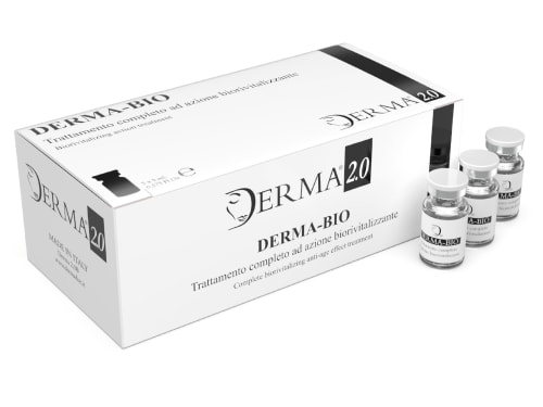
Discover the science behind Derma Bio, a mesotherapy product designed to combat signs of aging and achieve a brighter, smoother, and more youthful appearance.
-

The structural layer beneath the epidermis, containing fibroblasts, collagen, elastin, and the ground substance that together determine skin firmness, elasticity, and resilience.
-

The deeper layer of skin containing collagen, elastin, and blood vessels
-

The power of transformation: safe & effective tattoo removal at Creative Touch Rotherham. A personal approach with stunning results.