
Skin
Skin is the body’s largest organ, covering approximately 1.5 to 2 square metres in adults and accounting for around 15% of total body weight. It performs functions that no topical product or professional treatment can replicate independently: regulating water loss, providing mechanical protection, moderating temperature, conducting immune surveillance, synthesising vitamin D, and sustaining the microbial ecosystem that contributes to immune homeostasis. For clinical and aesthetic purposes, it is best understood as three structurally distinct but functionally interdependent layers – the epidermis, dermis, and subcutaneous tissue – each of which ages through its own mechanisms whilst being continuously influenced by the others.
The visible and tactile signs of skin ageing are not a single process but the accumulated surface expression of multiple concurrent biological changes: barrier lipid depletion in the stratum corneum, filaggrin suppression disrupting the protein scaffold of the outer epidermis, slowing keratinocyte renewal reducing the differentiation quality of new barrier cells, fibroblast senescence and mechanosensitivity loss reducing collagen and elastin synthesis in the dermis, ground substance depletion removing the hydrated volume and fibroblast support environment, and MMP-driven degradation of structural proteins outpacing repair. Understanding which of these is dominant in a given presentation (rather than treating the surface uniformly) is the foundation of the approach taken across Creative Touch’s treatment and skincare guidance.
The Epidermis: Production System for the Barrier
The epidermis provides the barrier that makes terrestrial life physiologically possible, preventing the desiccation that would occur without it and regulating the immune and chemical gradient between the body and its environment. Its functional quality is determined by the keratinocyte differentiation programme that builds it: a continuous production process originating in dividing basal cells and culminating in the formation of the stratum corneum’s lamellar lipid matrix and protein-dense corneocytes.
This programme has two critical dependencies that become clinically relevant in aesthetic practice. First, it requires the ceramide, cholesterol, and free fatty acid precursors that lamellar body secretion depends on; the synthesis of which is suppressed by the same inflammatory cytokines that commonly underlie barrier dysfunction, sensitised skin, and atopic-tendency presentations. Second, it requires the profilaggrin-to-filaggrin processing that produces both the corneocyte protein scaffold and the natural moisturising factor components that maintain intracellular hydration and the acid mantle pH on which ceramide-processing enzymes depend. Both dependencies, and the downstream cascade when either fails, are covered in depth in the Ceramides, Free Fatty Acids, Cholesterol, and Filaggrin entities within this knowledge base.
The renewal rate of this production system slows measurably after the age of 50, and epidermal thickness declines alongside the ceramide and structural protein changes of hormonal transition, meaning the quality of what the epidermis produces and the pace at which it is produced both deteriorate simultaneously at the same life stage. [7]
The Dermis: Structural Foundation and Ageing Target
The dermis provides the structural support, vascular supply, and cellular machinery that skin appearance and mechanical behaviour depend on. Its fibroblasts synthesise collagen Types I and III, the fibrillin scaffold that supports elastic fibre assembly, and the hyaluronic acid and proteoglycans of the ground substance. All of these decline through converging processes: reduced fibroblast activity from cellular senescence and mechanical slack as the matrix degrades; increased MMP-driven degradation from UV exposure, cortisol elevation, and oestrogen decline; and the fibre fragmentation and disorganisation of solar elastosis that progressively replaces functional elastic tissue with non-functional accumulated material. [1]
The dermis also provides the structural context the epidermis requires. The dermo-epidermal junction, the fibroblast-to-keratinocyte TGF-β signalling that drives differentiation quality, the rete ridge architecture that physically anchors the epidermis and enables nutrient transfer, and the ground substance environment that sustains both populations – all are dermal properties that influence epidermal function as directly as the differentiation machinery within the epidermis itself. Treating the epidermis and dermis as independent concerns is a clinical oversimplification that the depth of this cross-layer biology does not support. [2]
How the Layers Interact in Ageing
The most clinically useful insight about skin ageing is that the epidermal and dermal mechanisms do not run in parallel, they amplify each other. Dermal collagen loss creates the mechanical slack that reduces fibroblast synthesis activity, which further accelerates collagen loss. Barrier dysfunction in the stratum corneum allows allergen penetration that drives the type 2 inflammatory cytokine activity that suppresses ceramide synthesis and filaggrin expression, worsening the barrier dysfunction that started the cycle. Reduced HA in the ground substance impairs the fibroblast environment, reducing the TGF-β and fibronectin production that supports the DEJ signalling that sustains keratinocyte differentiation quality above it. These feedback loops are why skin quality can appear to decline rapidly once a threshold is crossed, and why addressing a single layer or mechanism whilst leaving others unaddressed produces slower and less durable results. [3]
The convergence is particularly pronounced during perimenopause, when oestrogen decline reduces ceramide synthesis, filaggrin expression, epidermal thickness, collagen production, tropoelastin synthesis, and hyaluronic acid content simultaneously through the shared PPAR and oestrogen receptor signalling networks. The frequently reported experience of skin changing more rapidly in the 40–55 age range than in the preceding two decades reflects this hormonal convergence arriving on top of cumulative UV and chronological change rather than a sudden biological failure. [6]
Skin Health as a System
The implication for skin health strategy is that effective care – whether through professional treatment, homecare, or internal supplementation – requires a system-level understanding rather than component-level interventions in isolation.
Professional treatments reach different biological targets at different depths: thulium fractional laser and CAP act at the epidermal and papillary dermal level; RF microneedling reaches the reticular dermis; polynucleotides and iPRF deliver signals across both dermal zones; skin boosters restore the ground substance environment that fibroblast mechanosensitivity depends on; exosomes delivered via microneedling channels restore the epidermal differentiation programme at the basal level and DEJ. The most complete treatment protocols combine modalities to address multiple layers and mechanisms simultaneously, rather than selecting a single treatment for a single visible concern.
Homecare addresses the ongoing maintenance environment that determines whether professional treatment outcomes are preserved or progressively lost. Topical ceramide, cholesterol, and free fatty acid formulations support the barrier lipid triad. Vitamin C provides the cofactor for collagen hydroxylation and simultaneously reduces the MMP activity that degrades what fibroblasts produce. Retinoids normalise the keratinocyte differentiation cycle and upregulate TGF-β signalling in fibroblasts. Broad-spectrum UV protection prevents the primary upstream driver of MMP-driven structural degradation. Together, these do not treat skin, they maintain the biological conditions in which the skin can function, repair, and respond to professional treatment at its best capacity. [5]
Internal supplementation addresses the synthesis supply side and the inflammatory environment that suppresses it: phytoceramides through the salvage pathway, omega-3 fatty acids through IL-4/ IL-13 modulation, vitamin C through collagen synthesis cofactor support, and L-histidine as an NMF precursor. None replaces topical or professional approaches, but each contributes to a biological environment in which the skin’s own production capacity is less constrained. [4]
References
Boraldi F, Lofaro FD, Bonacorsi S, et al. (2024). The Role of Fibroblasts in Skin Homeostasis and Repair. Biomedicines, 12(7) . doi.org/10.3390/biomedicines12071586
Chang H, Shen Q, Tan Y, et al. (2025). Red light promotes dermis-epidermis remodeling via TGFβ and AKT-mediated collagen dynamics in naturally aging mice. Zool Res, 46(5), 967-982 . doi.org/10.24272/j.issn.2095-8137.2024.405
Elias PM (2015). Stratum corneum acidification: how and why? Exp Dermatol, 24(3), 179-80 . doi.org/10.1111/exd.12596
Leo TK, Tan ESS, Amini F, et al. (2022). Effect of Rice (Oryza sativa L.) Ceramides Supplementation on Improving Skin Barrier Functions and Depigmentation: An Open-Label Prospective Study. Nutrients, 14(13) . doi.org/10.3390/nu14132737
Shin JW, Kwon SH, Choi JY, et al. (2019). Molecular Mechanisms of Dermal Aging and Antiaging Approaches. Int J Mol Sci, 20(9) . doi.org/10.3390/ijms20092126
Viscomi B, Muniz M, Sattler S (2025). Managing Menopausal Skin Changes: A Narrative Review of Skin Quality Changes, Their Aesthetic Impact, and the Actual Role of Hormone Replacement Therapy in Improvement. J Cosmet Dermatol, 24 Suppl 4(Suppl 4), e70393 . doi.org/10.1111/jocd.70393
Wang Z, Man MQ, Li T, et al. (2020). Aging-associated alterations in epidermal function and their clinical significance. Aging (Albany NY), 12(6), 5551-5565 . doi.org/10.18632/aging.102946
Anatomical Relationships
Structural Connections
- Produces Vitamin D Evidence: Vitamin D synthesis in the epidermis is a primary skin function.
- Connected to Fibroblast Evidence: Fibroblasts are the primary structural cell population of the skin dermis; entity text describes fibroblast senescence as central to skin ageing. Fibroblast locatedIn Skin already exists.
- Has sub-structure Dermis Evidence: kin comprises three layers: epidermis, dermis, subcutaneous tissue.
- Has sub-structure Epidermis Evidence: Skin comprises three layers: epidermis, dermis, subcutaneous tissue.
- Has sub-structure Hair follicle Evidence: Hair follicles are skin appendages embedded within dermal layers; entity text discusses follicles as integral skin components.
- Has sub-structure Papillary dermis Evidence: Papillary dermis is a named sub-zone of the dermis within skin; entity text references it as treatment target for CAP and thulium laser.
- Has sub-structure Reticular dermis Evidence: Reticular dermis is a named sub-zone of the dermis within skin; entity text references it as target for RF microneedling.
- Has sub-structure Sebaceous gland Evidence: Sebaceous glands are skin appendages within the dermis; entity text references sebum production in skin context.
- Has sub-structure Stratum corneum Evidence: Epidermis culminates in stratum corneum – terminal barrier layer within skin.
- Has sub-structure Subcutaneous tissue Evidence: Skin third layer is subcutaneous tissue; provides mechanical protection and thermal insulation.
- Part of system Integumentary system Evidence: Skin is the primary organ of the integumentary system; universally classified as such in anatomy.
- Dermatitis Evidence: Dermatitis involves skin inflammation affecting all layers; entity text discusses inflammatory cytokine activity in barrier dysfunction context.
- Hyperpigmentation Evidence: Hyperpigmentation is a pigmentary disorder of skin; Kovacs et al. (2016) Ann Dermatol 28(3):279 describe epidermal/dermal cross-talk in pigmentary disorders. doi:10.5021/ad.2016.28.3.279
- Perimenopausal skin changes Evidence: Perimenopause drives simultaneous epidermal, dermal, and barrier deterioration via shared PPAR/ER networks.
- Psoriasis Evidence: Psoriasis is a chronic inflammatory skin disease; entity text references cytokine disruption and MMP-driven degradation as shared skin mechanisms. Psoriasis -> associatedAnatomy -> Skin exists.
- Rosacea Evidence: Rosacea is a chronic inflammatory skin condition; Zheng et al. (2022) Life 12(5):725 use rosacea as disease model for cutaneous neuro-endocrine-immune dysfunction. doi:10.3390/life12050725
- Skin ageing Evidence: Skin ageing involves concurrent epidermis, dermis, and barrier mechanism decline
- Skin barrier dysfunction Evidence: Barrier dysfunction allows allergen penetration; IL-4/IL-13 suppress ceramide and filaggrin, worsening dysfunction.
- Cold Atmospheric Plasma therapy Evidence: Entity text explicitly states CAP acts at epidermal and papillary dermal level as a professional skin treatment. Tan et al. (2022) Front Oncol 12:918484 review CAP-dominated skin therapy. doi:10.3389/fonc.2022.918484
- Injectable Platelet-Rich Fibrin Evidence: iPRF delivers growth factor signals across both dermal zones; entity text explicitly names iPRF under professional treatments reaching dermis and papillary zones.
- LED therapy Evidence: Entity text references LED therapy reaching epidermal targets. Naharro-Rodriguez et al. (2024) Int J Mol Sci 25(8):4483 confirm LED photobiomodulation for skin rejuvenation. doi:10.3390/ijms25084483
- Mesotherapy Evidence: Mesotherapy delivers vitamins/minerals/hydrating ingredients directly into skin layers.
- Microneedling Evidence: Microneedling activates keratinocyte differentiation cascade; addresses corneocyte quality from production end.
- Platelet-rich plasma therapy Evidence: PRP is established for facial skin rejuvenation; entity text discusses growth factor delivery to both dermal zones. Masiello et al. (2024) Blood Transfus 22(4):265. doi:10.2450/BloodTransfus.730
- Polynucleotides Evidence: Polynucleotides deliver signals across papillary and reticular dermis; calm inflammation.
- Radiofrequency Microneedling Evidence: RF Microneedling targets reticular dermis for deep collagen stimulation.
- Skin Boosters Evidence: Skin boosters restore HA ground substance; support fibroblast mechanosensitivity in dermis.
- Thulium Fractional Laser Evidence: Thulium laser acts at epidermal and papillary dermal levels; improves keratinocyte differentiation.
Referenced in Conditions & Treatments
- this Contains Fibroblast PMID: 34297930 Evidence: Fibroblasts are the primary synthetic cells of the dermis
- this Affected by Cryolipolysis Evidence: Erythema, bruising, numbness affect skin post-treatment; protective gel membrane applied to skin surface during treatment. Entity text.
- this Affected by Deoxycholic acid Evidence: Skin necrosis documented as technique-dependent adverse event; bruising and swelling affect skin surface. Entity text; PMC9142263.
- this Affected by Paradoxical adipose hyperplasia Evidence: Visible palpable mass enlargement alters skin contour and surface appearance. Entity text; Nikolis 2020 PMC8279305.
- this Required by Microneedling Evidence: Microneedling requires intact skin as operational substrate; fine needles create micro-injuries specifically in epidermis and dermis to initiate wound healing cascade. Entity text.
- this Part of system Hypothalamic–pituitary–adrenal axis Evidence: Peripheral cutaneous HPA axis: skin expresses all HPA functional elements (CRH, POMC, ACTH, steroidogenic enzymes). PMC1839836
- this Part of system Integumentary system PMID: 29262154
- this Associated condition Dermatitis Evidence: Dermatitis is defined as inflammatory skin conditions; all three subtypes (atopic, contact, seborrhoeic) produce cutaneous manifestations (entity executive_summary).
- this Associated condition Paradoxical adipose hyperplasia Evidence: PAH presents as visible, palpable enlargement at skin level; skin contour change is the clinical diagnostic feature. Entity text.
- this Associated condition Perimenopausal skin changes Evidence: Perimenopausal skin changes manifest across all layers of skin simultaneously; skin is primary anatomical site of expression. Entity text; PMC12374573.
- this Associated condition Psoriasis Evidence: Psoriasis is defined as a chronic immune-mediated inflammatory skin condition; all plaque formation occurs on skin (PMC5796008).
- this Associated condition Topical steroid withdrawal Evidence: TSW is defined as a dermatosis with cardinal features of burning, erythema, and barrier dysfunction; all manifestations are cutaneous (PMC8481181; PMC11994697).
Learn More
This topic is discussed in 42 articles:
-

Getting adequate rest is the best investment you can make into your beauty. We take a look at some of your body’s natural skin rejuvenation processes that occur during sleep.
-

The body’s largest organ, comprising epidermis, dermis, and subcutaneous tissue. Barrier function, structural integrity, and cellular renewal all decline through distinct but interacting mechanisms.
-
The body’s largest organ, comprising epidermis, dermis, and subcutaneous tissue. Barrier function, structural integrity, and cellular renewal all decline through distinct but interacting mechanisms.
-

Discover how to restore and maintain your skin’s natural defences. From the causes of skin barrier damage to advanced treatments. Achieve healthy, radiant skin with our expert advice.
-

The body’s largest organ, comprising epidermis, dermis, and subcutaneous tissue. Barrier function, structural integrity, and cellular renewal all decline through distinct but interacting mechanisms.
-
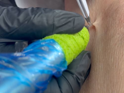
Not liking those pesky skin tags? Learn about causes, blemish removal options at Creative Touch, and prevention tips for smoother, bump-free skin.
-

The body’s largest organ, comprising epidermis, dermis, and subcutaneous tissue. Barrier function, structural integrity, and cellular renewal all decline through distinct but interacting mechanisms.
-
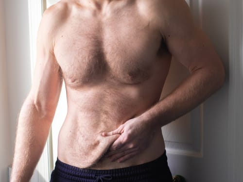
Don’t let loose skin overshadow your weight loss success. Learn why skin sags after significant weight loss and explore our advanced skin tightening and volume restoration treatments.
-
The science behind polynucleotides – a revolutionary treatment that offers a comprehensive approach to skin rejuvenation. How does it compare?
-
Largest organ of the body, highly responsive to hormonal changes during perimenopause